Medical Imaging of Children/Adolescents and Risk of Cancer (2025)

R Smith-Bindman et al. NEJM 2025; 393: 1269-1278. Medical Imaging and Pediatric and
Adolescent Hematologic Cancer Risk

Methods: This was a retrospective cohort of 3,724,623 children born between 1996 and
2016 in six U.S. health care systems and Ontario, Canada, until the earliest of cancer
or benign-tumor diagnosis, death, end of health care coverage, an age of 21 years, or December 31, 2017.

Key findings:

  • During 35,715,325 person-years of follow-up (mean, 10.1 years per person), 2961 hematologic cancers were diagnosed, primarily lymphoid cancers (2349 [79.3%]), myeloid cancers or acute leukemia (460 [15.5%]), and histiocytic- or dendritic-cell cancers
    (129 [4.4%]).
  • The excess cumulative incidence of hematologic cancers by 21 years of age among children exposed to at least 30 mGy (mean, 57 mGy) was 25.6 per 10,000
  • The authors estimated that 10.1% of hematologic cancers may have been attributable to radiation exposure from medical imaging, with higher risks from the higher-dose medical-imaging tests such as CT
Cumulative Incidence of Hematologic Cancer According to Attained Age and
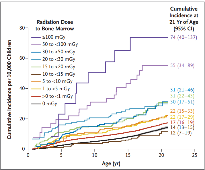
Radiation Dose to Bone Marrow among Children without Down’s Syndrome

Discussion Points:

  • “A 15-to-30-mGy exposure equivalent to one to two CT scans of the head was associated with an increased risk by a factor of 1.8”
  • “Although CT and other radiation-based imaging techniques may be lifesaving, our
    findings underscore the importance of carefully considering and minimizing radiation exposure during pediatric imaging to protect children’s long-term health”
  • “Research on Japanese atomic-bombing survivors showed that leukemia rates peaked 6 to 8 years after exposure, with excess risk lasting for more than five decades, particularly for acute myeloid leukemia”
  • This study tried to avoid concerns about reverse causation — in which imaging is performed because of existing cancer symptoms –by lagged exposures by 6 and 24 months
  • “The increasing use of low-value imaging in children and excessive radiation doses in CT are well documented…In many cases, reducing the imaging dose or substituting magnetic resonance imaging or ultrasonography may be more feasible than avoiding imaging altogether”

While the risks in aggregate appear quite substantial, the editorial (L Morton. NEJM 2025; 393; 1337-1339.Studying Cancer Risks Associated with Diagnostic Procedures –Interpret Wisely) makes the point that the risks for the individual are very small. “Fewer than 1% of youths in this study accumulated doses of 30 mGy or more from medical imaging and even at this exposure level, the excess cumulative incidence of hematologic cancers was low (25.6 per 10,000)…we need to ensure that all involved in medical imaging…wisely interpret the results…to understand the balance of the very small risks and the notable benefits of necessary imaging examinations to provide optimal patient care.”

My take: This study is a reminder to carefully evaluate the benefits, risks and alternatives when using ionizing radiation studies.

Related blog posts:

CT Imaging and Projected Cancer Risks: 2025 Analysis

Smith-Bindman R, Chu PW, Azman Firdaus H, et al.  JAMA Intern Med. Published online April 14, 2025. doi:10.1001/jamainternmed.2025.0505. Open Access! Projected Lifetime Cancer Risks From Current Computed Tomography Imaging

Methods: Lifetime radiation-induced cancer incidence and 90% uncertainty limits (UL) were estimated by age, sex, and CT category using National Cancer Institute software based on the National Research Council’s Biological Effects of Ionizing Radiation VII (BEIR VII) models and projected to the US population using scaled examination counts.

Key findings:

  • Ninety-three million CT examinations were performed in 61 510 000 patients in the United States in 2023, including an estimated 3,069,000 CTs (3.3%) in 2,570,000 children (4.2%) and 89,931,000 CTs (96.7%) in 58,940,000 adults (95.8%) 
  • In this risk model, the 93 million CT examinations performed in 62 million patients in 2023 were projected to result in approximately 103,000 future cancers
  • Estimated radiation-induced cancer risks were higher in children and adolescents, yet higher CT utilization in adults accounted for most (93,000) radiation-induced cancers
  • “If current practices persist, CT-associated cancer could eventually account for 5% of all new cancer diagnoses annually”

Discussion: “The projected number of radiation-induced cancers in this analysis is 3 to 4 times higher than the earlier assessment of CT exposure for several reasons”

  • CT use is 30% higher today than in 2007
  • Dose modeling in this study accounted for multiphase scanning
  • Substantially higher organ doses in this study were reconstructed using newer dosimetry methods
  • More granular CT categories reflecting imaging indications that have important dose differences
  • “Many of the model assumptions were conservative” and could underestimate the risk

My take (borrowed from authors): “Even very small cancer risks will lead to a significant number of future cancers given the tremendous volume of CT use in the United States…CT could be responsible for approximately 5% of cancers diagnosed each year. This would place CT on par with other significant risk factors, such as alcohol consumption (5.4%) and excess body weight (7.6%)”

Related blog posts:

Kiawah Beach, SC

ER Evaluations of Visits Related to Inflammatory Bowel Disease

A short communication (C Pant et al. JPGN 2015; 61: 282-84, ed 267-8) provides some insight into what is happening in the ER when patient with inflammatory bowel disease are evaluated.

The authors analyzed a national database, the Healthcare Cost and Utilizationa Porject Nationwide Emergency Department Sample (NEDS), from 2006-2010.

Key findings:

  • ED visits for children 5-19 years of age increased from 14,527 (2006) –>18,193 (2010)
  • Frequency of computerized tomography (CT) imaging increased 80.43% (P<0.01) for Crohn’s disease and 59.26% (P<0.01) for ulcerative colitis
  • Overall rate of hospital admissions decreased by 14.32% (P<0.01)

The associated editorial by Jennifer Dotson and Michael Kappelman explain the limitations of relying on data that are derived mainly for billing rather than research and comment on the “alarming increase” in CT  usage due to the risk of radiation exposure and the potential alternatives (eg. ultrasound, MRE).  With regard to limitations, they likely included inaccurate and incomplete entries.  In addition, the database included 950 hospitals, but may have under-sampled large freestanding children’s hospitals.  This could skew the data.  Finally, the data uses “visit-level” data rather than “patient-level” data; thus, one ‘cannot distinguish between a single patient who is seen on three separate visits or three unique patients.’

My take:  There has been increasing use of ER visits and CT scanning for pediatric patients with IBD.  One of my colleagues in town (Cary Sauer) has a humorous slide of a CT scanner as the entrance door to the ER.  The message from this study indicates that we should be working on changing practice by working with our ER colleagues along with radiologists to minimize CT scans in favor of more gently imaging.

Related blog posts:

Atlanta Botanical Gardens, Bruce Munro exhibit

Atlanta Botanical Gardens, Bruce Munro exhibit

Moving to MRE

A recent review (JPGN 2014; 59: 429-39) regarding imaging for inflammatory bowel disease reiterates the accepted view that magnetic resonance enterography (MRE) is typically the most useful imaging test for children with inflammatory bowel disease; in Table 5, MRE is listed for each indication, though CT scan is recommended “if emergent or after hours.”  The review reviews prior pediatric publications, radiation risks (with non-MRE studies), and alternative imaging.  The discussion on costs is minimized, though the authors note that MRE is the most expensive and can be compromised by motion artifact. As a practical matter, I think giving a typical charge (or range) for each of the imaging techniques would be helpful.  Also, another important issue is assuring that radiologists have the technical expertise to obtain quality imaging.

Another study (Clin Gastroenterol Hepatol 2014; 12: 1702-07) retrospectively looked at 1095 emergency room visits by 613 individuals (average age ~40 years) to determine if they could develop a model to limit unnecessary CT scans.  Of the 1095 CT scans, 24.8% were normal; 10.9% had either perforation or non-perianal abscess.  In their discussion, they note that the equation “no scan for ESR (mm/h) + 5*CRP (mg/dL) ≤10” would avoid 18.5% of CT scans.  Implementation of a more complex model could eliminate up to 43% of the CT scans.  The algorithm (Figure 2) suggested by the authors:

  • Assess for obstruction.  If suggestive symptoms, obtain abdominal X-rays.  If concerns for obstruction remain, consider CT scan.
  • If not concerned about obstruction, is there a high likelihood of perforation or abscess? If yes proceed with CT scan.  If not, consider anti-inflammatory therapy if CD symptoms present (without imaging).

Here’s the link to the abstract –supplementary materials can be obtained by those who log in.   http://dx.doi.org/10.1016/j.cgh.2014.02.036

Bottomline: Cross-sectional imaging is particularly helpful at determining whether complications are developing in patients with inflammatory bowel disease.  Increasing use of MRE will reduce radiation risks.

With regard to costs, a recent NPR story discussed “How Much Is That MRI, Really? Massachusetts Shines A Light.” While this story discussed costs related to a Massachusetts law which mandates that insurers reveal the costs of various tests, it did not relate any information regarding quality.  The study implied that an MRI at one institution would be equivalent to an MRI at another.  This is not the case.

Related blog posts:

Risk from CT scans -Best Data to Date

Thanks to Mike Hart for forwarding the following reference:

BMJ 2013; 346: f2360 doi: 10.1136/bmj.f2360

This study examines the risk of cancer among a cohort of nearly 11 million Australian children and adolescents since 1985.  Among this cohort, 680,000 (6.2%) pediatric patients were identified who had been exposed to a CT scan. This study was accomplished by analyzing CT scans funded by the Australian Medicare system which provides health services for all Australians.

One of the remarkable aspects of this study was the efforts the authors took to exclude reverse causation.  First of all, the data was analyzed with an exclusion period of a year “because of the possibility that the scan was part of the cancer diagnostic procedure…but we repeated the main analyses with lag periods of five and 10 years to explore the possibility of reverse causation.”  In addition, the authors analyzed all non-brain cancers in patients who had had cranial CT scans.  Despite all of the parameters, an increased risk of cancer was maintained among those who had prior CT and the risk was heightened by obtaining studies at younger ages and by having increased number of CT scans.

Key findings:

  • Almost 60% of CT scans were of the brain. Only 5% of CT scans were of abdomen or pelvis.
  • CT scan incidence increased over time.  Between 1985-89, 95,249 (14%) CT scans were ordered.  Whereas between 2000-2005, 266,971 (39%) were ordered.
  • The average CT dose was about 4.5 mSv per scan.
  • The increased relative risk (IRR) for brain cancers after a scan to a site other than the brain was 1.51 (confidence interval 1.19-1.91).
  • Each seivert (Sv) of effective dose was associated with 0.125 cancers; thus, by 2007, with average followup of 9.5 years, one cancer resulted from every 1800 CT scans.  This number is likely to climb with more time.
  • Among brain CT scans, the numbers are trickier due to the possibility of slow-growing tumors (which could trigger symptoms for imaging and still be difficult to detect).  However, up to one excess brain cancer would occur for every 4000 brain scans.
  • All solid cancers IRR 1.25, All lymphoid/hematologic cancers IRR 1.19, Brain cancers after CT IRR 2.44, Brain cancer after other scans 1.51.

There are several limitations to the study including the difficulty of knowing specific doses of radiation at various CT scanners, the possibility of CT scans funded outside the Australian Medicare system, or obtaining screening scans due to precancerous genetic conditions. Nevertheless, the magnitude of the cohort in this study along with its general agreement with a number of other studies provide ample evidence that these risks are real.

Take-home point: While CT scans have the potential for great benefits, they increase the risk of developing cancer; in many cases, an MRI or an ultrasound can provide similar information without this risk.  In this study, for lag (exclusion) period of one, five, and 10 years, the incidence rate for all cancers combined increased by 24%, 21%, and 18% respectively in the CT exposed group.  Eventual lifetime risk is likely to climb with longer followup.

Related blog posts:

“Beyond the bombs: cancer risks of low-dose medical radiation”

The catchy title comes from a Lancet editorial (Lancet 2012; 380: 455-57); the related article (Lancet 2012; 380: 499-505) details the radiation risk posed by CT scans.

While concerns about imaging radiation exposure have become commonplace, the evidence for the risk has been more in the theoretical realm rather than proven.  That is, the risk projection models were based on studies of survivors of the atomic bombs in Japan.

The retrospective study in Lancet examines 178,604 children who underwent CT between 1985-2002.  Typical followup was 10 years (maximum followup of 23 years).  None of these children had cancer at the time of CT.  The study determined the number of leukemias that developed more than 2 years following CT and brain tumors which occurred more than 5 years after CT.  This lag time was done to avoid any confounding of cancer that may have been present and not detected at time of CT.

Key results:

  • 74 patients developed leukemia and 135 developed brain tumors.  There was a dose-related risk: 0.036/mGy for leukemia and 0.023/mGy for brain tumors.  Thus, the relative risk of leukemia in patients who had at least 30 mGy was 3.18; whereas, brain cancer risk for a cumulative dose of > 50 mGy was 2.82. [1 mGy=1 mSv]
  • If typical doses of CT administered, 2-3 head CTs could triple the risk of a brain tumor and 5-10 head CTs could triple the risk of leukemia.
  • The absolute risk remains low.  In patients less than 10 years, one excess case of leukemia and one brain tumor would be expected for 10,000 head CT scans.

Goal with CT scans:

  1. ALARA: as low as reasonably achievable –for every study.  Newer protocols allow lower radiation doses while preserving good image quality.
  2. Think carefully about each CT.  It is estimated that 20-50% of CTs could be replaced with another type of imaging or not done at all.

For the skeptics about the risk of CT scans, the editorialist concludes that this study confirms “that CT scans almost certainly produce a small cancer risk…we must redouble our efforts to justify and optimize every CT scan.”

Related blog entries:

How much radiation from your CT scanner? | gutsandgrowth

More imaging needed? | gutsandgrowth

Magnetic resonance enterography for Crohn’s disease 

Additional references:

  • -AJR 2001; 176: 289-96. Estimated risks of radiation-induced fatal cancer from pediatric CT
  • -Br J Radiol 2012; 85: 523-28.  Justification of CTs -some not needed
  • -AJR 2010; 194: 868-73.  Lower CT radiation doses in pediatric patients.  ‘Image gently’
  • -Arch Intern Med 2009; 169: 2078-86.

Magnetic resonance enterography for Crohn’s disease

Magnetic resonance enterography (MRE) has increasingly been recognized as an effective way to characterize small bowel disease in Crohn’s disease (Inflamm Bowel Dis 2012; 18: 520-28).

In this retrospective pediatric study, 95 patients with Crohn’s disease underwent MRE from 2006-2009.  As expected, terminal ileal disease was the most common site of involvement with 54%; this was followed by ileum with 19%, and jejunum with 17%.  Other findings included solitary jejunal inflammation (3.7%), small bowel stenosis (1.9%) fistula (1%), and abscess (1%).  Specific evidence of inflammation included contrast enhancement, bowel wall thickening or derangement of bowel wall shape.  The images in the article are excellent.

The main advantage of MRE over CT enterography (CTE) is the lack of ionizing radiation.  In addition, MRE can better detect soft tissue contrast suggestive of bowel wall edema.  This helps distinguish acute from chronic inflammation.  Obtaining an MRE is technically more challenging and more costly.  The youngest patient in the study was seven; though the authors note that the youngest patient they have performed MRE was six.

Additional references:

  • More imaging needed?
  • -JPGN 2010; 51: 603.  MRE for suspected IBD.  Useful in pediatric Crohn’s disease.
  • -IBD 2009; 15: 1635. U/S compared favorably with endoscopy in correlating postoperative recurrence with Crohn’s.
  • -Clin Gastro Hepatol 2006; 3: 1221. MRI as accurate in evaluating ileocolonic disease with flareups as endoscopy.
  • -IBD 2004; 10: 452-61. U/S was very helpful in identifying disease and complications.

More imaging needed?

With new tools at our disposal in diagnosing inflammatory bowel disease, we need to decide how and when to use them.  Potential new modalities include stool inflammatory markers, video capsule endoscopy, CT enterography (CTE), and MR enterography (MRE).  Several studies have shown that the information that these studies yield may change management. The latest of these studies (Inflamm Bowel Dis 2012; 18: 219-25) looked at how the knowledge of CTE effected management and physician confidence with Crohn’s disease.

The authors prospectively assessed 273 patients with established or suspected Crohn’s disease.  In their analysis, 70 patients (48%) of established cases had altered management because of CTE and 69 (54%) of suspected cases.  These changes were considered to be  independent of clinical, serological or histologic findings.  Changes included medication modification in 45 (16.2%), excluding Crohn’s disease in 46 (16.8%), surgery referral in 10 (3.7%), alternate diagnosis established in 9 (3.2%), & canceling surgery in 7 (2.6%).  The authors considered excluding active small disease as an important management plan change; this occurred in 18 patients (6.6%).

The authors state that their current practice is to use MRE for serial imaging rather than CTE, to minimize risks from radiation; though CTE is often the initial imaging.

My take on this article is that information from imaging often increases the certainty about the diagnosis and gives a more complete picture of the severity.  It is likely that more information leads to more aggressive therapy.  At the same time, in pediatric gastroenterology, the trend towards using more effective therapy earlier in the course of the disease has developed even in the absence of extensive imaging (see previous: Only one chance to make first impression).  Whether more imaging in pediatric patients would be worthwhile is not known.

Additional references:

  • -JPGN 2008; 47: 31.  Capsule endoscopy may reclassify pediatric IBD
  • -NEJM 2010; 363: 1, 4. Safety of CT. Can have overdose of radiation and even standard doses could cause complications. Also, a big issue is downstream unnecessary testing due to incidental findings.
  • -Clin Gastro 2008; 6:283. Use of CT enterography.
  • -JPGN 2010; 51: 603.  MRE for suspected IBD.  Useful in Crohn’s disease.
  • -IBD 2004;10: 278-285.  WCE for Crohn’s (review)  Capsule can help differentiate UC from Crohn’s.