
T Ochaiai et al. Clin Gastroenterol Hepatol 2026; 24: 2308-2310. Open Access! Artificial Intelligence-Based Detection of Achalasia on Plain Chest Radiography
Methods: This retrospective study collected posteroanterior plain chest radiographs of
patients with and without achalasia. The training and validation datasets comprised 447
chest radiographs taken between January 2017 and March 2023.
Key findings:
- In the validation dataset, the area under the curve for identifying achalasia was 0.971, and using Youden’s index, the sensitivity, specificity, accuracy, and positive and negative predictive values were 0.950, 0.917, 0.932, 0.905, and 0.957, respectively
- In the temporal test dataset, the area under the curve for detecting achalasia was 0.964, and using Youden’s index, the sensitivity, specificity, accuracy, and positive and negative predictive values were 0.941, 0.891 , 0.901, 0.696 , and 0.983, respectively.
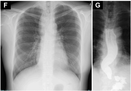
Discussion:
- It may help detect patients with early achalasia with mild symptoms who are unlikely to undergo EGD. However, the rarity of the condition may result in many false positives.
My take: In this cohort, AI was developed and validated to detect achalasia on chest radiographs. This is yet another example of how AI can yield additional information from routine testing. Previously, AI has been shown to potentially identify diabetes from routine CXR (Emory News 2023: AI model enables earlier detection of diabetes through chest x-rays).
Related blog posts:
- Dr. Jennifer Lee: AI for Peds GI
- AI for GI
- Artificial Intelligence in the Endoscopy Suite
- Rising Scientific Fraud: Threats to Research Integrity Plus One
- The Future of Medicine: AI’s Role vs Human Judgment
- ChatGPT4 Outperforms GI Docs for Postcolonoscopy Surveillance Advice
- Will Future Pathology Reports Include Likely Therapeutic Recommendations?
- Medical Diagnostic Errors
- Answering Patient Questions: AI Does Better Than Doctors


Pingback: AI Diagnosis of Achalasia on Plain Chest X-ray – Reviewer4you