D Rossi et al. J Pediatr Gastroenterol Nutr. 2026;83:185–207. Open Access! Updated European Reference Network for rare Inherited and Congenital Digestive and Gastrointestinal Anomalies guidelines for the management of rectosigmoid Hirschsprung’s disease 2025
These guidelines cover recommendations for diagnosis, pre- and postoperative care, poor functional outcomes, long-term follow-up, and Hirschsprung’s-associated enterocolitis.
Some specific recommendations:
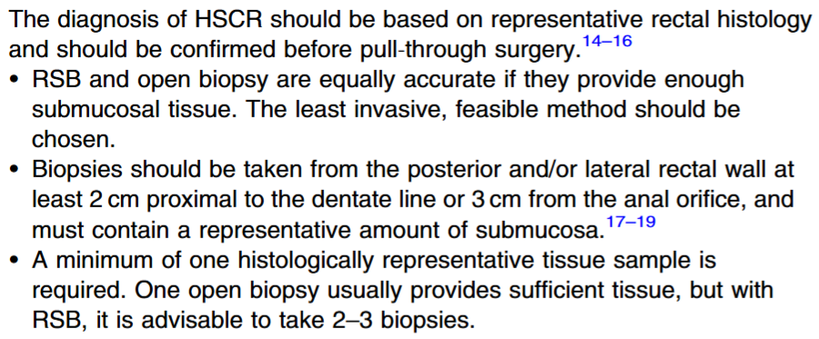
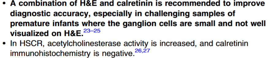
Diagnosis:
Preoperative Care:

- Routine screening for all patients with rectosigmoid Hirschsprung’s disease (HSCR))with ultrasound for congenital anomalies of kidney and urinary tract (CAKUT) and systematic assessment of nutritional status.
Operative Care:
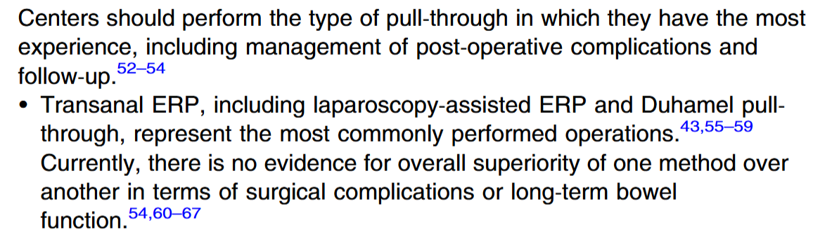
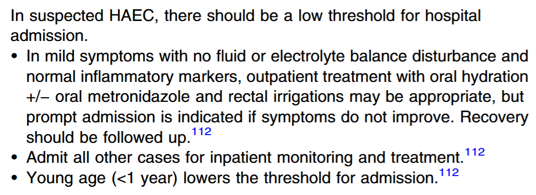
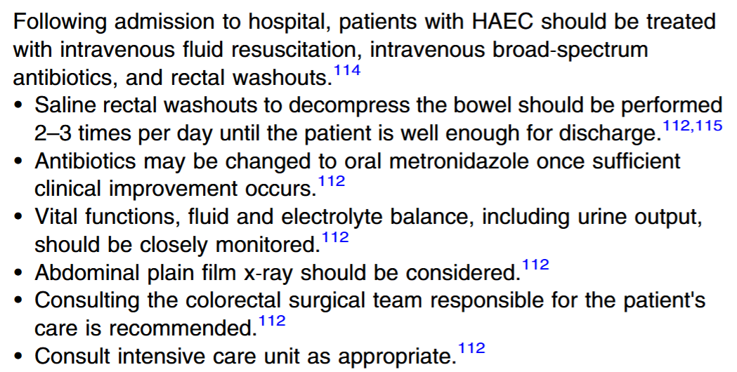
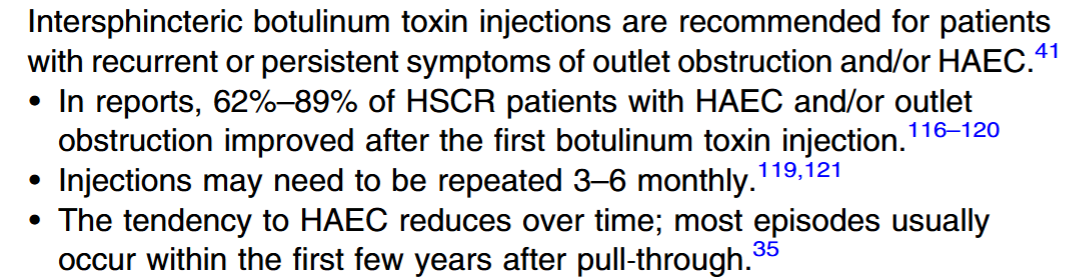
Hirschsprung’s-Associated Enterocolitis:
- Table 9 provides extensive advice for bowel management strategies/evaluation in children with fecal incontinence.
- Table 11 discussed genetic testing, noting that RET gene should be considered. Genetic counseling is recommended in those patients with a family history of Hirschsprung’s disease.
My take: This article provides good advice for optimizing care for patients with Hirschsprung’s disease.
Related blog posts:
- Pelvic AnoRectal Care Program (PARC)
- What is the Risk of Inflammatory Bowel Disease in Patients with Hirschsprung’s Disease
- Reducing Diagnostic Uncertainty in Hirschsprung’s Disease
- Rectal Suction Biopsies Less Accurate in Infants <40 days
- Image Only: Total Colonic Aganglionosis
- Position Paper: Pediatric Refractory Constipation Management
- More Cognitive Problems, Worse Outcomes with Hirschsprung’s Disease
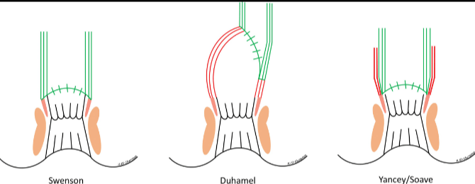
Diagrams of 3 common pull-through operations for Hirschsprung disease.
From left to right: full-thickness rectosigmoid dissection (Swenson), a recto-rectal pouch procedure (Duhamel), and an endorectal dissection (Soave). JPGN 2023; 76(4):533-546.