C Baker et al. J Pediatr Gastroenterol Nutr. 2026;82:1321–1324. Cyproheptadine: The secret sauce
Mechanism of action:
- A first-generation antihistamine with anti-cholinergic, anti-serotonergic, and local anesthesia properties; its precise mechanism of action is poorly understood.
Formulation/pharmocokinetics:
- Enteral tablet and syrup formulations appear to be well absorbed with peak serum levels 6–9 h after dosing in adults.2 However, anecdotally in pediatrics, the side effect of somnolence is often felt much more quickly.
Uses (off-label):
- Promote weight gain: “There is ample literature to support success in varying populations, including mild to moderately undernourished toddler-aged children and children with cystic fibrosis.4–6“
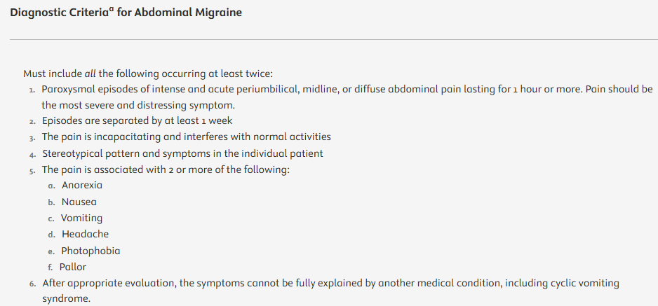
- Cyclic vomiting syndrome
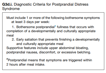
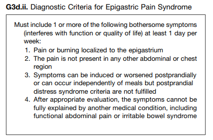
- Functional abdominal pain and dyspeptic symptoms
- Post-operative Retching After Fundoplication
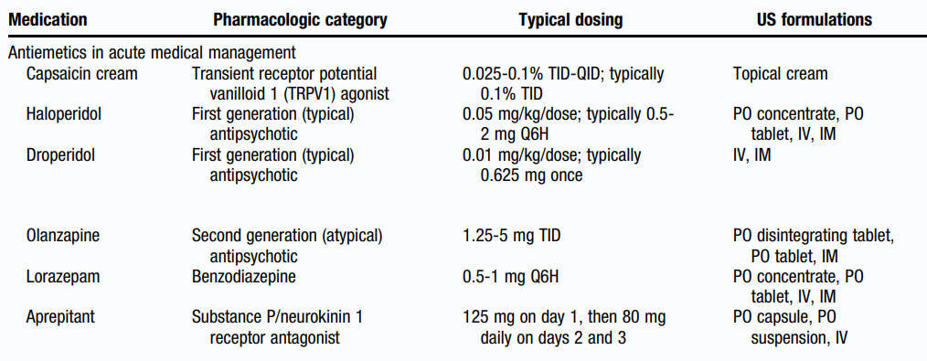
Dosing (by indication):
- “For cyclic vomiting syndrome prophylaxis, the recommended dose is 0.25–0.5 mg/kg/day divided every 8–24 h with a maximum dose of 12 mg a day.”
- “For appetite stimulation, the recommended dose is 0.25 mg/kg/day divided twice daily, with a maximum dose ranging from 12 to 32 mg depending on patient age.”
- “An effective dosing range for use in disorders of the gut–brain interaction (DGBIs) in pediatric patients (median age 9 years) of 0.13–0.2 mg/kg/day has been reported when used on average for 9 months, with a mean initial dose of 4.85 mg/day and final dose of 5.34 mg/day.1“
- “A large, systematic review from France identified the median dose for all pediatric clinical indications to be 0.25 mg/kg/day…Many clinicians use once-daily dosing to improve adherence.”
Contraindications, Drug Interactions, and Complications:
- “Cyproheptadine is contraindicated in newborns and premature infants due to the potential for CNS depression and a lack of evidence in its safety and efficacy.”
- “Cyproheptadine has potential for multiple drug-drug interactions, and dose adjustments may be required with concomitant therapies.”
- “Recent publications suggest that up to 30% of patients will experience mild and self-limited side effects while taking cyproheptadine.8 The most common side effects are somnolence, increased appetite, weight gain, and behavior changes.1, 8 Caution should be exercised in the use of cyproheptadine in children who are already considerably overweight…A rare side effect of adrenal insufficiency…has been documented in pediatric patients.”
My take: Because cyproheptadine is so useful for pediatric GI disorders, some of my colleagues have referred to it as “Vitamin P” (periactin). This review provides a lot of helpful data and guidance.
Related blog posts:
- Rome V Pediatric Upper Gastrointestinal Disorders of Gut-Brain Interaction (Part 2)
- 2025 Pediatric Cyclic Vomiting Syndrome Guidelines
- Ten-Year Trends in Pediatric Pharmacology for Gastroesophageal Reflux and Pediatric Feeding Disorders
- Dr. Neha Santucci: Management of DGBIs in the Post-Pandemic Era (Part 2)
- Dr. Praveen Goday: Tips on Managing Feeding Problems (Part 2)
- Cyproheptadine for dyspepsia

Disclaimer: This blog, gutsandgrowth, assumes no responsibility for any use or operation of any method, product, instruction, concept or idea contained in the material herein or for any injury or damage to persons or property (whether products liability, negligence or otherwise) resulting from such use or operation. These blog posts are for educational purposes only. Specific dosing of medications (along with potential adverse effects) should be confirmed by prescribing physician. Because of rapid advances in the medical sciences, the gutsandgrowth blog cautions that independent verification should be made of diagnosis and drug dosages. The reader is solely responsible for the conduct of any suggested test or procedure. This content is not a substitute for medical advice, diagnosis or treatment provided by a qualified healthcare provider. Always seek the advice of your physician or other qualified health provider with any questions you may have regarding a condition