
K Hawa et al. JPGN Reports 2022; doi: 10.1097/PG9.0000000000000255. Open Access! Postcolectomy Enteritis in a Pediatric Patient With Ulcerative Colitis
In this case report, the authors describe a 16 yo male with ulcerative colitis who on postoperative day 4 after colectomy developed an early onset of non-infectious enteritis. Treatment included corticosteroids “without significant improvement over 2 weeks. As his corticosteroid dose was tapered by 5 mg/day each week, ostomy output decreased, and abdominal pain and distension improved.” He continued to improve without further interventions. “6 weeks postoperatively, repeat upper endoscopy and ileoscopy demonstrated resolution of his duodenitis and ileitis grossly.”
“This is the first published case of a pediatric patient with PCE [postcolectomy enteritis], an entity previously only described in adults. PCE may be difficult to diagnose; in patients initially diagnosed with UC who develop small bowel inflammation following colectomy, the concern is often misdiagnosed Crohn’s disease.” The authors note that the “presentation is differentiated from Crohn’s disease based on timing [days to months after surgery], histology and diffuse pattern of mucosal involvement (3).”
My take: Rare cases PCE (a self-limited enteritis) occur and can be difficult to distinguish from Crohn’s disease. With PCE, if findings improve, this would suggest PCE whereas if symptoms persist, then this would suggest Crohn’s disease.
This case reminds me of the swimming test for a witch. Sinking to the bottom indicated that the accused was innocent while floating indicated a guilty verdict. Which is to say that we don’t have a great test to tell if someone has PCE at presentation.
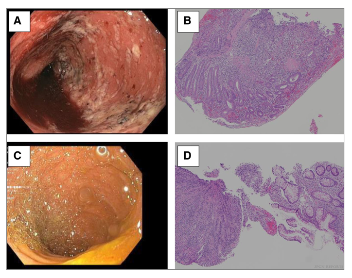
(A) Initial ileoscopy image—diffuse inflammation characterized by erythema, exudate, and friability. (B) Initial ileal histopathology—severe active ileitis, erosion, and focal crypt irregularity (magnification 100×).
Repeat ileoscopy image– (C) normal mucosa. (D) Repeat ileal histopathology—nonspecific changes including patchy lamina propria lymphoplasma cell infiltrate, eosinophilia, and spotty glandular and intraepithelial lymphocytosis (magnification 100×).
