
AS Patel et al. J Pediatr Gastroenterol Nutr. 2026;83:241–246. False positive autoimmune markers and elevated immunoglobulin G in genetically confirmed Wilson disease
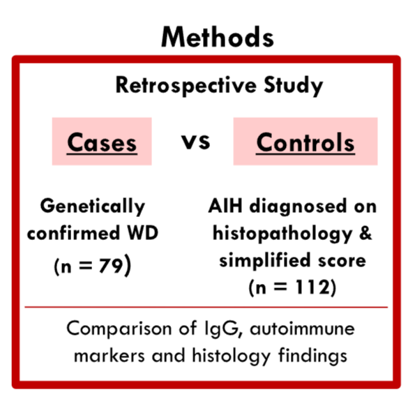
Key findings:
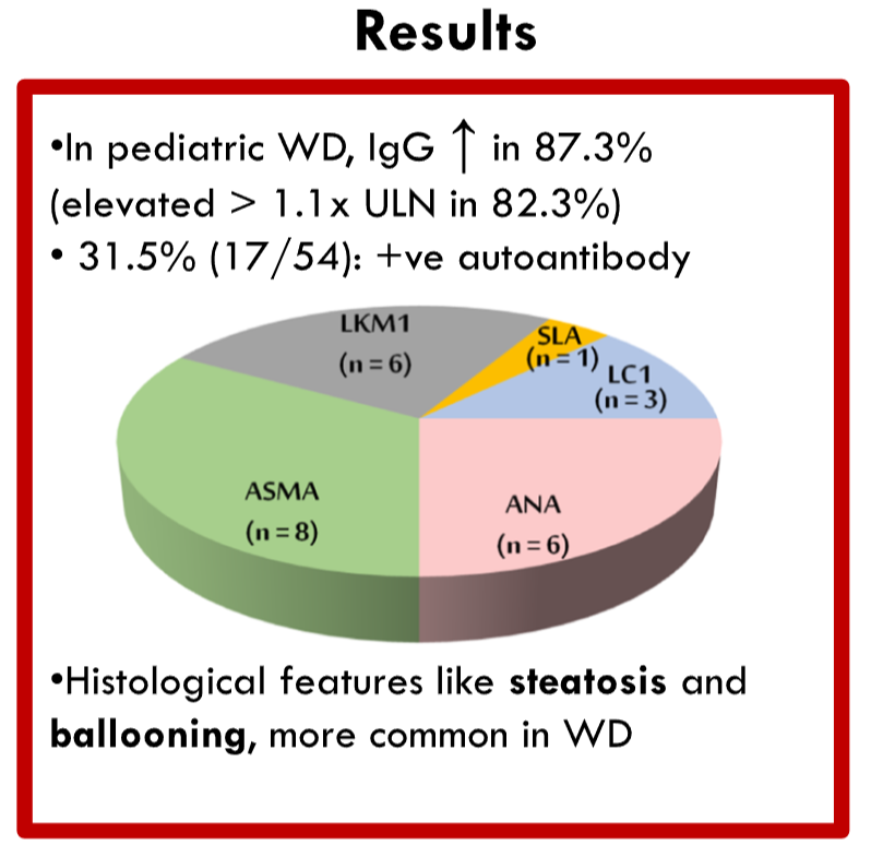
- Elevated IgG (>1.1× upper limit of normal) was observed in 82.3% of WD patients, while 31.5% tested positive for at least one autoantibody
- 20.2% of WD patients met the simplified diagnostic criteria for AIH
- Liver histology findings were available in 25 WD patients. Compared to AIH, WD patients were less likely to have interface hepatitis (40% vs. 89%; p < 0.001) and plasma cell infiltrates (32% vs. 72.6%; p = 0.009), while having a higher presence of glycogenated nuclei (24% vs. 6.2%; p = 0.009), ballooning degeneration (56% vs. 33%; p = 0.024), and steatosis (52% vs. 9.8%; p < 0.001). Copper quantification studies were not done
Discussion Points:
- “Reports of co-existence of WD and AIH in children further complicate the diagnosis.16, 17“
- “The presence of elevated IgG and autoantibodies, often interpreted as supportive diagnosis of AIH, may be misleading in WD.”
As an aside, I disagree with the authors introduction that “Wilson disease (WD) and autoimmune hepatitis (AIH) are the two most common treatable causes of chronic liver disease (CLD) in children.1, 2” Clearly, steatotic liver disease is the most common treatable chronic liver disease in children; in addition, both hepatitis B and hepatitis C are more common as well.
My take: A low threshold for testing of WD is needed in patients diagnosed with autoimmune hepatitis; this could include genetic testing and/or coppor quantification in liver biopsies..
Related blog posts:
- Wilson’s Disease –Pediatric Guideline
- Diagnosing autoimmune hepatitis | gutsandgrowth
- Mortality Risk With Autoimmune Hepatitis
- Rapid Genetic Testing in Liver Failure
- Current Practices and Wide Variation in Autoimmune Hepatitis Treatment Across Europe
- Autoimmune Hepatitis Associated with Anti-TNF Therapy
- Time to Adjust the Knowledge Doubling Curve in Hepatology


Pingback: Misleading Autoimmune Markers in Wilson Disease Diagnosis – Reviewer4you