Methods: The authors examined the predictive value of serum bile acids (sBA) patients (n=57) with biliary atresia who underwent a successful Kasai Procedure (KP), defined as a total serum bilirubin level of ≤25 μmol/L within 6 months after KP.
Key findings:
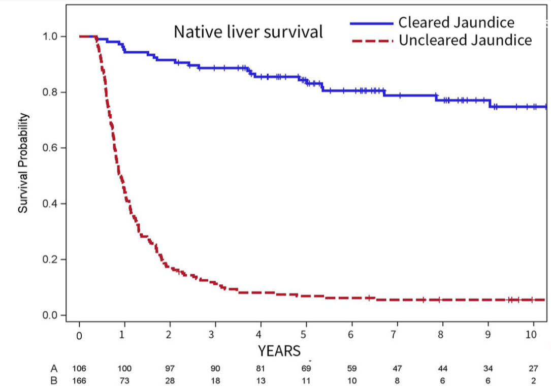
sBA thresholds of 56 and 30 μmol/L at a median time of 6 and 11 months after KP predicted HRV within 5 years after KP with a sensitivity of 100%.
No patient with sBA <18 μmol/L (between 4-9 months after KP) developed portal hypertension (PH) at 3 and 5 years after Kasai, whereas sBA >41 μmol/L predicted PH with PPV of 91% and 97% at 3 and 5 years after KP, respectively
From editorial on pages 1-2: “Persistent elevation f=of sBA likely reflects ongoing intrahepatic cholestasis and bile-acid mediated hepatocellular and fibrogenic injury, even in the setting of ostensibly adequate biliary drainage.”
My take: This study is in agreement with others regarding the predictive value of sBA. It suggests the current definition of a successful KP needs modification to incorporate sBA. Those with good bilirubin parameters but with persistent elevation of sBA could be considered partially-successful KP.
TL Mosher et al. J Pediatr Gastroenterol Nutr. 2026;82:765–769. Guidance adherence in the evaluation and management of hepatic hemangiomas in infants and children
Background: Hepatic hemangiomas are one of the visceral hemangiomas that can be seen and are categorized as infantile or congenital.1, 2Infantile hepatic hemangiomas (IHH) develop in the first weeks-to-months of life, reach peak size typically by 3 months and often spontaneously involute without medical management within 1–2 years.3Congenital hepatic hemangiomas (CHH) are present at birth and may rapidly involute, partially involute, or remain static for life.
Guidelines for screening with imaging and laboratory evaluation have been previously published and highlight differences between IHH and CHH.2 (Reference: Iacobas I, Phung TL, Adams DM, et al. Guidance document for hepatic hemangioma (infantile and congenital) evaluation and monitoring. J Pediatr. 2018; 203: 294-300 e2).” Screening with blood counts, fibrinogen, liver function tests, alpha-fetoprotein (AFP), thyroid function tests (TFTs), should be performed at diagnosis for both types of hemangiomas with frequency of repeat testing depending on whether IHH or CHH is diagnosed.2 Echocardiogram at diagnosis should be considered in both if patients have symptoms of cardiac failure. Serial monitoring with liver ultrasounds for at least 1 year or until stable size and vascularity twice in a row is recommended for CHH whereas continued monitoring with ultrasound until complete involution is recommended for IHH.2“
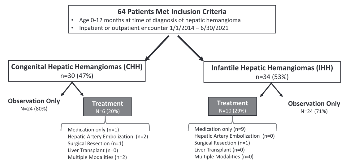
Methods: This study was a retrospective chart review with 64 patients
Key findings:
29 (46%) had CHH and 34 (54%) had IHH
In those 47 patients who were monitored clinically, 62% (n = 29/47) had ALT/AST collected whereas only 51% (n = 24/47) TSH/FT4, 55% (n = 26/47) AFP, and only 26% (n = 12/47) had an INR drawn. Only about one-third (28%, n = 13/47) had a screening echocardiogram performed during evaluation
When comparing across hemangioma type, patients with CHH were significantly less likely than those with IHH to have TSH/FT4 (26% vs. 75%, p < 0.001) or AFP (39% vs. 71%, p = 0.002) drawn
Sixteen patients (25%), 6 with CHH and 10 with IHH, required active management.
Among CHH patients, one (17%) received propranolol prior to transfer to our institution, two (33%) underwent embolization, one (17%) surgical resection, and two (33%) required multiple modalities (i.e., surgery plus embolization). Two patients with CHH receiving multiple treatment modalities developed multi-system organ failure and died.
Among IHH patients, most patients (90%) received propranolol and one (10%) underwent surgical resection.
Discussion Points:
“Current guidance recommends screening for the complications of hepatic hemangiomas, particularly in patients with diffuse or multifocal hemangiomas, as they are at increased risk for heart failure and hypothyroidism.”
“Current guidance recommends monitoring of hepatic hemangiomas until complete involution of the hemangioma is documented.2, 10 These recommendations are in place given the devastating sequelae that can develop in patients with proliferation of hemangiomas that may be missed when not evaluated with serial abdominal imaging.”
My take: This study shows that about 25% of these patients require medical intervention and is a good reminder of the recommended evaluations.
Disclaimer: This blog, gutsandgrowth, assumes no responsibility for any use or operation of any method, product, instruction, concept or idea contained in the material herein or for any injury or damage to persons or property (whether products liability, negligence or otherwise) resulting from such use or operation. These blog posts are for educational purposes only. Specific dosing of medications (along with potential adverse effects) should be confirmed by prescribing physician. Because of rapid advances in the medical sciences, the gutsandgrowth blog cautions that independent verification should be made of diagnosis and drug dosages. The reader is solely responsible for the conduct of any suggested test or procedure. This content is not a substitute for medical advice, diagnosis or treatment provided by a qualified healthcare provider. Always seek the advice of your physician or other qualified health provider with any questions you may have regarding a condition.
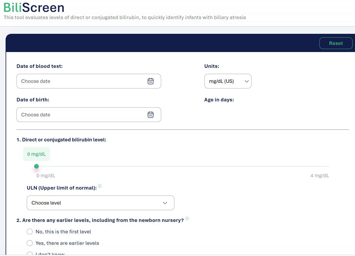
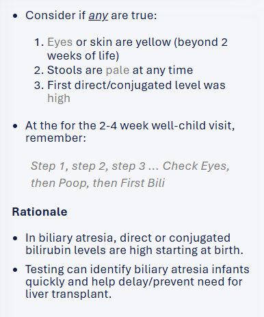
There is a new free screening tool, BiliScreen.org, which helps pediatricians triage patients for followup labs and/or hepatology referral. This site incorporates the recent AAP guidelines (see below) for evaluation.
Screenshots from website:
My take:
Overall, this is a quick easy tool which should be helpful & I recommend sharing this website with pediatricians/pediatric health providers
A potential improvement to the site would be a section with an explanation of other causes/consequences of cholestasis which could require more urgent evaluation (eg. metabolic diseases, vitamin K deficiency)
G Bora et al. JPGN Reports. 2026;7:1–5. Open Access! Maralixibat for the treatment of severe xanthomas in two children with Alagille syndrome: Case reports
Key findings:
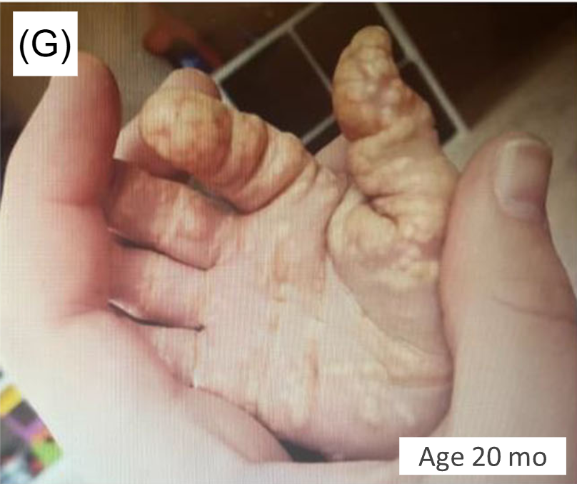
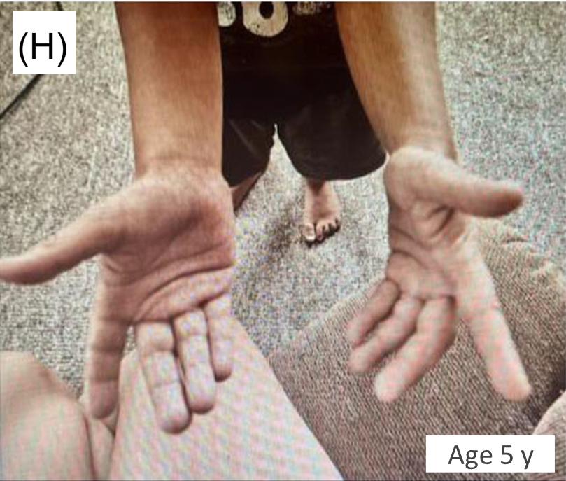
Maralixibat (MRX) resulted almost complete resolution of severe, debilitating xanthomas and clinically meaningful improvements in pruritus/serum bile acid levels.
Clinical scratch score in Case 1 dropped from baseline of 4 to 2 after MRX; in Case 2, it dropped from 3 to 1 after MRX
Serum bile acids in Case 1 dropped from baseline of 468 micromol/L to 206 after MRX; in Case 2 it dropped from 57 to 25 after MRX
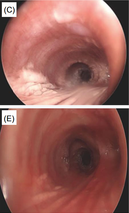
This case report involved two patients with ALGS and unusual manifestations of xanthomatosis, including one patient with airway xanthomas and a second patient with severe, diffuse xanthomas
From Figure 1 showing airway and skin changes in case 1:
Rigid bronchoscopy before (C) and then 18 months after maralixibat (E)
Discussion:
“The ICONIC study by Gonzales et al. showed significant improvements in xanthomas in participants treated with maralixibat by 48 weeks, based on overall reductions in the Clinician Xanthomas Score, with further improvements in patients treated for longer durations. Xanthoma reduction was associated with improved quality of life and levels of TC.4 This report reviewed real-world experience in two patients with ALGS and characterized the extent of their improvement after treatment with maralixibat.”
My take: This report provides convincing evidence that maralixibat was associated with reversing severe xanthomas in these two patients with Alagille syndrome.
Disclaimer: This blog, gutsandgrowth, assumes no responsibility for any use or operation of any method, product, instruction, concept or idea contained in the material herein or for any injury or damage to persons or property (whether products liability, negligence or otherwise) resulting from such use or operation. These blog posts are for educational purposes only. Specific dosing of medications (along with potential adverse effects) should be confirmed by prescribing physician. Because of rapid advances in the medical sciences, the gutsandgrowth blog cautions that independent verification should be made of diagnosis and drug dosages. The reader is solely responsible for the conduct of any suggested test or procedure. This content is not a substitute for medical advice, diagnosis or treatment provided by a qualified healthcare provider. Always seek the advice of your physician or other qualified health provider with any questions you may have regarding a condition.
MA Colak et al. J Pediatr Gastroenterol Nutr. 2026;82:358–365. Improvement in bile drainage after Kasai portoenterostomy with a tailored steroid protocol
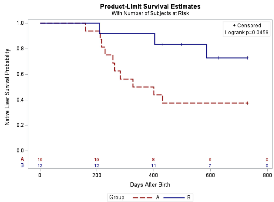
In this retrospective study, 28 infants underwent Kasai portoenterostomy (KPE) between 2015 and 2025. Group A had 16 infants managed without steroids between 2015 and 2021, while Group B included 12 infants managed under the new tailored steroid protocol between 2021 and 2025.
Determination of bile drainage: Postoperative stool color is monitored closely and collaboratively by hepatologists and surgeons according to the Japanese Tochigi Prefecture 3rd Edition stool card to assess bile drainage over the first five postoperative days.23 Patients with ≥50% of stools at color ≤3 are considered to have poor bile drainage, while those with >50% of stools at color ≥4 are considered to have good bile drainage.
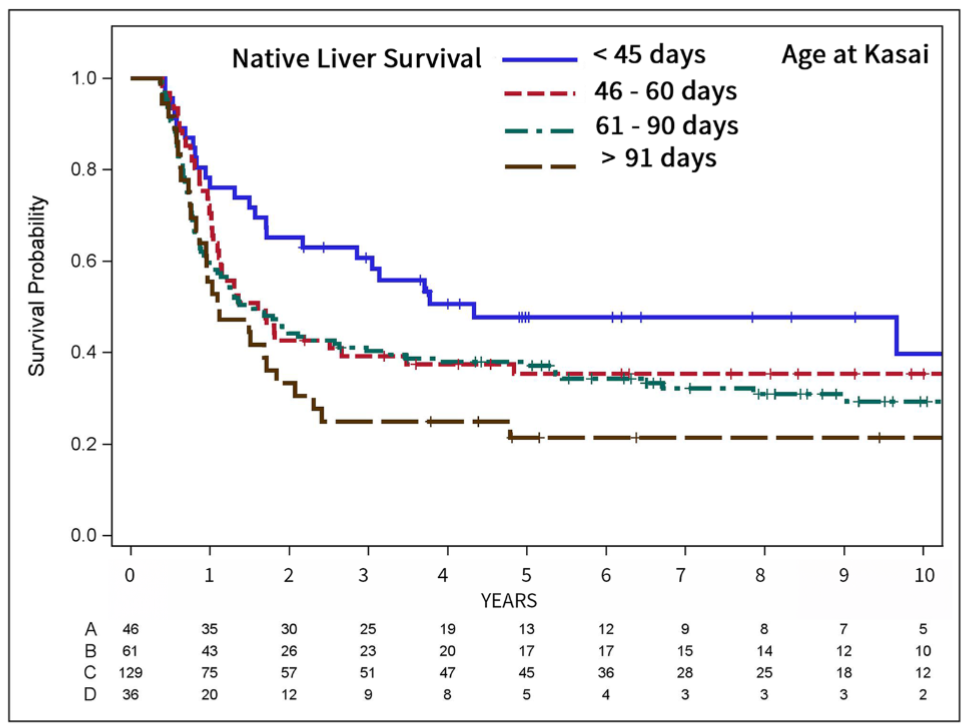
Tailored steroid protocol: “If patients have poor bile drainage, further management depends on age at time of operation. Patients ≤45 days old at operation are started on a combined steroid and antibiotic treatment immediately after bile leak is ruled out using abdominal ultrasound. Patients >45 days old at operation are started on the steroid and antibiotic treatment only if the liver biopsy obtained during operation demonstrated acute inflammation on histology.”
Key findings:
The 3-month post-KPE TB levels were significantly lower in Group B compared to Group A (0.9 [0.3, 1.9] mg/dL vs. 6.5 [0.6, 10.4] mg/dL, p = 0.036)
The 2-year native liver survival (NLS) was also significantly higher in Group B (72.9% vs. 37.5%, p = 0.046)
LOS, readmissions, reoperations, and complications in the 90-day postoperative period were not different between both groups
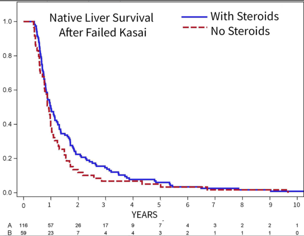
Kaplan–Meier curve of native liver survival at 2 years of age following Kasai portoenterostomy
In their discussion, the authors note that the “multicenter, placebo-controlled, double-blinded steroids in biliary atresia randomized trial (START) included 140 patients from the United States and assessed the effect of high-dose steroids (4 mg/kg/day).16 There was no significant difference in jaundice clearance at 6 months after operation (58.6% vs. 48.6%), nor significant difference in NLS at 2 years of age (58.7% vs. 59.4%) between the steroid and placebo groups.”
Subsequently, “similar to our study, Pandurangi et al. also reported a significant increase in the ratio of patients who had a TB level of <2 mg/dL at 3 months after operation in the customized steroid protocol cohort. However, although the steroid protocol cohort had greater 2-year NLS (68.8% vs. 50%), the difference did not reach statistical significance in their study.”
My take: The START study (n=140), which was powered to detect a 25% absolute treatment difference in TB levels, cannot exclude modest benefits from steroids. This current study, despite its limitations, showed that a tailored protocol for use of steroids may improve outcomes.
START Study: Steroids Not Effective For Biliary Atresia (After Kasai) In this study, the steroid intervention did not affect transplant-free survival which was 58.7% in the steroid group and 59.4% in the placebo group at 24 months of age. In addition, steroids were associated with an earlier onset of first serious adverse events.
Disclaimer: This blog, gutsandgrowth, assumes no responsibility for any use or operation of any method, product, instruction, concept or idea contained in the material herein or for any injury or damage to persons or property (whether products liability, negligence or otherwise) resulting from such use or operation. These blog posts are for educational purposes only. Specific dosing of medications (along with potential adverse effects) should be confirmed by prescribing physician. Because of rapid advances in the medical sciences, the gutsandgrowth blog cautions that independent verification should be made of diagnosis and drug dosages. The reader is solely responsible for the conduct of any suggested test or procedure. This content is not a substitute for medical advice, diagnosis or treatment provided by a qualified healthcare provider. Always seek the advice of your physician or other qualified health provider with any questions you may have regarding a condition.

O Ackermann et al. Gastroenterol 2026; 170: 188-198. The Natural History of Gastroesophageal Varices in Children With Portal Hypertension
Methods: Retrospective review of 1586 children with portal hypertension. 590 had two or more upper endoscopies (403 with biliary atresia).
“For the purpose of this study, and based on our previous experience in children,8,11 the endoscopic pattern associated with a high risk of bleeding (ie., HRV) included grade 3 esophageal varices as well as grade 2 esophageal varices with red color signs or gastric varices (cardia), or both.”
The authors developed a HRV [high risk of varices] score as a composite index calculated as follows: 1 point for grade 1 esophageal varices, 2 points for grade 2 varices, 3 points for grade 3 varices, and 1 point each for the presence of red color signs or GOV1 (HRV score range, 0–5). High-risk varices had an HRV score of 3 to 5.
Key findings:
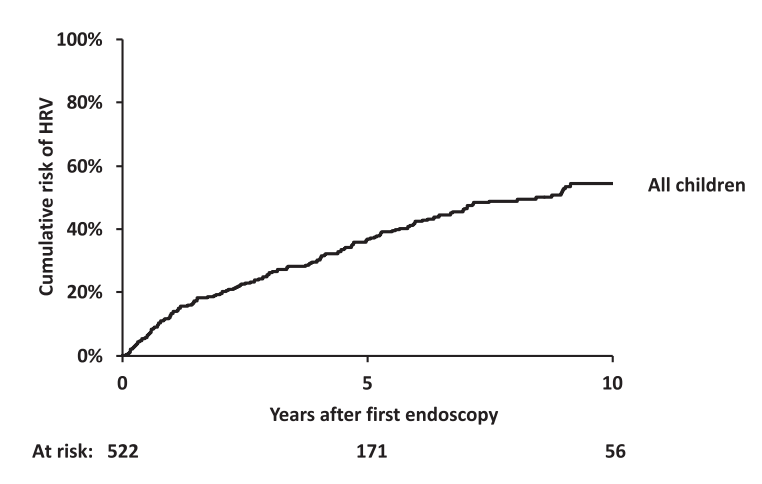
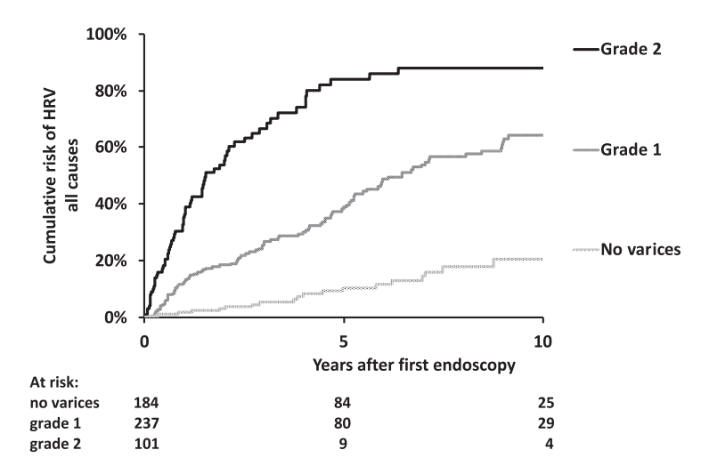
Worsening of the endoscopic pattern occurred in 58% of children over a mean 4-year interval
5- and 10-year probabilities of HRV emergence in initially HRV-negative children were 36% and 54%, respectively
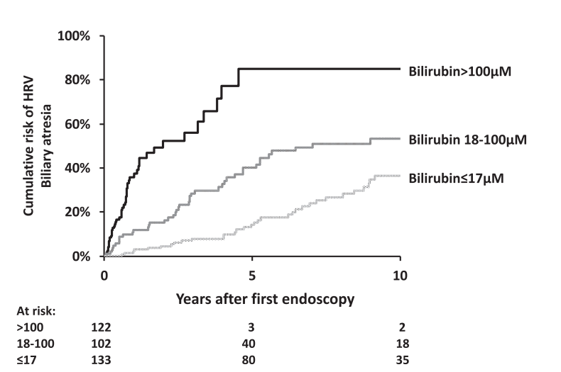
Infants with biliary atresia are at particularly high risk with correlation to the degree of cholestasis (see below)
Platelet count less than 150,000 as an indicator of HRV was mainly useful in older children. “A platelet count of ≥150,000/mm3 was recorded in 205 of the 629 children (32%) with HRV. Moreover, there was a decrease with age in the proportion of children with HRV and a platelet count of ≥150,000/mm3, falling from 62% in children aged <12 months to 2% in patients aged >10 year.” 16% of children 6-8 yrs, 12% of children 8-10 years of age with HRV had platelet count ≥150,000/mm3
“Gastrointestinal bleeding was recorded in 36 of 947 children (3.8%) who did not have HRV at their last endoscopy and in 270 of the 359 children (75%) with HRV at their last endoscopy who did not undergo endoscopic or surgical primary prophylaxis of bleeding.”
Bilirubin of 100 micoMol is equivalent to 5.8 mg/dL and 17.1 is eqivalent to 1 mg/dL
Discussion Points:
Variceal progression was much faster in infants and is is likely due to the severity of cholestasis and its impact on portal hypertension.
“It is notable that children with Alagille syndrome and those with genetic cholestasis with normal GGT have a lower rate of variceal progression and a lower mean HRV score than children with biliary atresia, despite comparably high levels of bilirubin. This suggests that different mechanisms of cholestasis … may have distinct consequences on intrahepatic portal vein branches resulting in varying degrees of portal hypertension.”
“In children with biliary atresia aged <12 months, grade 2 esophageal varices without red color signs or GOV1 (HRV score of 2) should be considered an indication for endoscopic primary prophylaxis.”
“Because the efficacy and safety of β-blockers have not been established in children, we suggest that this pattern—grade 1 varices with red color signs or GOV1—should prompt early repeat endoscopy to detect HRV in a timely manner…this repeat endoscopy could be recommended 6 months after the previous one.”
Limitations: High proportion of children with biliary atresia (limits conclusions with other disorders), and retrospective study since 1990
“Pending the results of future studies, the detection of palpable splenomegaly remains a simple and practical criterion for initiating screening endoscopy in children with portal hypertension”
My take: This is a very useful study providing important data to help improve decision-making in children with portal hypertension.
Disclaimer: This blog, gutsandgrowth, assumes no responsibility for any use or operation of any method, product, instruction, concept or idea contained in the material herein or for any injury or damage to persons or property (whether products liability, negligence or otherwise) resulting from such use or operation. These blog posts are for educational purposes only. Specific dosing of medications (along with potential adverse effects) should be confirmed by prescribing physician. Because of rapid advances in the medical sciences, the gutsandgrowth blog cautions that independent verification should be made of diagnosis and drug dosages. The reader is solely responsible for the conduct of any suggested test or procedure. This content is not a substitute for medical advice, diagnosis or treatment provided by a qualified healthcare provider. Always seek the advice of your physician or other qualified health provider with any questions you may have regarding a condition.
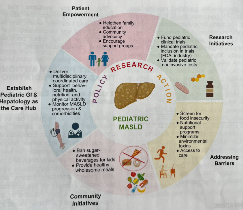
“Pediatric MASLD is still perceived as an indolent condition by many patients, families, and clinicians. In this Call to Action, we aim to raise awareness of pediatric MASLD as a public health crisis. Herein, we describe insufficient screening and disease staging practices, and a lack of accurate non-invasive tests and effective pharmacotherapy, both stemming from a paucity of multicenter clinical trials in pediatric MASLD.”
“GLP-1 RAs have not been formally investigated in pediatric MASLD.”
This was a retrospective study with 5 Latino adolescents obesity and MASLD who received semaglutide for at least 3 months. The range of BMI at the start of treatment was between 51 and 68.
Key findings:
There were clinically significant reductions in liver enzymes and APRI, a noninvasive marker of fibrosis. Specifically, mean ALT decreased by 38.4 IU/L (p < 0.01), mean AST decreased by 21.0 IU/L (p < 0.01), and mean APRI decreased by 0.128 (p = 0.01)
All 5 patients experienced weight loss with drop in BMI% from 2.3% to 14.2%
My take: This small study is consistent with others that show GLP1 RAs are likely to be an important tool for patients with MASLD. Current recommendations support use mainly in patients with comorbidities including obesity and T2DM.
Background: “Recent seminal studies such as the ESSENCE trial have demonstrated MASH resolution with improvement in fibrosis in adult patients with biopsy-proven steatohepatitis treated with semaglutide.18 This has led to the pivotal FDA approval of semaglutide for the treatment of MASH in adults.”
Methods: 42 patients with MASLD (see study for details). 71% of patients had a therapeutic indication for T2DM and 29% for obesity. Of the GLP-1RA medications, liraglutide was most frequently prescribed (44%), followed by semaglutide (27%), dulaglutide (25%), and exenatide (4%).
Key findings:
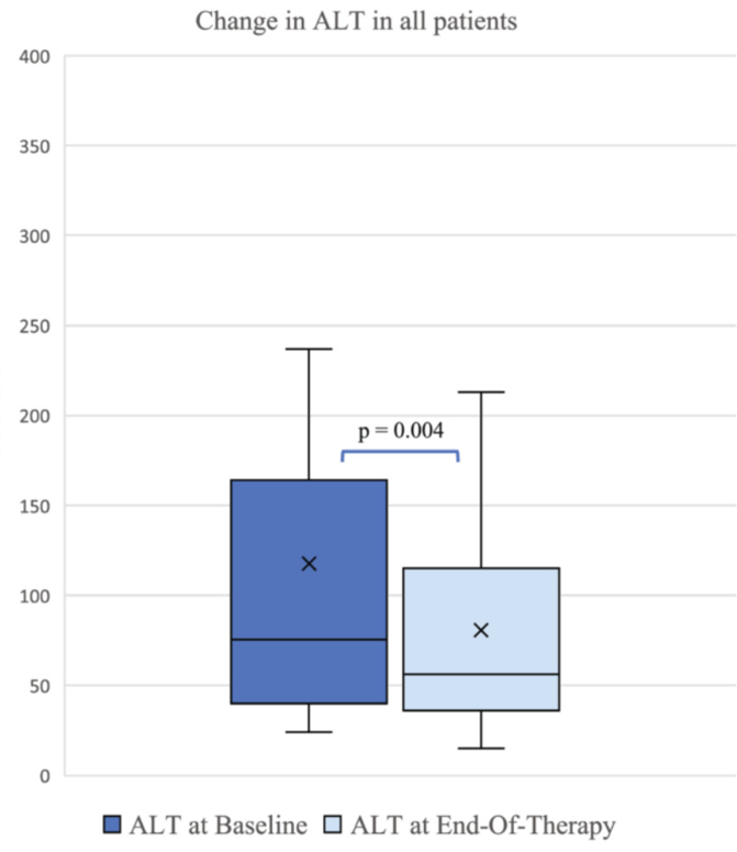
ALT improved by a mean of 56 U/L at 6 months (p = 0.04), and by 37 U/L at en-of-treatment (p = 0.004).
GLP-1RA use was associated with significant improvements in alanine transaminase (ALT) along with other cardiometabolic biomarkers even in the absence of improvements in body mass index percentile or z-score. There were reductions in GGT, AST, triglycerides, and HbA1C
In this study, there was not a significant decline in weight. Prior studies have shown less weight loss in patients with T2DM compared to those with obesity (w/o T2DM)
My take: It is helpful to have a pediatric study that shows that GLP1-RAs are effective specifically in those with MASLD.
The review of pediatric MASLD addresses epidemiology, pathophysiology, natural history, screening, diagnosis, treatment, comorbidity management, outcome monitoring, and transition of care. It also discusses the implications of the 2023 nomenclature revision, which emphasizes evaluating both hepatic steatosis and cardiometabolic risk factors.
Some key points:
Box 1 outlines numerous (32) research priorities, including the need for prospective longitudinal cohort studies.
“Globally, the estimated prevalence of MASLD in children is 7.6%, making it the most common cause of chronic liver disease in children”
Figure 4 describes the interplay between risk factors for MASLD included genetic predisposition, prenatal factors and environmental exposures
Figure 5 summarizes comorbid conditions which include obstructive sleep apnea, prediabetes/diabetes, cardiovascular disease (dyslipidemia, hypertension, left ventricular hypertrophy), anxiety/depression, reduced bone mineral density, renal dysfunction and polycystic ovarian syndrome. Table 6 summarizes evaluation and initial management with most of these conditions. Yearly screening for diabetes in children with MASLD is recommended.
ALT remains most common screening test with >26 U/L for adolescent males and >22 U/L for adolescent females having optimal sensitivity (>80-85%). We recommend “screen for MASLD in children aged 10 years or older with overweight and cardiometabolic risk factors or family history or obesity.” Annual screening recommended if at risk.
Table 2 provides a long list of medications which may promote weight gain. These include antihistamines, steroids, some contraceptives, anticonvulsants, antidepressants, antipsychotics, methotrexate, and doxycycline
Diagnostic evaluation:
Diagnosis of MASLD requires confirmation of steatosis (by imaging or biopsy) in addition to the presence of at least one cardiometabolic risk factor. ALT elevation with a cardiometabolic risk factor is insufficient.
“Consider liver biopsy in cases where there is uncertainty, especially if ALT levels are persistently elevated (>2 times the ULN)”
Table 3 lists inborn errors of metabolism and monogenetic diseases which may cause childhood-onset steatotic liver disease. Evaluation of inborn errors of metabolism should be considered if atypical signs or symptoms, such as early onset (<3 yrs), rapidly progressive, absence of obesity, or other organ involvement (especially neurological)
Table 4 summarizes imaging modalities to assess steatosis and fibrosis in children. Only MRI-PDFF has been validated in children (for steatosis)
Table 5 describes BMI classification in children (WHO and AAP)
Lifestyle treatments are detailed including diet (reduction of added sugars, Mediterranean diets) and exercise
Emerging medications are reviewed. However, practice statement notes “No pharmacotherapies are currently recommended or approved as specific treatments for MASLD or MASH in children…Medications approved for use in children ages 12 years and older to treat obesity or type 2 diabetes may be considered for children with MASLD.”
My take: This is a comprehensive practice guidance. It emphasizes an extensive diagnostic evaluation. The threshold for liver biopsy is relatively low in this guidance. As more data emerges, it is likely that more emphasis will be placed on the use of pharmacotherapies.
Disclaimer: This blog, gutsandgrowth, assumes no responsibility for any use or operation of any method, product, instruction, concept or idea contained in the material herein or for any injury or damage to persons or property (whether products liability, negligence or otherwise) resulting from such use or operation. These blog posts are for educational purposes only. Specific dosing of medications (along with potential adverse effects) should be confirmed by prescribing physician. Because of rapid advances in the medical sciences, the gutsandgrowth blog cautions that independent verification should be made of diagnosis and drug dosages. The reader is solely responsible for the conduct of any suggested test or procedure. This content is not a substitute for medical advice, diagnosis or treatment provided by a qualified healthcare provider. Always seek the advice of your physician or other qualified health provider with any questions you may have regarding a condition.
“A rising tide lifts all boats” has been used to express the sentiment that a good economy is beneficial to all. However, this has been criticized as not all boats are lifted equally and some boats are a lot nicer than others. I was thinking about this expression with these recent publications. The articles indicate that the availability of living donor liver transplant (LDLT) is clearly beneficial to the recipients but also is helpful, in a lesser way, to others on the transplant list as well.
Researchers analyzed data from 474 pediatric candidates listed for liver transplants at a single center from 2001 to 2023 (Toronto).
Key findings:
The pLDLT group had a higher likelihood of receiving a liver transplantation (adjusted HR: 1.38) a lower risk of dying without a transplant (adjusted HR: 0.11)
Survival rates from the time of listing were significantly better in the pLDLT group compared to the pDDLT (on live donor) at 1—(98.6% vs. 87.6%), 5—(96.6% vs. 84.4%), and 10—(96.6% vs. 83.1%) years
Having a potential live donor was linked to a 72% reduction in mortality risk (adjusted HR: 0.28)
The waiting time for deceased donation shortened. This correlated with increased LDLT utilization, suggesting LDLT not only improved outcomes but also shortened wait times even for pDDLT patients
From the associated editorial:
“LDLT continues to be underutilized in the United States with only 15% of all pediatric LTs being LDLTs.1… In 2024, only 6 pediatric centers across the United States performed 5 or more LDLTs.6…”
“Black and African-American and Hispanic candidates and those with public insurance are half as likely to undergo LDLT compared with Caucasian candidates and those with private insurance.7,8“
“In a survey of over 200 parents of pediatric candidates and recipients of LT, only 72% reported knowing the steps to gain access to LDLT, and only 69% knew that donor costs were covered by the recipient’s insurance.7“
The authors recommend collaboration between centers offering LDLT and those that don’t so that more patients could benefit
My take: More use of LDLT will result in better outcomes.