
A Reunamo et al. Gastroenterology, 2026; 171, 553-555. Open Access! Three-Dimensional Micro-Computed Tomographic Imaging Reveals Early Mucosal Remodeling in Individuals With Potential Celiac Disease
Background: “A subset of individuals, characterized as having potential CeD (PCeD), present with positive transglutaminase 2 or endomysial antibodies, or both, but lack histologic findings that meet the diagnostic criteria.1 Importantly, only a subgroup of individuals with PCeD progress to CeD during follow-up.1“
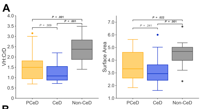
Methods: The authors “recently developed a 3-dimensional (3D) x-ray micro-computed tomographic (micro-CT) imaging and analysis procedure for duodenal biopsy specimens.6,7 The method enables the precise histomorphometric quantification, including measurements of villus height–to–crypt depth ratio (VH:CrD) and mucosal surface area, with excellent reproducibility.7” This study used micro-CT imaging to investigate the early-stage mucosal changes in individuals initially diagnosed with PCeD. This study enrolled 20 patients with PCeD who progressed to CeD, 5 patients who continued with PCeD, and 16 control individuals.
Key findings:
- Micro-CT imaging revealed a significantly reduced VH:CrD at both the PCeD and CeD stages compared with the controls, but the difference between PCeD and CeD did not reach significance
- Only 7/20 with PCeD had normal biopsy VH:CrD values >2.0 across all sites
Discussion:
“Most of the PCeD cases were already below the commonly used VH:CrD cutoff when assessed using micro-CT cross-sections, implying that histologic thresholds may lack sufficient sensitivity to capture signs of early CeD.”
My take (borrowed in part from the authors): This study strengthens the “concept that enteropathy in CeD represents a progressive continuum.5 In fact, pinpointing the exact moment when the condition becomes “CeD” is difficult, especially because what is considered normal can vary individually and depending on the biopsy site.” This study may help explain why many symptomatic patients with PCeD respond to a gluten-free diet.
Related blog posts:

