Discordinate Recommendations for Celiac Screening with Down Syndrome

VN Dargenio et al. J Pediatr Gastroenterol Nutr. 2026;83:304–313. A systematic review of celiac disease recommendations for children with Down syndrome

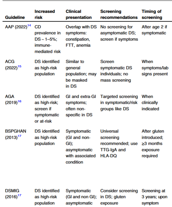
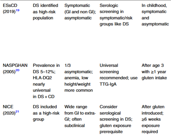
Background: “CD [celiac disease] prevalence in DS is higher than in the general population.10 The overlapping symptoms between CD and DS, such as growth failure, fatigue, GI disturbances, and, less frequently, neurological and behavioral problems, pose significant diagnostic challenges, as these are often attributed to underlying DS comorbidities, leading to delays in recognizing CD.411 

Guidelines:

The authors note wide variation in the prevalence of CD in DS in various studies. “A meta-analysis of 31 studies including 4383 individuals found a pooled prevalence of biopsy-confirmed CD of 5.8% (95% CI 4.7%–7.2%), with slightly higher rates in children (6.6%) compared to mixed-age samples (5.1%)31…these findings align with the 5%–13% prevalence range documented in earlier European studies2

The authors recommend the following strategy:

“Given the high prevalence of asymptomatic and atypical presentations, reliance on symptoms alone is insufficient. A pragmatic strategy should initiate with universal serological screening for all children with DS after gluten introduction (12–24 months of age), followed by periodic re-screening every 2–3 years, using tTG-IgA with total IgA assessment, supplemented by IgG-based assays in the context of the high rate of selective IgA deficiency in DS.”

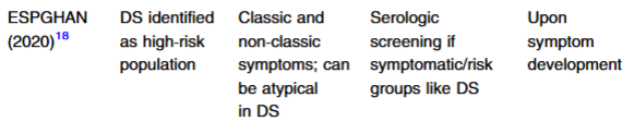
My take: There are wide discrepancies in the recommendations for screening for celiac disease in asymptomatic individuals with Down syndrome.

Related blog posts:

Should the Gluten Threshold Dose in Celiac Disease Change Based on Immune Activation?

Background: Interleukin 2 (IL2) rises acutely after gluten ingestion, allowing the definition of threshold doses that trigger immune activation.

Methods: This was a randomized double-blind, placebo-controlled adaptive dose–response trial in adults with biopsy-proven celiac disease on a gluten-free diet for >2 years. Participants (n = 51; median age, 52 years; 69% were female) underwent 3 oral gluten (1–1000 mg) or placebo challenges at 4-week intervals. Eliciting dose (EDp, ie, the dose at which p% of people respond) was estimated by interval-censored survival analysis.

Key findings:

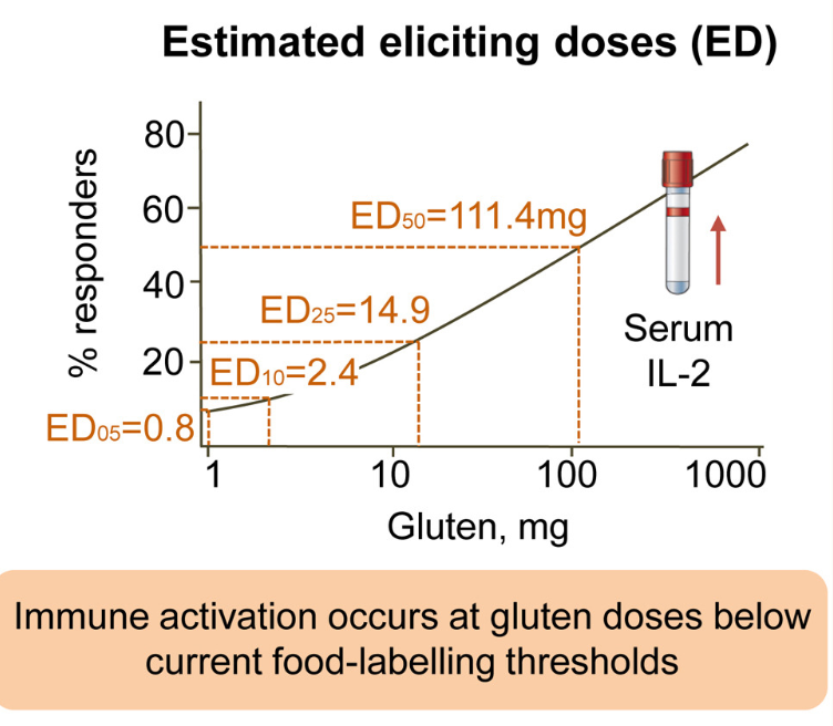
  • Gluten induced dose-dependent IL2 elevations, with ≥2-fold increases in 83% at 1000 mg, 83% at 610 mg, 36% at 90 mg, 17% at 13 mg, 27% at 8 mg, and 17% at 3 mg; none responded to 5 mg, 2 mg, 1 mg, or placebo.  
  • Estimated ED50 was 111 mg, ED10 was 2.4 mg, ED05 was 0.8 mg, and ED01 was 0.1 mg

From the editorial:

“To date, the few investigators who addressed this question defined tolerance in relation to quantifiable histologic damage (intestinal morphometry)…Bearing in mind that the typical Western diet contains 10–15 g of gluten per day, these studies found that the protracted, daily ingestion of 1000, 200, or 50 mg of gluten elicits significant damage at the mucosal level.2,3 A daily dose of 10 mg was also associated with minimal architectural changes in some patients…

In 2007, the Food and Agricultural Organization/World Health Organization Codex Alimentarius Commission4 applied these findings to establish that commercially available gluten-free food must not contain more than 20 mg/kg gluten (20 part per million [ppm]), as this would ensure that daily gluten intake does not exceed 10 mg…Not a single case of proven intolerance to 20 ppm gluten-free food has been described…in some 20-ppm countries, most labeled gluten-free products already contain much less than 20 ppm of gluten, usually <10 ppm5

The authors concluded that the daily gluten threshold should be decreased because acute IL2 release occurred at 3 mg, which is less than is allowed by current food-labeling rules for gluten-free products…

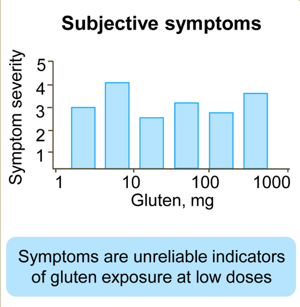
Because symptoms did not correlate with the result of the IL2 test for gluten doses <1 g, further prolonged microchallenge studies remain the only reliable strategy to investigate the correlation between the IL2 test and the gold standard of CeD activity—histologic damage of the small intestinal mucosa.10,11

My take: This study is intriguing that there is evidence of immune activation at lower gluten exposures; however, it does not prove that changing the gluten threshold is beneficial.

Related blog posts:


Camp Weekaneatit 2026

Last Sunday, along with my colleagues, Jeff Lewis, and Nirav Patel, I helped check in kids for Camp Weekaneeatit! (glutenfreecamp.org). This is a gluten-free camp for youth with ​Celiac Disease and ​Gluten Intolerance. This year’s staff shirt was “S’more Fun Without Gluten;” though, I elected to wear my T-shirt from 2018.

The camp was started more than 15 years ago by my partners, Dr. Jeff Lewis and Dr. Bill Meyers.

The camp is located just north of Atlanta at Ft Yargo (Winder, Georgia). The ~125 participants come from all over the U.S including Texas, Michigan, Ohio, Massachusetts, Colorado, Arizona, Florida, South Carolina, and Alabama. There was one kid, whose father is a marine, who flew in from Japan!

Most of the campers have come several times and have had a great experience. Here is an excerpt from a letter from a camper’s parent:

My daughter was at camp with you this summer.  I can’t tell you how much fun she had.  She was diagnosed with celiac when she was just 2, so she has never known a world where she could just relax and be like everybody else.  Camp gave her so much freedom, and she grew so much in just one week.  Today is her birthday and her first day of school.  Her camp friends have already texted to wish her happy birthday!…This is the first time she has had friends with celiac disease — I can only imagine how much more supported that makes her feel…  I asked her what it was like to not have to ask a million questions before every bite she ate.  She said, “I felt like I didn’t even have celiac disease all week. I felt like a normal person.” 

Last year enrollment started in the middle of November for summer 2026. Space is limited!

Related blog posts:

Low Risk of Solid Organ Transplantation with Celiac Disease

JB Doyle et al. Clin Gastroenterol Hepatol 2026; Risk of solid organ transplantation in individuals with celiac disease: a nationwide cohort study

Background: “Celiac Disease (CeD) is associated with immune-mediated diseases of the liver, including autoimmune hepatitis, primary biliary cholangitis, primary sclerosing cholangitis. Individuals with CeD are also at increased risk for non-autoimmune diseases of the liver, including metabolic dysfunction-associated fatty liver disease…CeD has also been associated with chronic kidney disease and cardiovascular disease. Multiple studies have demonstrated that individuals with CeD have increased rates of end-stage renal disease than the general population, with particularly strong associations between CeD and
autoimmune nephropathy, diabetic nephropathy, and systemic lupus erythematosus (19-22).
Population-level data also suggests that individuals with CeD have an increased risk of
cardiovascular disease and ischemic heart disease>”

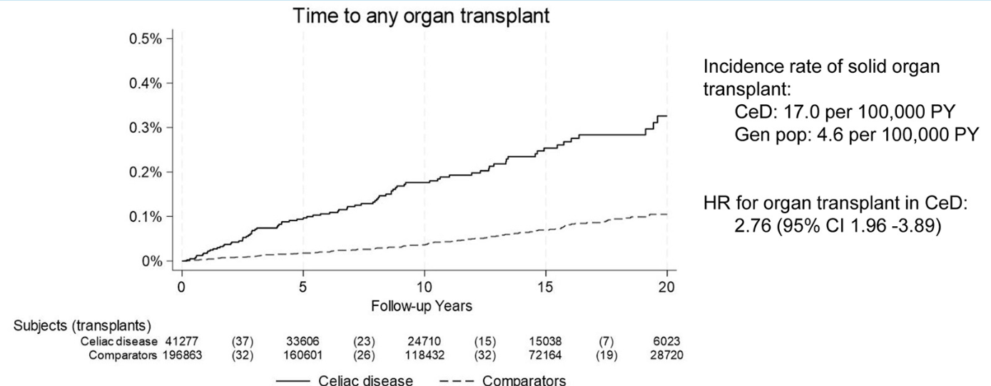
Methods: Population-based matched cohort study in Sweden. We identified individuals with biopsy-proven CeD diagnosed between 2000-2023 using the nationwide histopathology cohort ESPRESSO. We calculated the incidence of solid organ transplantation (liver, heart, kidney, and lung) in CeD patients and estimated the risk relative to the general population. There were  41,277 individuals with CeD and 196,863 age- and sex-matched comparators with a mean follow-up of 12.1 years.

Key findings:

  • There were 85 solid organ transplantations in patients with CeD (17.0 per 100,000 person-years) and 111 in matched comparators (4.6 per 100,000 person-years). This corresponded to an adjusted hazard ratio (HR) of 2.76 
  • The highest relative risk was for liver transplantation: HR 7.26; for kidney HR 1.85. For heart, HR was 2.35 did not reach statistical significance (CI: CI 0.84-6.61)

Discussion:

“Shared genetic susceptibility may explain physiological links between CeD and autoimmune liver disease, especially since we also detected an increase in liver transplantation prior to CeD diagnosis…owever, CeD patients in our analysis had an increased risk of liver transplantation relative to their nonaffected siblings, suggesting that CeD itself may be a risk factor beyond genetic predisposition”

My take: While nearly triple the incidence compared to the general population, the absolute increased risk of needing a solid organ transplant was about 1 in 10,000. For comparison, the risk of dying in a bicycle accident in one’s lifetime is about 1 in 5,000. Nevertheless, it may be worthwhile to screen for CeD in those with end-organ disease. Additionally, checking liver tests periodically in patients with CeD would be reasonable.

Related blog posts:

Should We Adopt Mass Screening for Celiac Disease?

MG Stahl et al. Gastroenterol 2026; 170: 240-245. Open Access! Mass Screening of Celiac Disease: A Crossing Point Between Secondary and Primary Prevention?

The authors make several good arguments for mass screening for celiac disease.

Key Points:

  • “The worldwide prevalence is high, affecting an estimated 1% to 3% of the general population…One study showed that two-thirds of children remain undiagnosed.” (Dig Liver Dis. 2023; 55:608-613). Thus, screening would identify many cases that would otherwise go undiagnosed and untreated
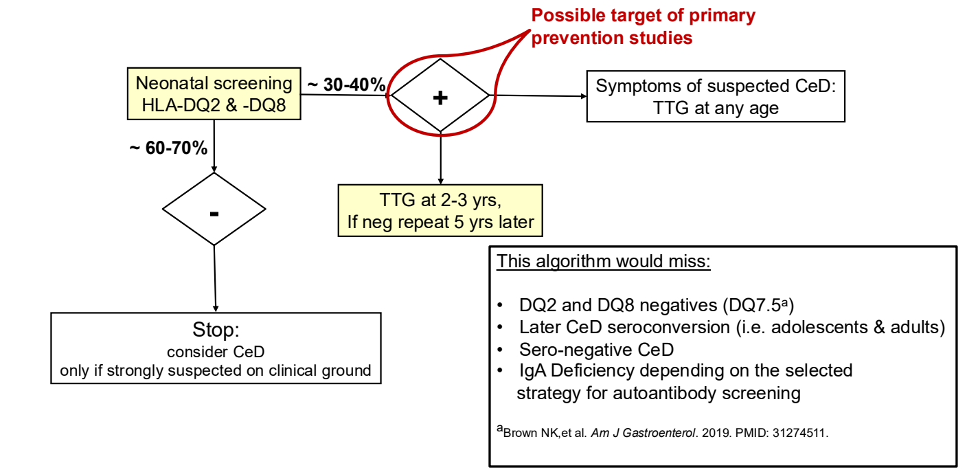
  • ” As one of the most HLA-restricted diseases, nearly all patients carry HLA-DQ2.5, DQ2.2, or DQ8 (and very rarely DQ7.5), and their absence makes CeD extremely unlikely. Although 30% to 40% of the general population carry these alleles, only approximately 3% will go on to develop CeD”
  • “The Environmental Determinants of Diabetes in the Young (TEDDY) study showed that although there was some early anxiety reported, there was no long-term psychological harm in disclosing genetic risk to parents of children that were tested and most parents adapted over time”
  • “Children identified as highest risk at birth through HLA-DQ typing also represent an ideal cohort to test interventions such as dietary modifications or microbiome targeted therapies during the predisease phase, during the key “window of opportunity,” ideally before seroconversion (primary prevention).” There are no currently proven primary prevention interventions.
  • Some of the drawbacks to screening: 1. “Evidence on health benefits of treating asymptomatic CeD is limited.” 2. “The potential psychosocial effects of both newborn genetic testing and a strict gluten-free diet in individuals diagnosed through screening need to be considered.” 3. “HLA-DQ typing may be cost prohibitive in some regions of the world.” 4. “Most children identified to be at risk for CeD based on HLA-DQ will not develop CeD.”

My take: I am skeptical about the benefits of screening at birth and how it would work in our current health care system. We have plenty of examples in our field in which early screening is not followed up well (eg. Hepatitis C, Biliary Atresia). If more evidence emerges on the benefits of primary prevention, then more widespread screening at birth may be worthwhile. For example, there is “a clinical trial underway in Sweden, the GRAin study (NCT04593888) that aims to investigate whether eating a gluten reduced diet (<2 g of gluten per day) may reduce the risk of CeD in children with genetic risk.”

Related blog posts:

Outcomes in Children with Celiac Disease Presenting with Constipation

A Almallouhi et al. J Pediatr Gastroenterol Nutr. 2025 DOI: 10.1002/jpn3.70316. Clinical outcome of constipation as the presenting symptom in children with celiac disease

Background: “It is not clear if CeD prevalence is higher in children with refractory and chronic constipation or not.1115 The current guidelines from the American Gastroenterological Association (AGA) and the North American Society for Pediatric Gastroenterology, Hepatology, and Nutrition (NASPGHAN) do not consider constipation an indication for CeD testing in the absence of suggestive family history, growth, or developmental delay.”

Methods: This was a retrospective study (1994-2024) of children (<18 years) who presented with constipation and then diagnosed with celiac disease (CeD). There were 248 children with CeD, 177 (71%) had biopsy-confirmed CeD, and 56 (23%) were diagnosed with serology-only criteria

Key findings:

My take:

  1. It is unclear if having constipation increases the risk of celiac disease
  2. Many children with celiac disease also have functional disorders like irritable bowel and constipation that often continue despite a gluten-free diet

Related blog posts:


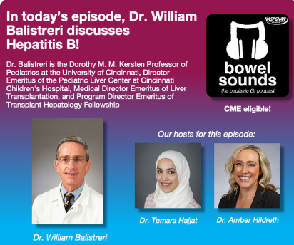
Also, Dr. Balistreri gives a Bowel Sounds Podcast on Hep B. Here’s the link: https://t.co/BmEUoC9YQt

Related blog posts:

Likelihood of Celiac Disease with Conflicting Serology Results

R Mandile et al. J Pediatr Gastroenterol Nutr. 2025;81:1482–1487. Advantages of anti-endomysial evaluation in children with low titers of anti-transglutaminase antibodies: A retrospective study

This was a single center retrospective study examining children (n=202) undergoing EGD (2022-2024) to evaluate for celiac. Among those with low anti-TTG IgA titers, Group 1 (n=25) was EMA negative and Group 2 (n=100) was EMA positive.

Key findings:

  • The finding of discordant serology (anti-transglutaminase [anti-TG] positive and EMA negative) is infrequent (12% cases, 25 out of 202), and all patients with discordant serology had anti-TG positive at low titer (<4 times the upper limit of normality).
  • Group 1 (N = 25) had a mean anti-TG titer of 1.86× ULN and villous atrophy (VA) in only 8% (2/25). Group 2 (N = 100) had VA in 35% (35/100)
Percentage of patients with villous atrophy between EMA positive and EMA negative children.

Discussion Points:

  • The diagnosis of CD still requires performing an EGD in at least half of the cases
  • This “study suggests that patients with low levels of anti-TG but EMA positive antibodies should anyway receive an EGDS in the next 6 months, since in around one-third of the cases a duodenal atrophy will be detected”
  • In those with low anti-TG but EMA negative, ” it could be reasonable to initially follow-up patients over time with clinical and serological monitoring (in particular of anti-TG titer), postponing the EGDS to a later stage, when the disease is more advanced and the chance of finding a concomitant VA (and thus the need to start a GFD) is higher”

My take: In patients with minimal symptoms and low level anti-TG, my strategy has been to follow with serological monitoring and if repeatedly abnormal, proceed with endoscopy. This study suggests that obtaining EMA early may influence choice to proceed earlier with endoscopy.

Related blog posts

Shared Decision-Making in Celiac Disease Diagnostic Approach

Y Sunkoy, S Talathi. Am J Gastroenterol 2025; 2190-2193. Utility of the ESPGHAN Biopsy-Sparing Guidelines for Celiac Disease in Children

Thanks to Ben Gold for this reference.

Methods: Retrospective study of patients (n=2942 children) who had celiac serologies and duodenal biopsies

Key findings:

  • Prevalence of CD in this cohort was 9% (226 of 2942 patients)
  • In those with a high titer (>10xULN), 106 of 107 patients (99%) had celiac disease
  • In this cohort, even in those with with >7XULN, had a Sensitivity of 55.3%, a specificity of 99% and a PPV of 97%

In their discussion, the authors note that “we did not obtain an EMA in a second sample, which is recommended in the ESPGHAN guidelines.”

Associated commentary: Erica Brenner, American Journal of Gastroenterology 120(9):p 1985-1986. The No-Biopsy Approach for Pediatric Celiac Disease: Ready for Prime Time in North America?

  • “Shiha et al (8) found that the PPV ranged from 65% for a 1% CD prevalence to a 99% for a 40% prevalence. As the 9% CD prevalence in the study by Sunkoj et al (4) exceeds the 0.81$-1.4% prevalence in the United States (9), the reported PPV may overestimate reality.” (Related post: No-Biopsy Approach to Celiac Disease Diagnosis and Positive Predictive Value (Based on Population)
  • “Children with type 1 diabetes and trisomy 21 have a higher risk of false-positive serology and therefor may not be appropriated candidates for a no-biopsy approach.”

My take: A larger recent study (Chang et al. Pediatrics. 2025;156(3):e2025070897) found that the no-biopsy approach had a significantly lower PPV in their cohort (94.9% overall, and 95.7% in those without T1DM). Thus, in cohorts with lower prevalence of CD, the no-biopsy approach could lead to 2-4% of children being placed unnecessarily on a gluten free diet. As such, it would be good practice to discuss making a diagnosis via endoscopy vs. the no-biopsy approach as part of shared decision-making.

Related blog posts:

When Celiac Disease Symptoms Continue Despite a Gluten Free Diet

A Kruegger et al. JPGN 2025; 81:596–605. Open Access! The prevalence and predictive factors of overlapping disorders of gut–brain interaction and celiac disease in children

Methods: Single-center, retrospective study of children (4–21 years old, n=191) with biopsy-proven Celiac disease (CeD) who were evaluated for DGBI based on Rome IV criteria. Patients who were adherent to a GFD, demonstrated tissue transglutaminase immunoglobulin A (TTG IgA) decline, and had at least one visit 9–24-months after diagnosis with a pediatric gastroenterologist. For this study, sustained TTG IgA decline required at least two declining TTG IgA values, a 90% decline from baseline, or normalization of TTG IgA.

Key findings:

  • 43% (n = 83) met Rome IV DGBI diagnostic criteria.
  • Functional constipation (27/83, 33%) and functional abdominal pain (24/83, 29%) were the most common DGBI
  • Abdominal pain, constipation, and vomiting at initial presentation as well as comorbid joint hypermobility, headaches, and chronic musculoskeletal pain increased risk of developing DGBI after serological decline

Discussion Points:

  • “The prevalence reported here is similar to a study of adults with CeD who were adherent to a GFD that reported over 50% met criteria for a functional gastrointestinal disorder19 and is higher than previously reported pediatric prevalence rates”
  • “The majority of patients who met DGBI criteria did so through having the persistence of the same gastrointestinal symptoms that were present at CeD diagnosis. This raises the question as to whether the symptoms at presentation were due to CeD, DGBI, or both”
  • “Clinicians could consider discussing that while symptoms related to CeD should improve on a GFD, some symptoms may persist, especially if they have an increased likelihood of having a comorbid DGBI. Such counseling may prevent the misattribution of persistent symptoms to ongoing gluten exposure and mitigate hypervigilance”
  • “Having complete villous blunting on diagnostic biopsy increased the likelihood of having a DGBI. Intuitively, it is possible that complete villous blunting can lead to greater nerve sensitization and subsequently higher rates of DGBI. It is also possible that complete villous blunting is slower to recover”

My take: Given the overlap of DGBI symptoms with CeD, diagnosing DGBI in patients with CeD can be challenging. However, DGBI is much more likely to contribute to lingering symptoms than refractory CeD.

As a practical matter, the high frequency of ongoing GI symptoms despite use of a GFD provides another drawback to relying on a no-biopsy diagnosis. A no-biopsy diagnosis introduces greater uncertainty in the diagnosis and does not allow for a histologic comparison if a subsequent evaluation is needed.

Related blog posts:

Claude Monet, Bridge over a Pond of Water Lilies at The Metropolitan Museum of Art

How to Diagnose Celiac Disease in Patients Already Receiving a Gluten Free Diet

Open Access: Evaluating for Celiac Disease in Patients on a Gluten-Free Diet: A Practical Approach

Algorithm -Figure 2:

Figure 3 lists the content of several common foods -some noted below

While gluten exposure increases the diagnostic yield of currently available tests, there are novel tests being developed “which may aid in the diagnosis of CeD regardless of diet, with a particular focus on immune-based assays. One such innovation involves the use of tetramer-based assays, which enable the direct detection of gluten-specific T cells in the blood. These tetramers, designed to bind to HLA-DQ2 molecules, can help identify T cells that have been activated by gluten exposure. This presents a highly specific immune marker for CeD. Even for those on a GFD, sensitivity (97%) and specificity (95%) have been impressive.”

My take: This article provides practical advice for evaluating whether celiac disease is present in those already consuming a GFD.

Related blog posts:

And news from The Onion 8/26/25: Hummingbird Feels Like Fucking Idiot After Seeing Other Bird Gliding