Celiac Risk Among First-Degree Relatives of Index Case
S Karimzadhagh, et al. The American Journal of Gastroenterology 2025; 120(7):p 1488-1501. Global Prevalence and Clinical Manifestations of Celiac Disease Among First-Degree Relatives: A Systematic Review and Meta-Analysis

Methods: Of 8,764 studies screened, 34 studies involving 10,016 first-degree relatives (FDRs) of patients with Celiac Disease (CeD) were included

Key findings:

  • The pooled estimates for seroprevalence and the biopsy-confirmed CeD prevalence in FDRs were 11% and 7%, respectively
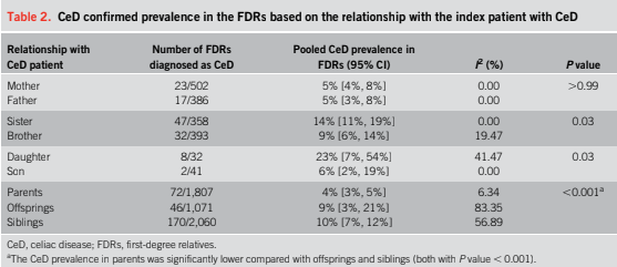
  • Daughters and sisters had the highest prevalence rates at 23% and 14%, compared with sons and brothers at 6% and 9%, respectively. Mothers/fathers prevalence rates were 5%. It is noted, however, that the stud only included 32 daughters and 41 sons, making these estimates less reliable
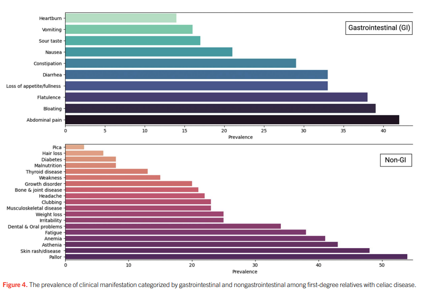
  • Abdominal pain (42%), bloating (39%), and flatulence (38%) were the most common gastrointestinal symptoms, while 34% of FDRs with CeD were asymptomatic

Discussion points:

Discrepancy between serology and biopsy: “First, not all individuals who tested positive through serological screening underwent a confirmatory duodenal biopsy. Second, some individuals with positive anti-tTG Ab may have false-positive results, or the disease process is still in the early stages of the disease, where intestinal damage is not yet detectable. This highlights that relying solely on serological screening without follow-up evaluations and intestinal biopsy can lead to overestimating the true prevalence of CeD.”

Limitations: “Some included studies only screened the siblings of indexed patients with CeD. For example, one study reported a prevalence of 22% among siblings. Given that genetic factors play a pivotal role in the pathogenesis of CeD and that the prevalence of CeD among siblings is often higher than that of other FDRs, this selective screening approach could potentially introduce selection bias into the overall prevalence of CeD in FDRs.”

My take: This study supports routine screening of first-degree relatives of patients with Celiac Disease, especially as many are asymptomatic.

Related blog posts:

Celiac Studies: Lower Rates of Undiagnosed Celiac Disease in Norway, and Lower Rates of Celiac with Early Dietary Fiber

P Lukina et al. Clin Gastroenterol Hepatol 2025; 23: 1143-1151. The Prevalence and Rate of Undiagnosed Celiac Disease in an Adult General Population, the Trøndelag Health Study, Norway

Methods: The study used the fourth Trøndelag Health Study (HUNT4), conducted in 2017–2019, where 56,042 adult (aged >20 years) residents of Nord-Trøndelag County, Norway, participated. Serum samples from 54,505 participants were analyzed for anti-transglutaminase 2 IgA and IgG.

Key findings:

  • The rate of CeD seropositivity was 2.0% (1107/54,505).  
  • The total biopsy-confirmed prevalence of CeD was 1.5% (853/56,042).
  • The study confirmed a new CeD diagnosis after participation in HUNT4 in 470 individuals and a known CeD diagnosis before participation in HUNT4 in 383 individuals.
  • The ratio of new, previously undiagnosed CeD cases (after HUNT4) to known, previously diagnosed CeD cases (before HUNT4) was 1.2:1 (470/383).

My take: This study showed that the ratio of undiagnosed cases to diagnosed case of CeD was improved from previously in Norway.

EM Hård af Segerstad, et al. Gastroenterol 2025; 168: 1185-1188. Open Access! Early Dietary Fiber Intake Reduces Celiac Disease Risk in Genetically Prone Children: Insights From the TEDDY Study

Methods: This study examined the quantity and timing of dietary fiber intake in children up to 5 years of age who were at genetic risk for celiac disease, assessing its relationship with their subsequent risk of developing the condition. The analysis included 6520 children carrying the HLA-DQ2 and/or DQ8 risk haplotypes who were prospectively followed for a total 61,669 person-years to age 13 years in The Environmental Determinants of Diabetes in the Young (TEDDY) study.

Key findings:

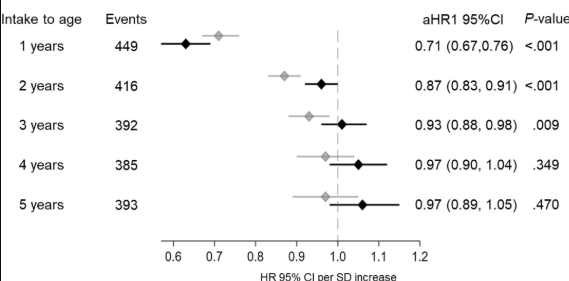
  • Dietary fiber intake from 6 months to 3 years of age was inversely associated with the subsequent risk of celiac disease in models unadjusted for the concurrent gluten intake and to 2 years of age in models adjusted for gluten intake (Figure 1A). In contrast, no association was observed between dietary fiber intake to 4 and 5 years of age and the risk of celiac disease regardless of whether models accounted for the gluten intake. 

My take: Higher dietary fiber intake during the first 2 years of life was associated with a lower risk of celiac disease in children at genetic risk. Although this effect was modest, it was independent of gluten intake and other components found in fiber-rich foods in the child’s diet.

Related blog posts:

Navigating Celiac Disease: Tips for Pediatric to Adult Transition

L Kievela et al. Clinical Gastroenterology and Hepatology,2025; 23: 908 – 911. Transition of Care in Celiac Disease: A Chance to Advance

This clinical practice article offers some useful advice in transitioning patients with Celiac disease into adult provider care. Some of their recommendations for adult provider care:

  • Regular visits
  • Assessment of gluten-free diet adherence with low thresholf for dietician referral
  • Checking serum tissue transglutaminase antibody levels
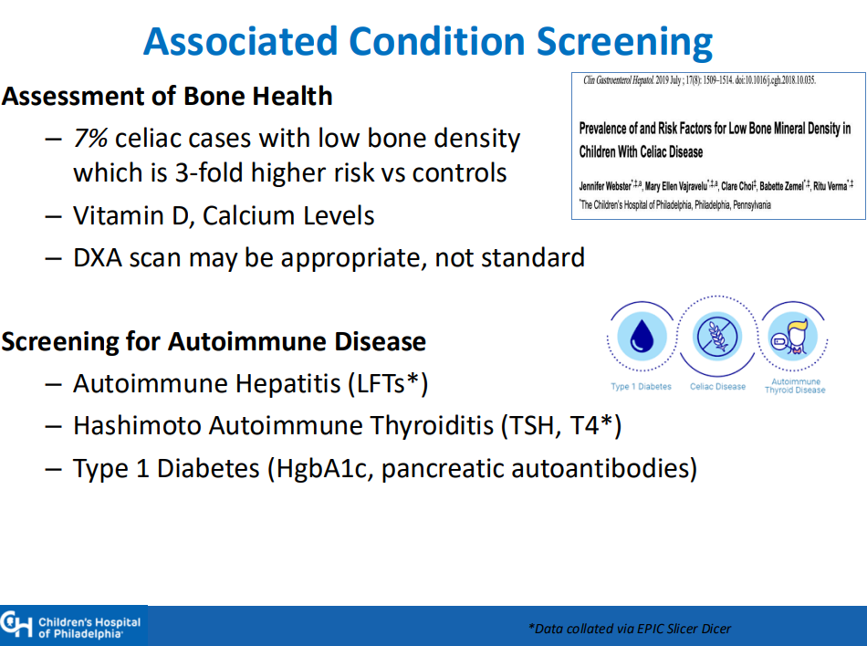
  • Screening for other autoimmune conditions
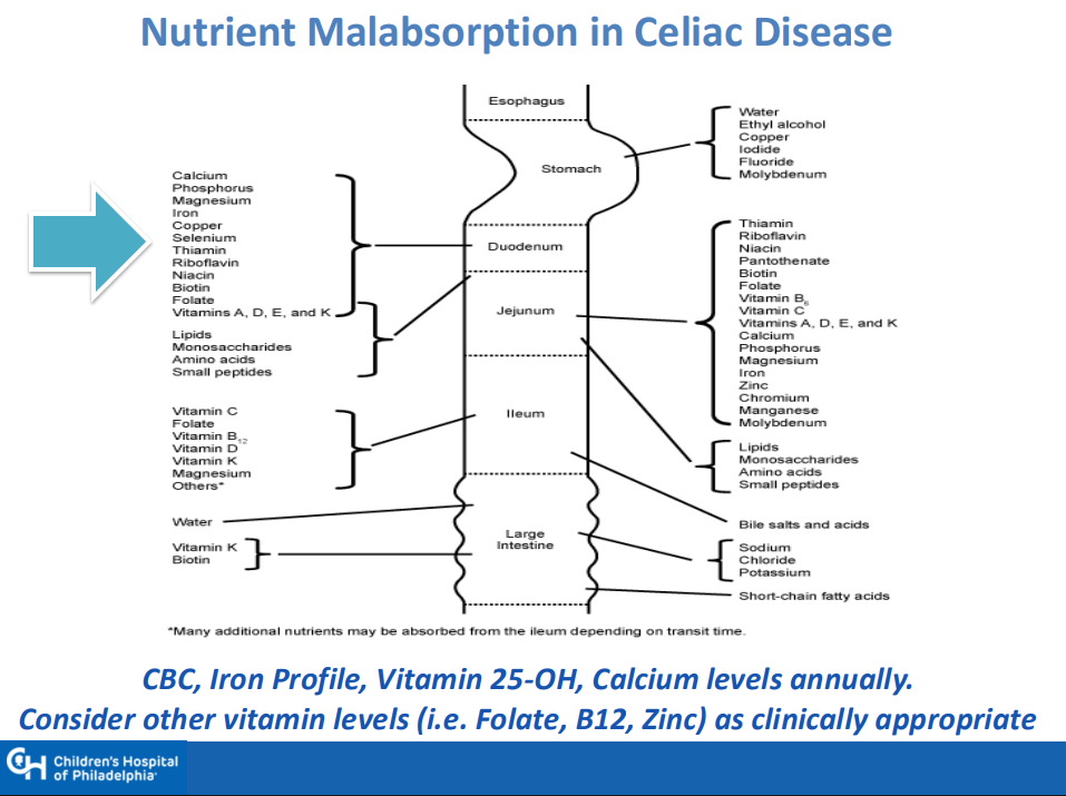
  • Consider additional lab tests (eg. CMP, CBC, Ferritin, ALT, Albumin, Folate, Zinc, Vit D, Vit B12, and TSH) and DEXA scan based on clinical situation
  • Recommend screening family members
  • Check vaccination status
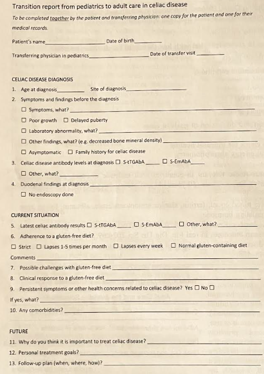
Figure 1 provides suggestions for a transition report

Related information that is not in the article:

College/school resources:

Dr. Arun Singh: Tips and Tricks to Managing Celiac Disease

Recently Dr. Arun Singh gave our group a terrific update on Celiac Disease. I have taken some notes and shared some slides. There may be inadvertent omissions and mistakes in my notes.

Key points:

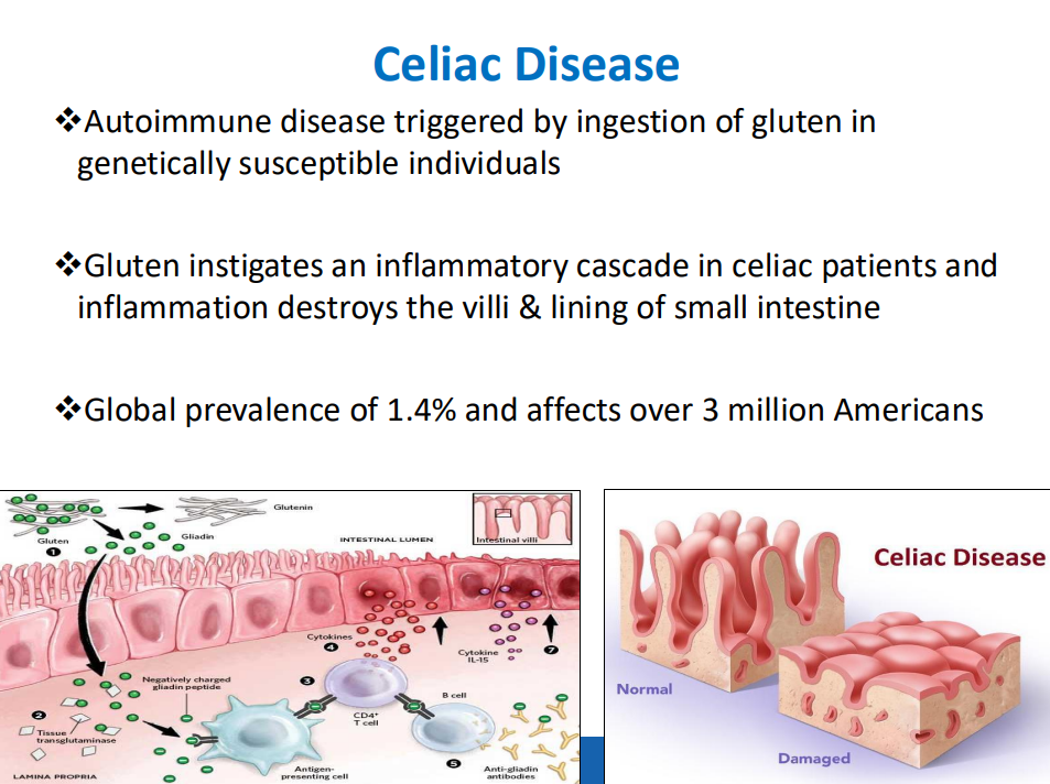
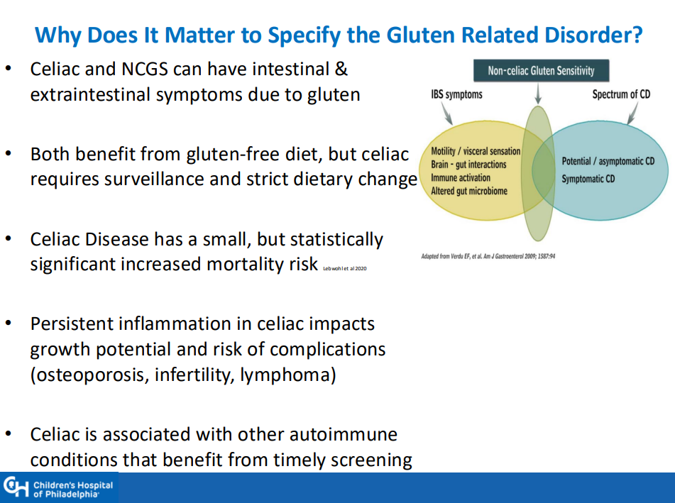
  • Celiac disease (CD) global prevalence is about 1.4%, though there are ‘pockets’ with much higher rates (~3% prevalence in Colorado). This equates to more than 3 million Americans with CD
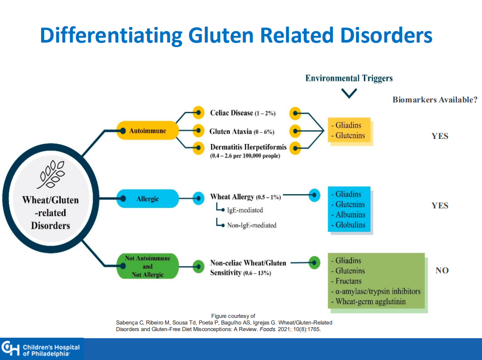
  • There is a huge gluten free diet market of ~$7 billion. This market includes CD, nonceliac gluten sensitivity (NCGS) and those with wheat allergies
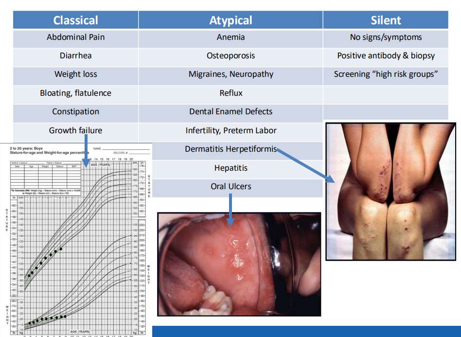
  • Many patients have atypical symptoms which can include ADHD, brain fog, headaches, and elevated LFTs. Many have silent CD with no symptoms
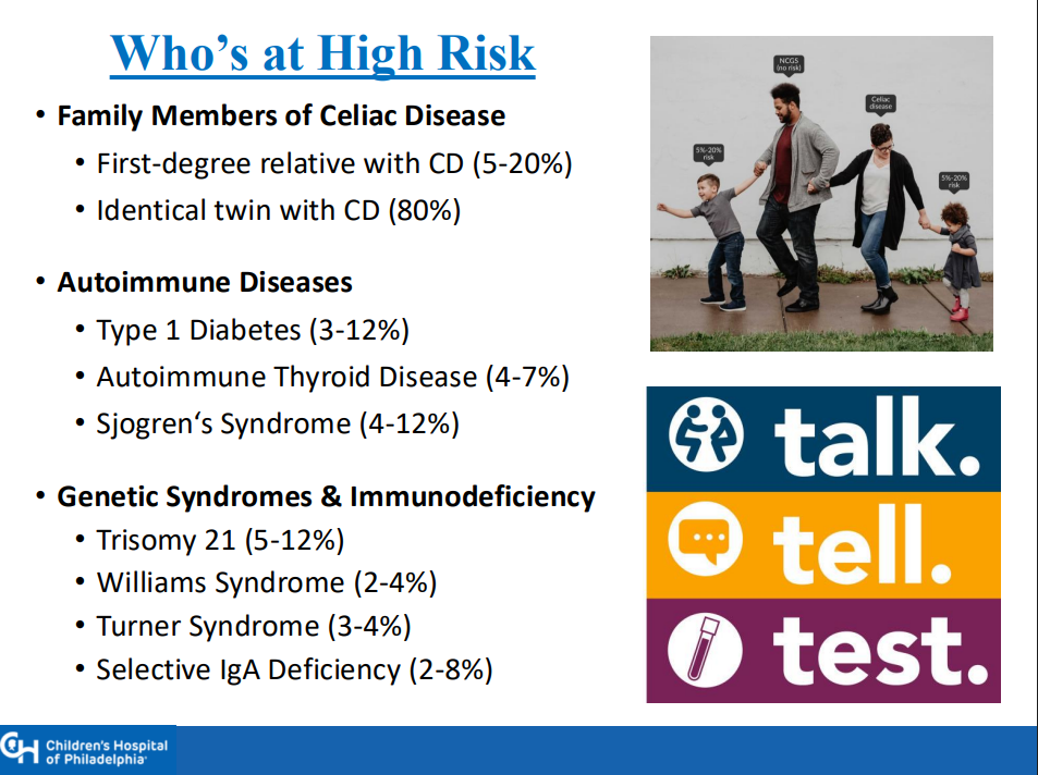
  • Higher risk groups include family members (~10% risk for 1st degree, ~80% risk for identical twin), autoimmune diseases (thyroid, diabetes, others), and genetic disorders (Down syndrome, Williams syndrome, Turner syndrome)
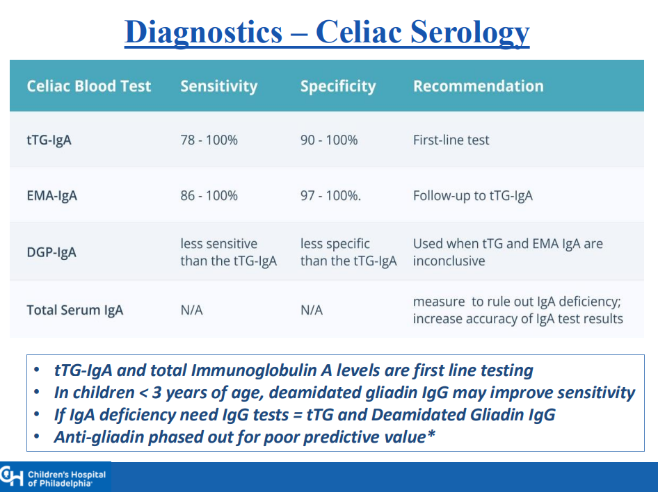
  • Transient elevation of TTG IgA is common. In TEDDY study, 19% had TTG IgA spontaneously normalize. Thus, a single abnormal lab is not reliable
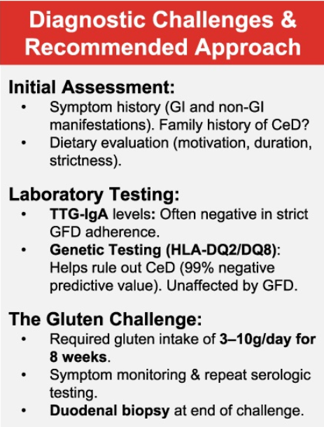
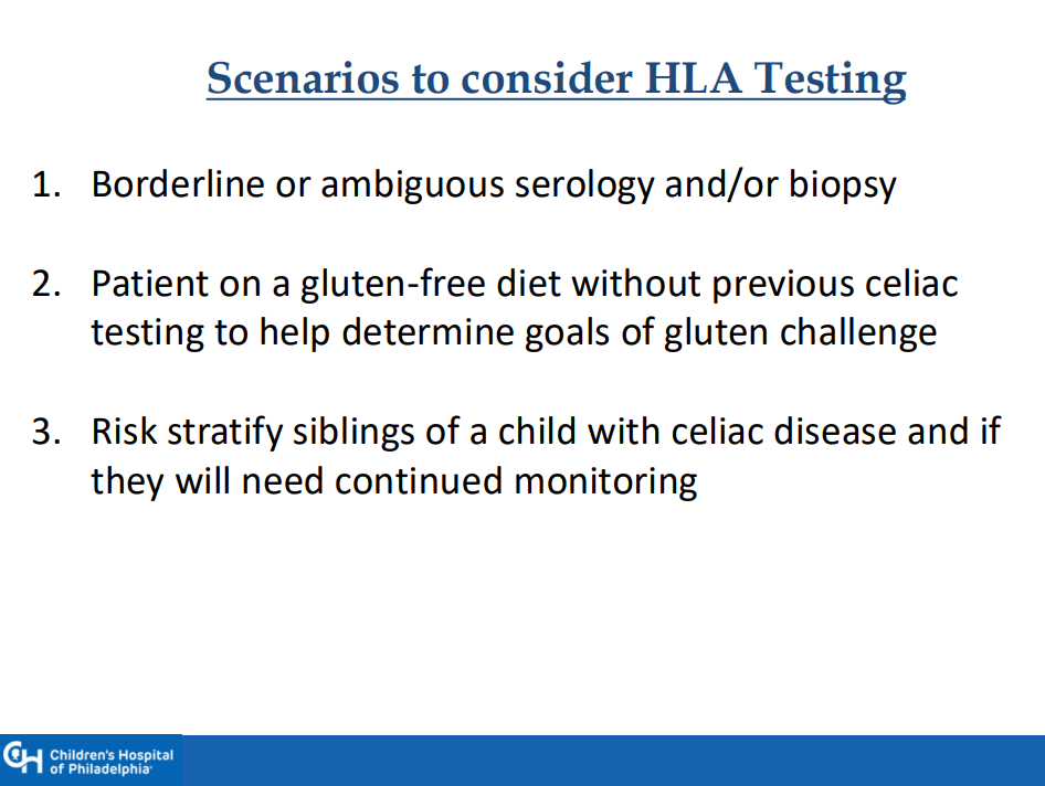
  • Genetic testing can be a useful adjunct in a few specific situations, including prior to instituting a gluten challenge
  • 1-3% of those with celiac disease may be negative for HLA-DQ2 and HLA-DQ8
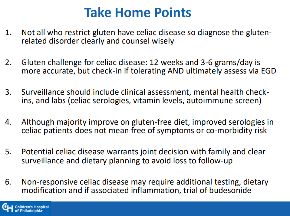
  • If a gluten challenge is needed, 12 weeks is ideal. However, if poorly tolerated, then consider endoscopy earlier and Dr. Singh recommends checking in with family 4-6 weeks into the challenge
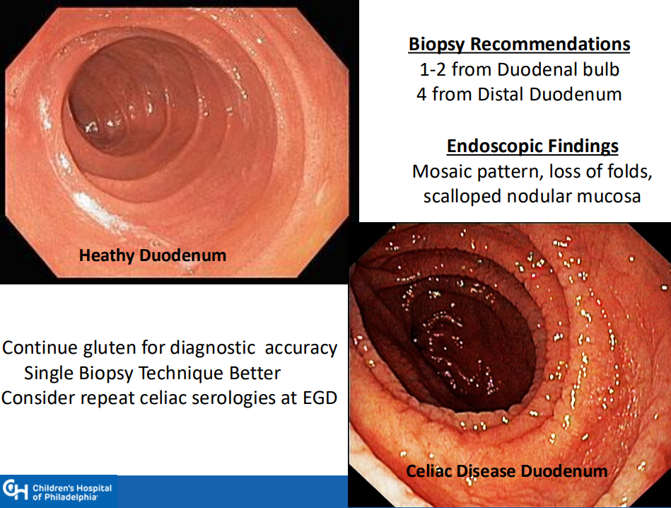
  • Endoscopy recommendations: Taking a single biopsy per pass can improve orientation when obtaining duodenal biopsies (bulb and distal portion, 5-6 in total)
  • NASPGHAN has not updated comprehensive guidelines for CD in 20 years
  • A survey of NASPGHAN members indicated that ~40% utilize a “no-biopsy” approach in patients. Dr. Singh noted that the accuracy of this approach, based on data from North America, may be about 96%
  • Drug trials for CD require a biopsy-confirmed diagnosis
  • Surveillance practice is quite variable. Important to follow growth and serology. CHOP approach includes surveillance for type 1 DM
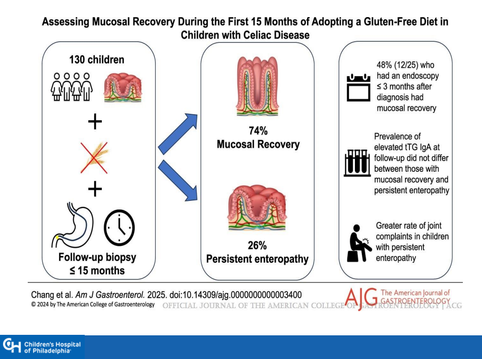
  • Followup endoscopy to assess mucosal healing is not the current standard of care but could be helpful in some patients
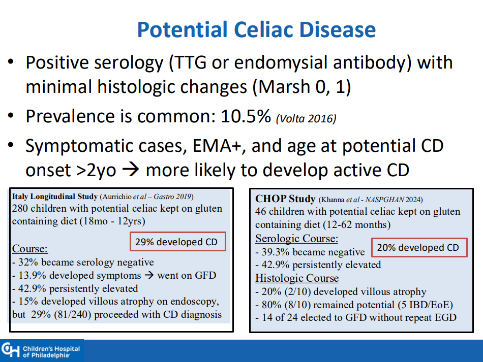
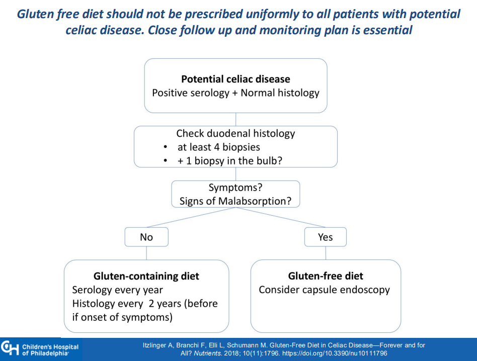
  • Among patients with potential CD, about 30% develop CD over time. Thus, these patients should be monitored (yearly labs, f/u scope after 2 years)
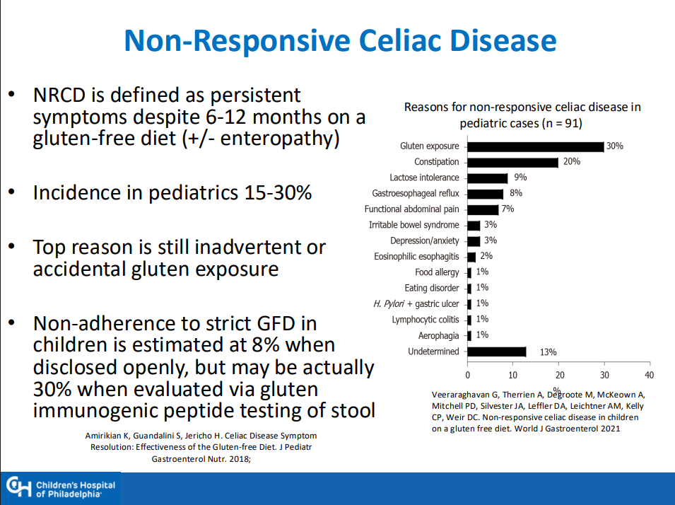
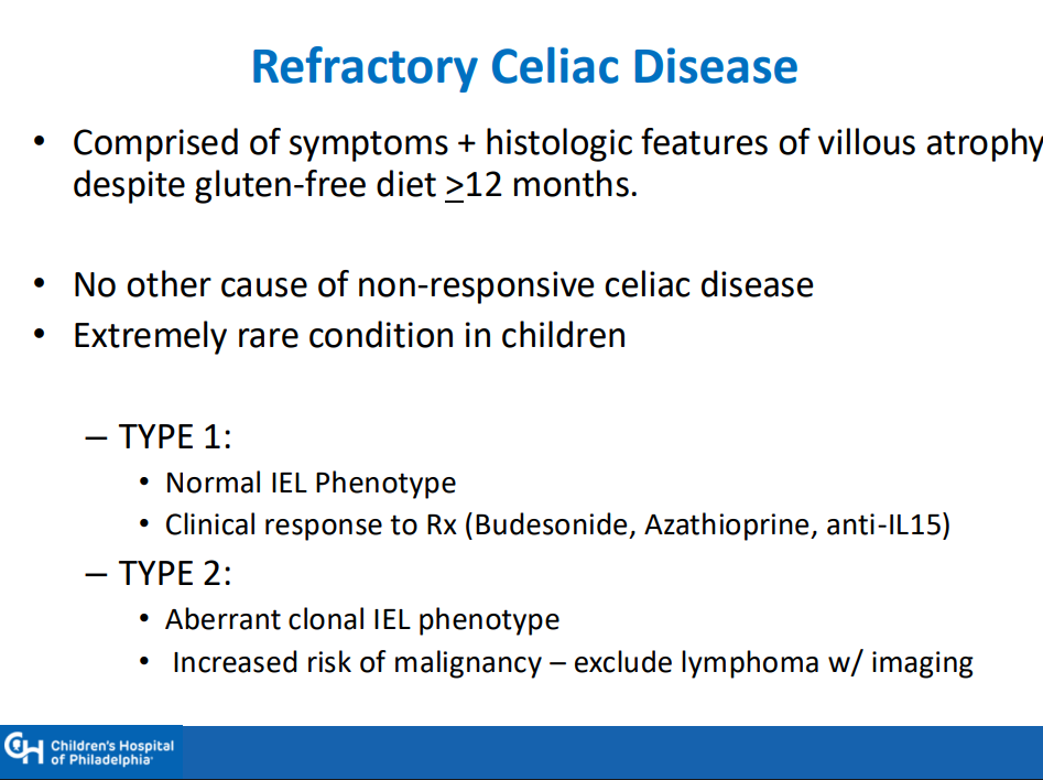
  • Nonresponsive/refractory CD: start with nutrition assessment, often needs a f/u scope before consideration of budesonide therapy (9 mg x 12 weeks) or gluten contamination elimination diet

Related blog posts:

Safe for Patients with Celiac Disease to Kiss after Partner’s Gluten Ingestion

An excerpt from News Medical:

Researchers recruited 10 couples, each with one partner who has celiac disease, for a two-part study. In each session, the non-celiac partner ate 10 saltine crackers, and then the couple kissed for 10 seconds. In one session, the partners waited five minutes before the kiss, and in the other, they drank 4 ounces of water before kissing…

Although gluten was still found in saliva after kissing a partner who had consumed gluten and then had a glass of water, in all cases the amount was less than 20 parts per million, the level allowed in gluten-free products, which is considered safe.

“Patients with celiac disease can be more relaxed, knowing that the risk of gluten cross-contact through kissing a partner who has consumed gluten can be brought down to safe levels if food is followed by a small glass of water.”

From NBC article:

In the first scenario — waiting five minutes before kissing — two of the celiac participants had more than 20 parts per million of gluten in their saliva sample. 

In the scenario in which non-celiac partners drank 4 ounces of water before the kiss, everyone’s saliva tests contained fewer than 20 ppm of gluten

My take: Sounds like a fun study. Best to drink water before kissing your partner who has celiac disease.

Reference: Anne Lee. DDW Abstract Mo1242, 5/5/25: “Assessing gluten transfer via kissing; a prospective study of celiac-discordant couples”

Celiac Disease: Lower TTG-IgA Titers Associated with Isolated Duodenal Bulb Presentation

QY Wang et al. JPGN 2025; 80:678–685. Open Access! Low TTG-IgA associated with isolated bulb pathology in pediatric celiac disease: Implications in a no-biopsy approach era

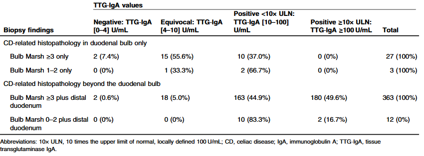
Methods: There were 405 cases included in this retrospective study (mean age = 9.6 years). TTG-IgA values were considered negative if <4 U/mL, equivocal between 4 and 10 U/mL inclusively, and positive if >10 U/mL. At the authors’ institution, TTG‐IgA ≥10× ULN corresponds to ≥100 U/mL.

Key findings:

  • Bulb-restricted CD was present in 7.4% of cases
  • TTG-IgA was negative or equivocal in 60.0% of bulb-restricted CD, compared to 5.3% of distal duodenal CD (odds ratio [OR] = 26.6
  • No bulb-restricted CD cases attained TTG-IgA ≥10× ULN, compared to 48.5% of distal duodenal CD

My take: This study confirms and quantitates what most clinicians have experienced. Isolated duodenal bulb pathology is associated with lower celiac titer abnormalities.

Related blog posts:

Gorgeous stained glass by Margaret Vetter at Piedmont Arts Festival

Celiac Disease: Pro Tips (Part 3)

In June 2024 (special issue), Gastroenterology published an entire issue (193 pages) focused on celiac disease. There was a lot of useful information on almost every aspect of this disease. Below I have summarized some of the points.

ME Robert et al. Open Access! Opportunities for Improving Biopsy and Non–Biopsy-Based
Diagnosis of Celiac Disease

This article has the most pragmatic advice in the entire issue.

Key points regarding the Non-Biopsy Approach should be applicable in adults:

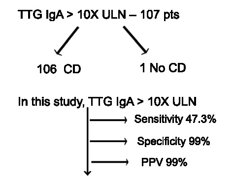
  • “Retrospective studies in adults found that, in different clinical settings, TTG IgA raised to 10 or more times the upper normal value predicts the presence of mucosal atrophy from 95.2% in low-risk populations to 100% in high-risk populations. There is evidence that the magnitude of the TTG increase correlates with the risk of more severe damage at histology.”
  • “A subsequent prospective multicenter, international study added further evidence for a non-biopsy approach. This study confirmed that a 5-fold and a 10-fold increase of TTG in a high-risk population predicted mucosal atrophy in 97.4% and 97.5%, respectively, when the biopsy was interpreted locally. However, after a central expert pathologist re-evaluated local histology, the PPV of a 10-fold TTG elevation was 99.4%…the only patient [without initial diagnosis of CeD] was eventually diagnosed as having CeD.”
  • The authors discount the rationale for endoscopy in those with TTG IgA >10-fold ULN. 1. Missed CeD-related diagnosis (eg. EoE, lymphocytic gastritis): “The concern about missed endoscopic diagnoses in a biopsy-free approach appears to be a theoretical concern without empiric data.” 2. Concern for CeD complications: “Ulcerative jejunitis and enteropathy-associated T cell lymphoma are rare and, generally, not detected by the initial endoscopy” 3. Overdiagnosis (identification of Potential CeD):”PCeD is rarely found in cases of very high levels of TTG…Moreover, concern about the long-term implications of untreated PCeD may lead to a recommendation for a GFD, rendering moot the need for a biopsy.”
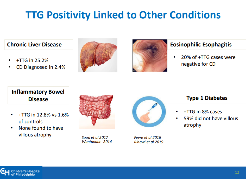
  • Certain populations like those with type 1 diabetes should undergo a biopsy as the serology has a “lower specificity” in this group.
  • There is concern that widespread adoption of a biopsy-free approach may lead to an over-reliance on serologies that fall short of the criteria that would lead to an accurate diagnosis.

Key points about Biopsy-Based Approach:

  • At present, there is broad consensus that among individuals with an elevated TTG IgA that falls short of a 10-fold elevation, a biopsy-based approach is necessary.
  • Obtain appropriate biopsies: guidelines recommend 2 biopsies from the first portion of duodenum and 4 biopsies from the distal duodenum (1 biopsy per pass recommended)

Related blog posts:

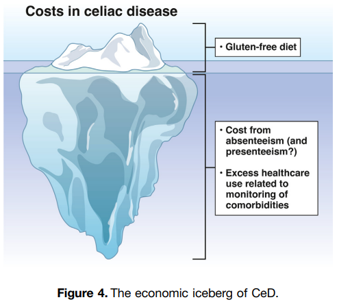
SR Bozorg et al. Open Access! The Economic Iceberg of Celiac Disease: More Than the Cost of Gluten-Free Food

Key points:

  • Long-term data from Sweden have revealed a persisting excess use of health care, with health care costs estimated to be 1.79 times higher than in reference individuals up to 5 years after diagnosis. Similar observations were made by Violato et al, who found UK primary health care costs in patients with incident CeD to be approximately 1.9 times higher than in reference individuals from 5 to 10 years after diagnosis.
  • Data collected from the Swedish national social insurance register showed that working-age patients with prevalent CeD had 1.49 times more work loss than matched reference individuals (42.5 days vs 28.6 days), equivalent to $2800 in lost productivity in 2015

Related blog post: Work Disability with Celiac Disease

Celiac Disease: Pro Tips (Part 2)

In June 2024 (special issue), Gastroenterology published an entire issue (193 pages) focused on celiac disease. There was a lot of useful information on almost every aspect of this disease. Below I have summarized some of the points.

V Disepolo et al. Open Access! How Future Pharmacologic Therapies for Celiac Disease Will Complement the Gluten-Free Diet

  • This article focuses on the emerging pharmacologic treatments
One of the treatment strategies has been to try to sequester gluten. This has included using enzymes to degrade dietary gluten. Other approaches include genetically modifying diet, tight junction modulation, immune modulation and tolerance induction.
Some therapies being developed in adults may have unacceptable risk profiles for children. This could include blocking cytokine signaling (anti–IL-15, anti–IL-15/IL-21) and blocking intestinal T-cell recruitment

Related blog posts:

G Malamut et al. Advances in Nonresponsive and Refractory Celiac Disease

Celiac Disease: Pro Tips (Part 1)

In June 2024 (special issue), Gastroenterology published an entire issue (193 pages) focused on celiac disease. There was a lot of useful information on almost every aspect of this disease. Below I have summarized some of the points.

F Zingone et al. Open Access: Celiac Disease–Related Conditions: Who to Test?

  • The authors detail disorders with increased risk for CeD and which merit screening (see Table 2 below). Some disorders that merit screening that are more obscure include idiopathic pancreatitis, autoimmune hepatitis, delayed menarche, and chronic fatigue.
  • They note that type 1 diabetes mellitus could require serial screening. “Because CeD can manifest at any time and with greater frequency during the initial 5 years, conducting additional screenings in CeD-negative T1DM patients 2 and 5 years after T1DM diagnosis and those who later develop gastrointestinal (GI) symptoms may be advisable.” In addition, it is important to recognize that CeD serology testing is less reliable in patients with T1DM.

S Gatti et al. Patient and Community Health Global Burden in a World With More Celiac Disease, describes the worldwide burden of celiac disease and how to improve detection.

  • They note that the worldwide prevalence is between 0.7% and 2.9%. In this issue, most authors estimate the prevalence to be about 1% with more than 50% undetected.
  • There are many places with higher rates. In U.S. “children in Colorado had a 2.5-fold higher risk compared to Washington State…similar regional differences were seen …in Sweden, Finland, and Germany.”
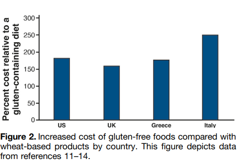
  • They note the burden before and after diagnosis. Before diagnosis/undetected, there can be persistent symptoms, complications (eg. osteoporosis, decreased fertility) and impaired quality of life. Afterwards, there are increased costs of a GFD and psycho-social burden of GFD.
  • In terms of generalized screening compared to case-finding, the authors note that given the number of at-risk groups, the case-finding approach could entail screening >50% of the population.

V Abadie et al. New Insights on Genes, Gluten, and Immunopathogenesis of Celiac Disease, reviews the intricate details of genetic, biochemical, and immunologic studies, which together have revealed mechanisms of gluten peptide modification and HLA binding, thereby enabling a maladapted anti-gluten immune response.

  • What I was most interested in was the mechanisms behind ‘potential’ celiac disease (PCeD) in which patients have autoimmunity (+serology) but normal histology. In potential CeD, the anti-CD4+ T-cell response is present but decoupled from tissue cytotoxicity. However, notably, IL-21, a cytokine produced by gluten-specific CD4+ T cells in active CeD, is not up-regulated in potential CeD…In addition, patients with potential CeD lack the presence of an epithelial stress response associated with IL-15, HSP70, and HSP27 upregulation in epithelial cells.” “The presence of epithelial stress is a crucial prerequisite for the development of tissue damage.”

Increased Risk of Irritable Bowel Before and After the Diagnosis of Celiac Disease

K Marild et al. Clin Gastroenterol Hepatol 2024; Open Access (PDF)! Association Between Celiac Disease and Irritable Bowel Syndrome: A Nationwide Cohort Study

Methods: Using Swedish histopathology and register-based data, we identified 27,262 patients with CD diagnosed in 2002–2017 and 132,922 age- and sex-matched general population comparators.

Key Findings:

  • During an average of 11.1 years of follow-up, 732 celiac patients (2.7%) were diagnosed with IBS vs 1131 matched general population comparators (0.9%).
  • Compared with siblings (n= 32,010), celiac patients (n = 19,211) had >/= 2-fold risk of later IBS (aHR, 2.42)
  • Compared with celiac patients with mucosal healing, those with persistent villus atrophy on follow-up biopsy were less likely to be diagnosed with IBS (aHR, 0.66)

Interpretation of findings:

“We found celiac patients with persistent villus atrophy on follow-up biopsy less likely to be
diagnosed with IBS than those with mucosal healing. Traditionally, physicians have hesitated to diagnose IBS in patients with an organic gastrointestinal disorder (eg, CD), possibly underestimating the observed IBS risk in CD. This reluctance to diagnose IBS may be particularly true for celiac patients who have not achieved mucosal healing, because persistent villus atrophy may indicate that ongoing symptoms are due to gluten exposure
instead of IBS.”

Surveillance bias is another challenge of studies associating IBS with CD. From the outset of diagnosing and managing these conditions, they are often mutually excluded (eg, CD-specific serology tests are often part of the workup of IBS-like symptoms). Consequently, the
strength of the association between these conditions may be overestimated.” This is why the authors focused on IBS events beyond the first year of CD diagnosis and there continue to be an increased risk of IBS 10 years of follow-up.

Another limitation of this study: “a large proportion of IBS patients are cared for in
primary care or never seek care at all, and hence our study may have had a low sensitivity for IBS, particularly mild IBS.”

My take: While recurrent symptoms in patients with CD could indicate ongoing gluten exposure, recurrent symptoms can also be due to IBS which can occur even with mucosal healing.

Related blog posts: