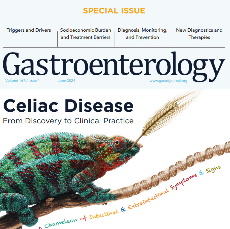
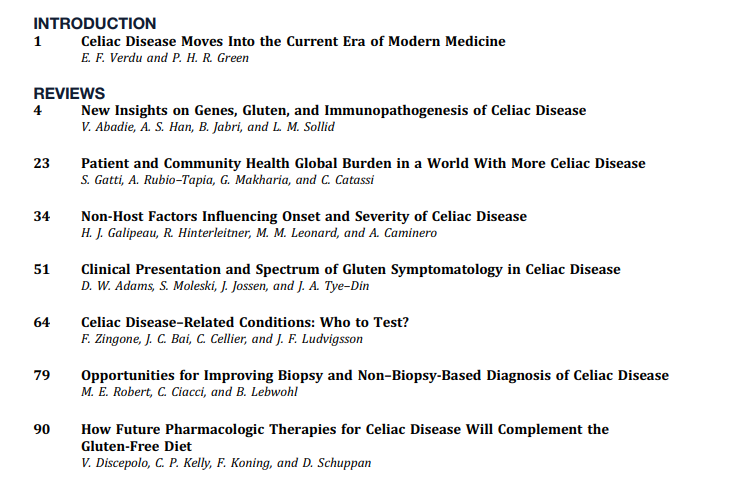
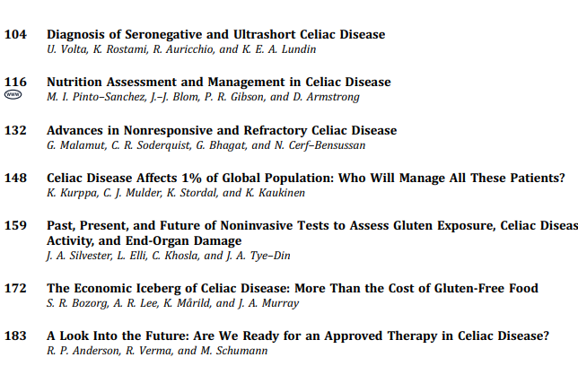
In June 2024 (special issue), Gastroenterology published an entire issue (193 pages) focused on celiac disease. There was a lot of useful information on almost every aspect of this disease. Below I have summarized some of the points.
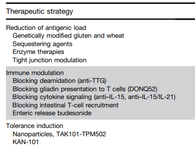
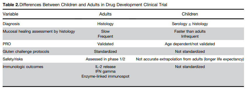
This article focuses on the emerging pharmacologic treatments
One of the treatment strategies has been to try to sequester gluten. This has included using enzymes to degrade dietary gluten. Other approaches include genetically modifying diet, tight junction modulation, immune modulation and tolerance induction.Some therapies being developed in adults may have unacceptable risk profiles for children. This could include blocking cytokine signaling (anti–IL-15, anti–IL-15/IL-21) and blocking intestinal T-cell recruitment
This article addresses nonresponsive CeD and refractory CeD. In the pediatric population, nonresponsive CeD is mainly related to persistent gluten exposure. Refractory CeD is rare in children.
In June 2024 (special issue), Gastroenterology published an entire issue (193 pages) focused on celiac disease. There was a lot of useful information on almost every aspect of this disease. Below I have summarized some of the points.
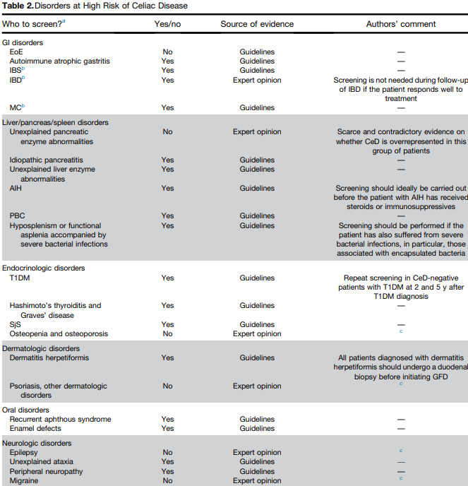
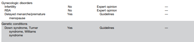
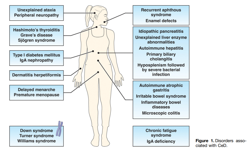
The authors detail disorders with increased risk for CeD and which merit screening (see Table 2 below). Some disorders that merit screening that are more obscure include idiopathic pancreatitis, autoimmune hepatitis, delayed menarche, and chronic fatigue.
They note that type 1 diabetes mellitus could require serial screening. “Because CeD can manifest at any time and with greater frequency during the initial 5 years, conducting additional screenings in CeD-negative T1DM patients 2 and 5 years after T1DM diagnosis and those who later develop gastrointestinal (GI) symptoms may be advisable.” In addition, it is important to recognize that CeD serology testing is less reliable in patients with T1DM.
They note that the worldwide prevalence is between 0.7% and 2.9%. In this issue, most authors estimate the prevalence to be about 1% with more than 50% undetected.
There are many places with higher rates. In U.S. “children in Colorado had a 2.5-fold higher risk compared to Washington State…similar regional differences were seen …in Sweden, Finland, and Germany.”
They note the burden before and after diagnosis. Before diagnosis/undetected, there can be persistent symptoms, complications (eg. osteoporosis, decreased fertility) and impaired quality of life. Afterwards, there are increased costs of a GFD and psycho-social burden of GFD.
In terms of generalized screening compared to case-finding, the authors note that given the number of at-risk groups, the case-finding approach could entail screening >50% of the population.
V Abadie et al.New Insights on Genes, Gluten, and Immunopathogenesis of Celiac Disease, reviews the intricate details of genetic, biochemical, and immunologic studies, which together have revealed mechanisms of gluten peptide modification and HLA binding, thereby enabling a maladapted anti-gluten immune response.
What I was most interested in was the mechanisms behind ‘potential’ celiac disease (PCeD) in which patients have autoimmunity (+serology) but normal histology. In potential CeD, the anti-CD4+ T-cell response is present but decoupled from tissue cytotoxicity. However, notably, IL-21, a cytokine produced by gluten-specific CD4+ T cells in active CeD, is not up-regulated in potential CeD…In addition, patients with potential CeD lack the presence of an epithelial stress response associated with IL-15, HSP70, and HSP27 upregulation in epithelial cells.” “The presence of epithelial stress is a crucial prerequisite for the development of tissue damage.”
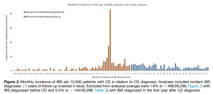
Methods: Using Swedish histopathology and register-based data, we identified 27,262 patients with CD diagnosed in 2002–2017 and 132,922 age- and sex-matched general population comparators.
Key Findings:
During an average of 11.1 years of follow-up, 732 celiac patients (2.7%) were diagnosed with IBS vs 1131 matched general population comparators (0.9%).
Compared with siblings (n= 32,010), celiac patients (n = 19,211) had >/= 2-fold risk of later IBS (aHR, 2.42)
Compared with celiac patients with mucosal healing, those with persistent villus atrophy on follow-up biopsy were less likely to be diagnosed with IBS (aHR, 0.66)
Interpretation of findings:
“We found celiac patients with persistent villus atrophy on follow-up biopsy less likely to be diagnosed with IBS than those with mucosal healing. Traditionally, physicians have hesitated to diagnose IBS in patients with an organic gastrointestinal disorder (eg, CD), possibly underestimating the observed IBS risk in CD. This reluctance to diagnose IBS may be particularly true for celiac patients who have not achieved mucosal healing, because persistent villus atrophy may indicate that ongoing symptoms are due to gluten exposure instead of IBS.”
“Surveillance bias is another challenge of studies associating IBS with CD. From the outset of diagnosing and managing these conditions, they are often mutually excluded (eg, CD-specific serology tests are often part of the workup of IBS-like symptoms). Consequently, the strength of the association between these conditions may be overestimated.” This is why the authors focused on IBS events beyond the first year of CD diagnosis and there continue to be an increased risk of IBS 10 years of follow-up.
Another limitation of this study: “a large proportion of IBS patients are cared for in primary care or never seek care at all, and hence our study may have had a low sensitivity for IBS, particularly mild IBS.”
My take: While recurrent symptoms in patients with CD could indicate ongoing gluten exposure, recurrent symptoms can also be due to IBS which can occur even with mucosal healing.
In this Swedish cross-sectional cohort study with 162 children who had been diagnosed with celiac disease (CD) between 2013-2018, the authors examined the outcomes of children who had continued follow-up compared to those who had not been seen in 24 months. The average disease duration of study participants was 5.3 years.
Key findings:
Similar rates of TTG IgA normalization: 94% vs 91% for those without and those with follow-up respectively
Similar rates of very good dietary adherence 65% vs 72% for those without and those with follow-up respectively
Lack of follow-up was not significantly associated with growth, symptom scores, or HRQoL.
It is possible that there is a selection bias in that patients without symptoms may be less likely to followup.
My take: Based on this study, it looks like good education after diagnosis is crucial and that regular follow-up is less important in achieving good outcomes.
In 2022, a study in JPGN showed that the rate of Hepatitis B virus (HBV) vaccination and immunity was similar in individuals with and without celiac disease (CD). In addition, there was no increased risk of HBV infection detected in CD patients. Thus, routinely checking hepatitis B status in all patients with CD was no longer justified. (See: Celiac Disease, Hepatitis B and Paul Harvey).
The same researchers in this study expand their findings to inflammatory bowel disease (IBD) and CD. In this retrospective cohort (2000-2019), using the Rochester Epidemiology Project which includes data from 162,847 residents. Key findings:
1264 incident cases of IBD/CD, only 6 HBV infections were diagnosed before the index date; 5 of the 6 had risk factors including IV drug use or living in endemic region.
No new HBV infection developed in any of 1258 patients with IBD/CD during a median follow-up of 9.4 years
The proportion of patients with HBV-protective titers (≥10 mIU/mL) decreased with time before plateauing, with protective titer rates of 45% at 5 up to 10 years and 41% at 15 up to 20 years after the last HBV vaccination. The control population with protective titers also decreased similarly with time though was consistently higher than the levels of patients with IBD/CD within 15 years after the last HBV vaccination
Context/Discussion:
Only 16% of vaccine recipients have measurable protective titers by age 18 years, according to the CDC.32
“Time-related waning of Ab levels to HBV after vaccination has unclear clinical significance. Although screening persons for HBV immunity by using anti-HBs titers is widely accepted, prior study results have shown that cellular immunity can also provide long-term HBV protection, even in the setting of nonprotective titers.”
Reactivation of HBV is a well-documented complication of immunosuppression in patients with IBD, and screening for dormant infection is of paramount importance at diagnosis
Limitations: the study population had a low rate of HBV acquisition; thus, the study findings may not apply to areas with higher risk for HBV.
My take: This study shows that treating low hepatitis B surface antibody levels with reimmunization is likely NOT needed in either the IBD or the celiac disease population, except perhaps in those at high risk. Checking HBV status prior to immunosuppressive therapy, though, is still needed to prevent reactivation of HBV in those at risk.
Disclaimer: This blog, gutsandgrowth, assumes no responsibility for any use or operation of any method, product, instruction, concept or idea contained in the material herein or for any injury or damage to persons or property (whether products liability, negligence or otherwise) resulting from such use or operation. These blog posts are for educational purposes only. Specific dosing of medications (along with potential adverse effects) should be confirmed by prescribing physician. Because of rapid advances in the medical sciences, the gutsandgrowth blog cautions that independent verification should be made of diagnosis and drug dosages. The reader is solely responsible for the conduct of any suggested test or procedure. This content is not a substitute for medical advice, diagnosis or treatment provided by a qualified healthcare provider. Always seek the advice of your physician or other qualified health provider with any questions you may have regarding a condition.
In this study, the authors analyzed laboratory testing results from pediatric patients newly diagnosed with celiac disease (2018-2021) to determine the usefulness of each test derived from recommended guidelines (J Snyder et al. Pediatrics 2016; 138: e20153147). Screening protocols in their center resulted in an estimated cost of approximately $320,000 during the study. Tests at diagnosis included hemoglobin, alanine/aspartate aminotransferase, ferritin, iron, TSH, Free T4, and vitamin D screening. These screening tests were done in ~80% of 468 patients.
Key findings:
Ferritin was abnormal in 29%, hemoglobin was abnormal in 12%, and iron was abnormal in 22%. Abnormal ferritin captured all patients in this cohort with an abnormal iron. If ferritin was used as an isolated screen with reflective iron testing, this would have reduced costs by about $12,000
AST and ALT were abnormal in 2% and 11% respectively
25-OH Vitamin D was abnormal in 14%. Recent data indicated that low Vit D levels are similar among patients with and without celiac disease (R Ahlawat et al. JPGN 2019; 69: 449-454)
TSH and Free T4 were abnormal in 7% and 0.3% respectively. For thyroid disease, TSH and free T4 testing did not lead to any new diagnosis of thyroid disease (7 carried a preexisting diagnosis). There were 19 additional patients with abnormal lab values who had more testing due to initial abnormalities. If TSH alone were used for screening, costs savings would be about $29,000. If no thyroid testing were done, this would have reduced costs by about $40,000.
Hepatitis B immunity was NOT present in 69%. However, recent studies have shown similar levels of immunity in those with and without celiac disease. In addition, it is not clear that a low level hepatitis B surface antibody always indicates a lack of immunity. Eliminating hepatitis B screening would have reduced costs by about $63,000.
The authors note that the cost savings by adopting their recommendations would have saved about $104,000 (out of $320,000).
My take: This is a very useful study and indicates that curtailing initial testing for celiac disease could reduce costs substantially and without compromising care. This would include notchecking a serum iron, a free T4, or hepatitis B studies. The authors note that the value of Vit D testing is also questionable but may be worthwhile due to increased risk of bone disease in individuals with celiac disease.
This retrospective study of patients with trisomy 21 (T21) who underwent EGD between 2000-2020. Key findings:
Among 836 patients with T21, 419 (50.1%) of whom had duodenal histologic abnormalities.
290 of 419 had villous atrophy (VA) and of those, 172 of 290 met celiac disease (CD) diagnostic criteria, while 118 of 290 did not meet CD criteria (nonspecific VA).
Only one of the 118 with nonspecific VA had markedly abnormal celiac serology (TTG IGA >10 times ULN) but had no IELs on biopsy and did not respond to GFD. Four patients with nonspecific VA were later diagnosed with celiac disease over a median of 2.5 years with conversion to abnormal celiac serology.
Among the 129 with duodenitis without villous atrophy, 38 (29%) had increased intraepithelial lymphocytes and two patients were diagnosed with CD many years later (development of VA and positive celiac serology.
The authors offer an algorithm (Figure 2) to assist clinical approach based on biopsy results in this population.
In those with no villous atrophy, if Marsh score of 0/1, biopsies not consistent with celiac disease. In those with Marsh score of 2 (which is rare), consider as consistent with celiac disease (see next bullet point).
In those with villous atrophy consistent with celiac disease, assess response to gluten free diet (GFD). In those without a response, consider RD consult to evaluate exposures and HLA-typing as next steps.
In those with villous atrophy NOT consistent with celiac disease, obtain serology (TTG IgA, EMA, IgA, DGP IgG). If serology is negative, consider peptic duodenitis or SIBO treatments and assess/discontinue medications for potential for mucosal damage. If serology is positive, consider HLA-typing, review pathology slides, assess for competing disorders, and could need f/u endoscopy.
My take: Overall, ~20% of patients with T21 undergoing endoscopy, will have pathology consistent with celiac disease; this represents ~40% of those with abnormal histology. In the other 60% with abnormal histology, many have alternative explanations for the histologic findings (like peptic duodenitis). Some will evolve to meeting the diagnostic criteria for CD with time indicating need for ongoing monitoring. This study highlights the diagnostic uncertainty in those with potential for seronegative celiac disease.
At the start of my training in pediatric gastroenterology, the serological testing was not reliable and as a result, very few cases of children who did not have a ‘classical’ phenotype (eg. abdominal distention, poor growth, anemia) were diagnosed. The main exception was the diagnosis in children already recognized as high risk (eg. children with type 1 diabetes).
This recent retrospective study indicates that even with improvement in celiac serology, there are cases of seronegative celiac disease (SN-CD) that are difficult to diagnose. In this study, SN-CD diagnosis required clinical correlation and either confirmatory genetics or follow up endoscopy on a gluten-free diet. Key findings:
Of the 424 patients who met celiac disease (CD) criteria, 4.7% (n = 20) fulfilled the criteria for SN-CD
Nearly 65% of SN-CD were IgA sufficient compared with 98.4% in the seropositive group
All SN-CD patients were symptomatic whereas 82% of seropositive group was symptomatic
The discussion notes that it has been understood that the sensitivity of TTG IgA is about 95% and specificity about 96%. However, the authors caution that this may be “largely overestimated due to failure to account for verification bias. Only 3.6% of IgA-ATTG negative individuals were referred for biopsy” in Hujoel et al meta-analysis (J Clin Gastroenterol 2021; 55: 327-334); thus, the sensitivity could be as low as 57% based on this meta-analysis. In addition, gluten restriction prior to serological testing can further reduce the sensitivity of serological tests.
An important limitation of the study is proving that SN-CD was in fact SN-CD and not one of the mimics for CD (eg. inflammatory bowel disease, autoimmune disorders, medication effect). However, they noted that their cohort had followup over 6-9 years and with symptom resolution with a gluten free diet.
My take: Identifying SN-CD is difficult since so many children have similar digestive symptoms unrelated to celiac disease. Most children with vague digestive complaints do not need to undergo endoscopy; as such, SN-CD can be easily overlooked.
Methods: A retrospective cohort study was performed using the Military Healthcare System database. N=968,524 children with 1704 cases of celiac disease (CD) in this group (from 2001 to 2013) with prescription for PPIs, H2RAs or antibiotics in first 6 months of life.
Key findings:
PPIs (HR, 2.23; 95% CI, 1.76-2.83), H2RAs (HR, 1.94; 95% CI, 1.67-2.26), and antibiotics (HR, 1.14; 95% CI, 1.02-1.28) were all associated with an increased hazard of CD.
The risk is increased by use of multiple categories of these medications and/or if acid suppression medications are used for longer periods
There have been previous studies indicating an increased risk of CD in patients given acid suppression (Lebwohl et al. Dig Liver Dis 2014; 46: 36-40) and conflicting data regarding the use of antibiotics. With regard to acid suppression, recent studies have indicated that these medications in infancy may increase the risk of food allergies as well. The authors speculate in their discussion that the increased risk for CD could be related to changes in protein degradation, mucosal permeability, microbiome changes, and immune reactivity. The authors note that their dataset did NOT show an increased risk of CD associated with C-section delivery.
One of the limitations of this study is that early presentations of CD could lead to prescriptions of agents to to help reduce symptoms rather than the medications increasing the risk of developing CD. However, this is unlikely as gluten introduction is often later in infancy.
My take: Better stewardship of antibiotics and acid blockers is needed. Use of acid suppression medications is associated with an increased risk of celiac disease as well as food allergies.
In this retrospective study, 49 patients were screened due to an affected first-degree relative with celiac disease. They were compared to 178 patients who were screened for other clinical indications. Key findings:
Although 51% of patients screened due to an affected first-degree relative were asymptomatic, their disease histology and TTG levels were as severe as those screened for symptoms suggestive of celiac disease (in the comparison group 16% were asymptomatic).
Comments:
“Previous studies have shown that asymptomatic adolescents and those diagnosed with CD by serologic screening are less likely to adhere strictly to a GFD when compared to younger children and adults diagnosed because of classical symptoms” (Dig Dis Sci. 2008 Jun; 53(6): 1573–1581).”
Some individuals who are thought to be asymptomatic, clinically improve with a gluten free diet (GFD). In one study, “the GFD group also had reduced indigestion (P=.006), reflux (P=.05), and anxiety (P=.025), and better health, based on the visual analog scale (P=.017), than the gluten-containing diet group” (Gastroenterology 2014 Sep;147(3):610-617).
My take: In this study, being asymptomatic (identified due to affected first-degree relative) was NOT associated with milder celiac disease based on serology or histology.