Dr. Steve Erdman: Perplexing Polyposis Patients: a Case-Based Discussion

Recently, Dr. Steve Erdman gave our group a great update on polyposis disorders.  My notes below may contain errors in transcription and in omission. Along with my notes, I have included many of his slides.

Key points:

  • There has been breath-taking progress in understanding of polyposis disorders. It is important to have genetic counselors participate to optimize testing and evaluation
  • In patients with suspected polyposis syndromes, a genetic diagnosis is very important and can help guide management
  • Family history is very important. If several family members have had GI or other cancers at a young age, more aggressive interventions are usually indicated. However, individual family members can have a wide variation in presentation
  • In patients with many polyps, it is worthwhile to alert family to the fact that some polyps can be missed on colonoscopy and to contact medical team if there are recurrent symptoms like rectal bleeding
  • Some disorders, like juvenile polyposis syndrome (JPS), the connection between polyp presence and cancer risk is not clear. The increased risk for colon cancer my remain after polypectomy.
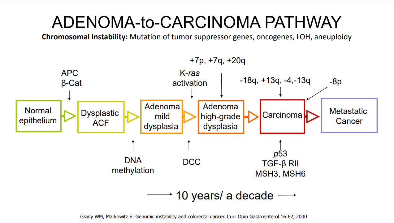
  • For most individuals with adenomatous polyps, removal of the polyps prevents cancer development (in the GI tract) as there is a well-described adenoma-to-cancer sequence that typically takes 7-10 years  to progress from adenoma to colon cancer
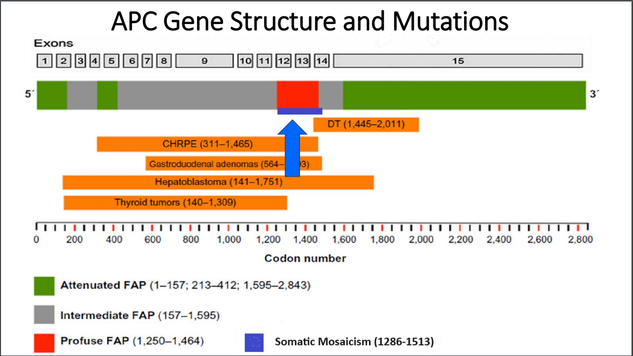
  • With FAP, the severity is related in part to the specific mutation. Mutations in the mutation cluster region are associated with an aggressive phenotype and mutations causing attenuated FAP are less aggressive
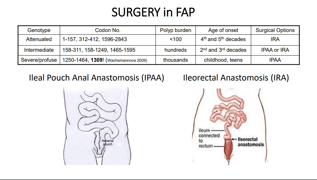
  • For FAP, timing of potential colectomy involves factors including severity as well as social factors. In teenagers in which there is a concern about being lost to follow-up, this is a factor that could influence earlier intervention
  • Many times a 2nd opinion in pathology can be helpful, especially if colon cancer is reported. However, histologic dysplasia can be tricky as well
  • Isolated CHRPE usually does not require evaluation. Dr. Erdman noted that sometimes genetic testing is offered to a family for reassurance. He discouraged colonoscopy in this setting unless a genetic diagnosis has been established or symptoms like rectal bleeding are present.  The penetrance of APC mutations (development of polyps) can be  quite variable (especially with attenuated form)
  • For JPS, after all polyps have been removed, consider surveillance every 3-5 years or for active symptoms (related post: ESPGHAN Juvenile Polyposis Syndrome in Children –Position Paper)

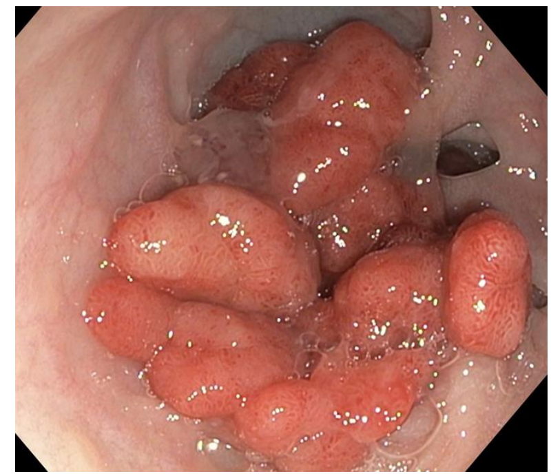
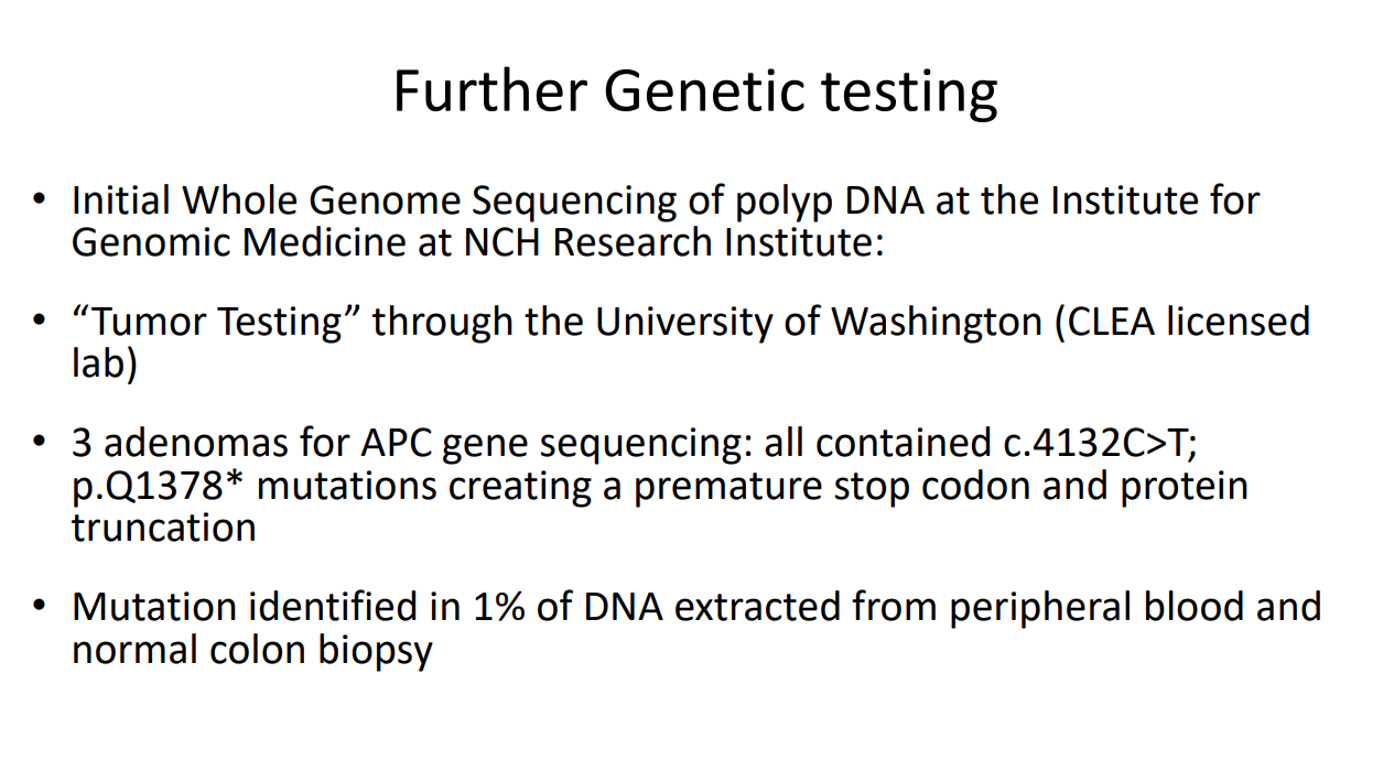
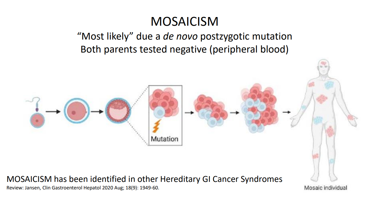
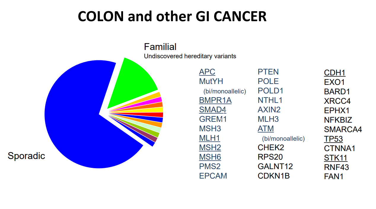
Case #1 presented a 14 yo with 50+ multilobulated pedunculated polyps which histologically were tubulovillus adenomas. Initial diagnosis was elusive despite extensive testing

Case#2 presented 8 yo twins. Aggressive management was indicated as 5 family members developed colorectal cancer prior to age 20 years.

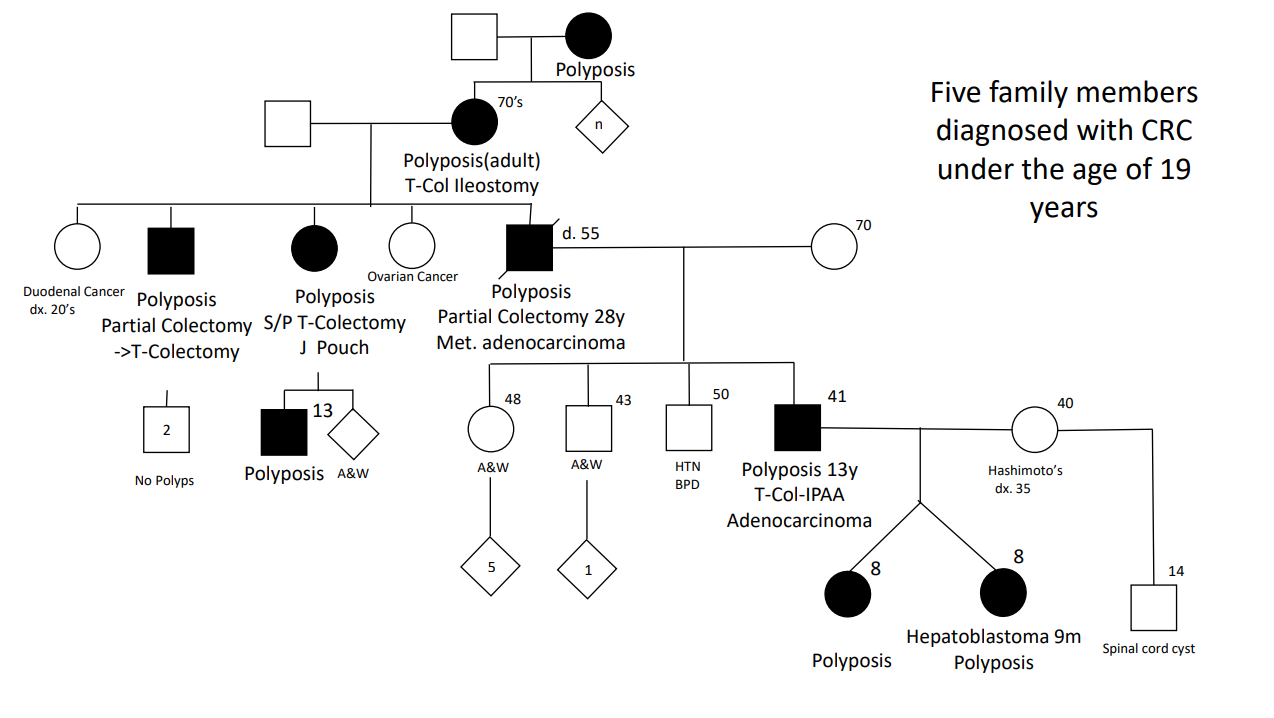
Case#2 Improvements in testing allowed identification of a point mutation in the 1B promoter region of the APC gene

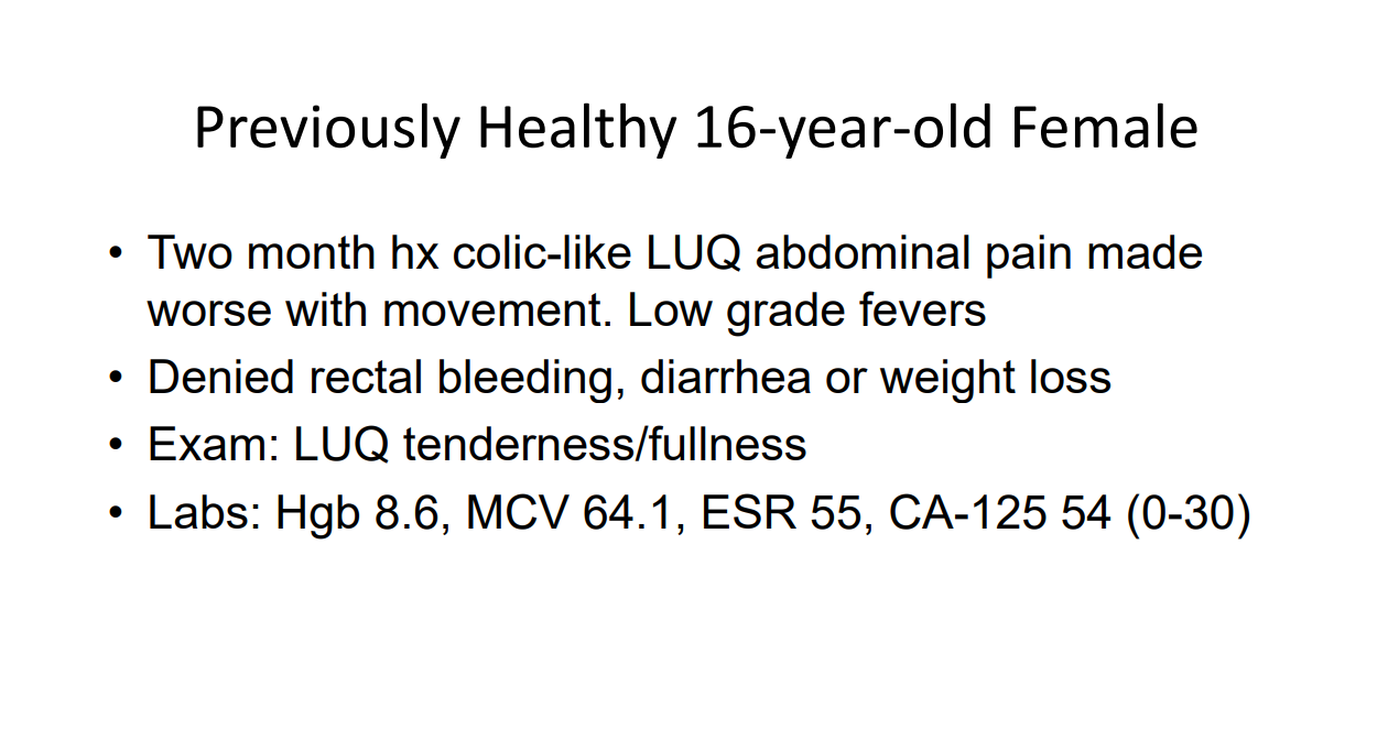
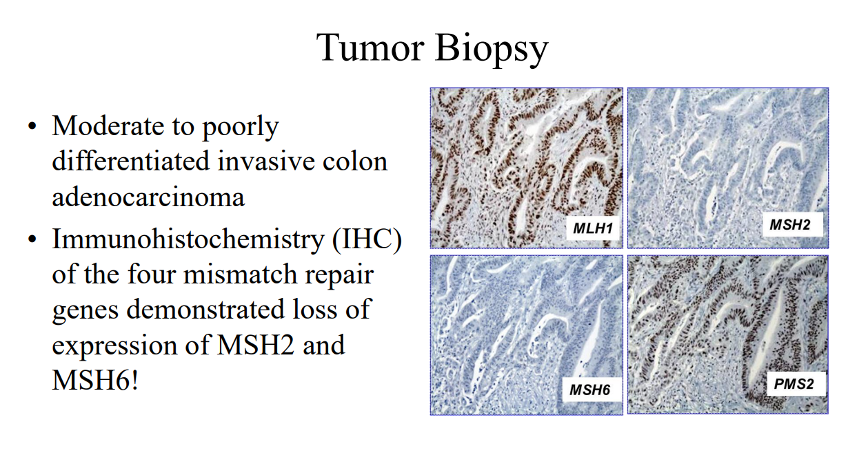
Case#3 presented a 16 yo with anemia and pain who was found to have a colonic mass related to mismatch repair mutation. Dr. Erdman indicated that obtaining adequate tissue for a diagnosis (“dig a hole”) is important. (As an aside, other colleagues have had the experience of tumors which were highly vascular and it is important to keep this possibility in mind)

Amsterdam II Criteria for Lynch Syndrome

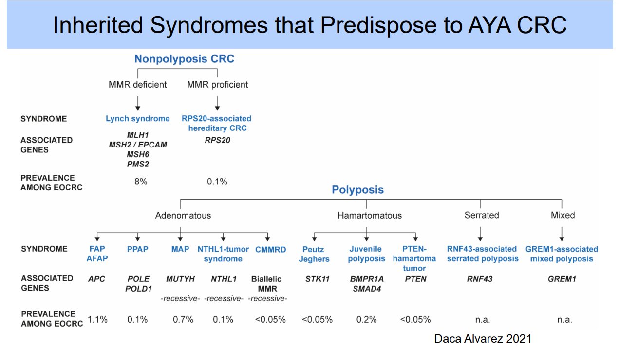
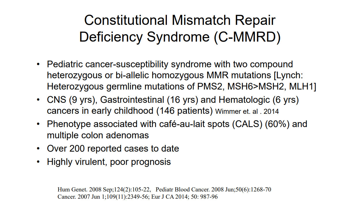
Case#4 presented a 12 yo with neurofibromatosis (NF-1) who developed CRC and ultimately diagnosed with CMMR-D. This is a highly aggressive cancer susceptibility disorder with a very poor prognosis (see post: Are you familiar with CMMR-D?)

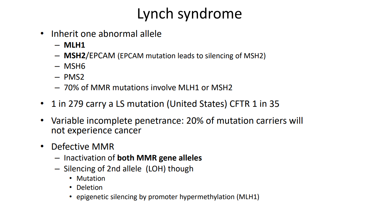
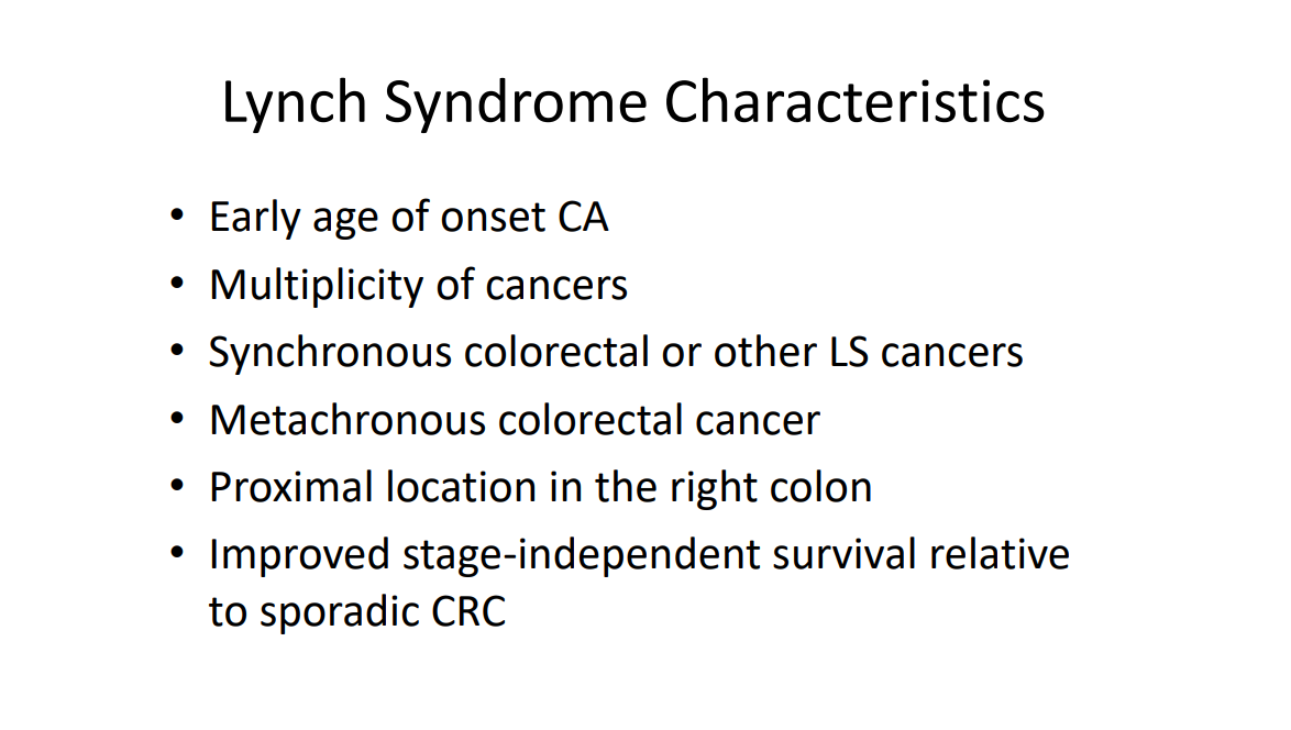
  • Case#5 presented two siblings (13 yo, 17 yo) who had half-sibling who died from CRC at age 25 yrs. This case illustrated “genetic anticipation” as each generation in this family with Lynch syndrome tended to develop CRC earlier in life. Amsterdam criteria can be helpful in identifying Lynch syndrome

Related blog posts:

Disclaimer: This blog, gutsandgrowth, assumes no responsibility for any use or operation of any method, product, instruction, concept or idea contained in the material herein or for any injury or damage to persons or property (whether products liability, negligence or otherwise) resulting from such use or operation. These blog posts are for educational purposes only. Specific dosing of medications (along with potential adverse effects) should be confirmed by prescribing physician.  Because of rapid advances in the medical sciences, the gutsandgrowth blog cautions that independent verification should be made of diagnosis and drug dosages. The reader is solely responsible for the conduct of any suggested test or procedure.  This content is not a substitute for medical advice, diagnosis or treatment provided by a qualified healthcare provider. Always seek the advice of your physician or other qualified health provider with any questions you may have regarding a condition.

Endoscopy Module -Postgraduate Course Notes

Advances in Hemostasis for Upper GI Bleeding Brad Barth, MD, MPH  (page 77)

Upper GI Bleeding

Effect of IV PPI on patients with UGI bleeding PRIOR to EGD

  • 6 trials including 2223 patients
  • No significant difference in mortality, rebleeding or need for surgery compared to controls
  • DID significantly reduce rates of high risk stigmata identified on EGD
  • DID significantly decrease the need for endoscopic therapy
  • Reference: Sreedharan A, Martin J, Leontiadis G, et al. Proton pump inhibitor treatment initiated prior to endoscopic diagnosis in upper gastrointestinal bleeding. Cochrane Database of Systematic Reviews 2010


Upper GI Bleeding – Proton Pump Inhibitors/Prokinetics

  • Omeprazole 1 mg/kg q 12 hours  (Solana, et al. J Pediatr 2013:162:776-82)
  • Proposed PPI drip dose: 1 mg/kg bolus followed by 0.1 mg/kg/hour infusion
  • IV erythromycin or metoclopramide; infuse 20-120 minutes prior to endoscopy in patients with acute UGIB; decreased need for repeat endoscopy to determine cause and site of bleeding. Prokinetic did NOT affect transfusion requirements, duration of stay, need for surgery. Reference: Barkun et al. Prokinetics in acute upper GI bleeding:a metaanalysis. GIE 2010;17:126-132

Upper GI Bleeding —Other points:

  • Epinephrine alone is RARELY enough
  • Non bleeding adherent clot has 8-35% chance of rebleeding in adults. Consider removing it CAREFULLY!
  • A conservative transfusion strategy is usually appropriate

Useful References

Surveillance Endoscopies: The established, the debated, and the unknown –Mitchell Shub, M.D. (page 85)

“Beware of false knowledge; it is more dangerous than ignorance.” —George Bernard Shaw

Familial Adenomatous Polyposis

Surveillance protocol: Age of initial evaluation/Type of procedure/Frequency

  • Colon 10 – 12 y of age (Sooner: family h/o aggressive disease) -Flex sig or Colonoscopy, 1 – 2 y
  • Upper GI tract 20 – 25 y or at initial colonoscopy
  • EGD and side viewing scope, 1 – 3 y
  • Post-colectomy (pouch) 6 – 12 mo. after surgery, Flex sig 1 y (6 mo. If retained rectum)
  • Small bowel: capsule or MRI, frequency unknown

Peutz-Jeghers Syndrome: begin screening at age 8 years or when symptomatic with colonoscopy, EGD, and small bowel imaging (?capsule vs alternatives); then every 2-3 years

Juvenile Polyposis Syndrome: begin screening at age 10-15 years or when symptomatic with colonoscopy, EGD, and  possibly small bowel imaging (?capsule vs alternatives); then every 1-3 years

Discussed guidelines for IBD cancer surveillance and for Barrett’s esophagus

  • For UC, start surveillance 8-10 years after diagnosis.
  • For Crohn’s with ~1/2 colon (or more) involvement, follow same guidelines
  • For coexisting PSC, annual surveillance
  • Barrett’s esophagus in children: adenocarcinoma very rare, evidence lacking to develop surveillance schedule

Expanding the view: Update on Upper GI StricturesMark A. Gilger, M.D. (page 95)

Why balloons for kids (for dilatation)?

You can see what you’re doing

  • Blind pouches
  • Abnormal mucosa
  • Caustic injury
  • Epidermolysis bullosa
  • Already requires general anesthesia
  • Ability to wire through narrow strictures
  • Ability to use radiographic assistance

Tip: Can use vegetable spray (eg. Pam) to make advancement of balloon catheter easy

How to do balloon dilation

  • Inflate balloon to ½ desired initial atmospheres & re‐check placement
  • Begin dilation at to 1‐2 mm more than initial estimated stricture diameter
  • Hold for 1 minute/dilation
  • •ove balloon catheter in and out during dilation;  if balloon moves freely, increase diameter by 1mm.  If stricture moves with the balloon, hold x 1 minute, then done
  • Oh, oh, there’s blood! Good! No blood, no dilation.
  • After dilation, carefully advance endoscope through the stricture; if resistance stop, can try cork‐screw maneuver
  • Document everything; especially stricture location (CM from incisors), dilation diameters (to help you next time)

Adjunct therapy for recalcitrant strictures  –adjunct therapy to sustain dilation needs further study

  • Oral & intravenous corticosteriods
  • Injectable corticosteroids – Thins the mucosa, OK 1‐2 times, but not repeated
  • Mitomycin C
  • Acid reduction
  • Stents

Endoscopy in the high‐risk patient: Keeping your patient safeJenifer R. Lightdale, MD, MPH (page 63)

Safety of Pediatric GI Procedures

  • Peds‐CORI data from >10,000 procedures
  • Overall rate of complications 2.3%: risk of hypoxia 1.5%; risk of bleeding 0.3%

Examples of pediatric populations at increased risk for perforation

  • History of caustic ingestion
  • Esophageal atresia/tracheo‐esophageal fistula
  • Severe duodenitis
  • Severe ulcerative colitis
  • Patients with multiple co‐morbidities (i.e. Type I diabetes, cerbrovascular disease, peripheral vascular disease, renal insufficiency, liver disease)
  • Ehlers‐Danlos Syndrome (Vascular Type)

Pre‐procedure Assessment –lends itself to a checklist 

Thrombocytopenia -Current recommendations

  • EGD ok if platelets >20,000/mL
  • Biopsies ok if platelets >50,000/mL

Bleeding –discussed high risk conditions

Decreasing Risk of Perforation:

  • Avoiding excessive pressure
  • Avoiding premature cutting of a polyp – Coagulate before cutting
  • Avoiding blind intubation of the lumen

Decreasing Risks of Infection

  • SBE Prophylaxis– generally NOT indicated in diagnostic procedures. Congenital heart disease is complex & may be needed on a case‐by‐case basis
  • Single‐dose cephalexin has been shown to decrease peristomal infection during PEG placement
  • Prophylactic antibiotics recommended for cirrhotic patients admitted with GI hemorrhage

Postgraduate Course Syllabus (posted with permission) with complete slides of above lectures: PG Syllabus

Related blog references:

Disclaimer: These blog posts are for educational purposes only. Specific dosing of medications (along with potential adverse effects) and specific medical management interventions should be confirmed by prescribing physician.  Application of the information in a particular situation remains the professional responsibility of the practitioner.