NASPGHAN Postgraduate Course 2017 (Part 1): Strictures, GI Bleeding, Pancreatic Fluid Collections

Over the next 2 weeks or so, I am posting my notes/pictures from this year’s annual meeting.  The first few days will review the postgraduate course.  For the most part, I find the postgraduate course reassuring that I have kept up with current approaches; there is usually not a lot of new information but a solid review of the topics.

Here is a link to postgraduate course syllabus: NASPGHAN PG Syllabus – 2017

This blog entry has abbreviated/summarized these presentations. Though not intentional, some important material is likely to have been omitted; in addition, transcription errors are possible as well.

Strictures beyond the esophagus

Petar Mamula, Children’s Hospital of Philadelphia

Some useful points:

  • Fluoroscopy very useful with most strictures –may improve safety and effectiveness. Helps define anatomy
  • Reviewed strictures in stomach –rare. May be due to caustic ingestion, Crohn’s disease or chronic granulomatous disease
  • Intestinal/colonic strictures (or narrowing): duodenal webs -can be treated with needle knife, Crohn’s disease strictures -can be balloon dilated, Short gut syndrome, Graft versus host disease

GI Bleeding Update

Diana Lerner  Medical College of Wisconsin

Useful points

Upper GI Bleeding:

  • IV PPIs reduce risk of transfusion and reduce risk of re-bleeding
  • IV PPI BID treatment has been shown to be noninferior to continuous drip
  • Conservative transfusion therapy
  • Erythromycin can be helpful
  • Lecture had good videos with review of techniques: clipping, heater probe, epinephrine injection (not recommended as monotherapy), argon plasma coagulation, and bipolar electrocautery

Cleveland et al. World J Pediatr 2012

Lower GI Bleeding:

  • Etiologies include the followiing: Post-polypectomy, Solitary Rectal Ulcer syndrome, Blue Rubber Bleb syndrome, anastomotic ulcer bleeding, Meckel’s diverticulum
  • Lower GI evaluation is best after prep –much higher yield

Management of Pancreatic Fluid Collections

Matt Giefer Seattle Children’s Hospital

Key points:

  • Imaging in first 7 days of diagnosis may miss the development of fluid collections
  • With necrotizing pancreatitis, fluid collections are either ANC: acute necrotic collection (<4 weeks) or WON: walled off necrosis (>4 weeks); Bryan et al. Radiographics 2016; 36: 675
  • With interstitial edematous pancreatitis, fluid collections are either acute peripancreatic fluid collection (<4 weeks) or Pseudocyst: >4 weeks,
  • Fluid collections do not preclude feeding patients
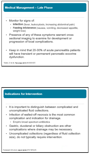
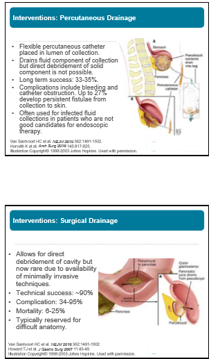
  • Drainage often needed if fluid collection becomes infected or if fluid collection causes obstruction
  • Endoscopic drainage is first-line approach: equally effective as surgery, fewer complications, equal efficacy, and lower cost


The Story Behind a 30 Year Esophagitis Study

A recent retrospective study ( SS Baker et al. JPGN 2015; 61: 538-40) reported on changes in esophagitis over a 30 year period at one center.  While the authors focus on the fluctuating percentage of esophagitis noted during three periods, in my opinion, they miss the opportunity to discuss more relevant findings.

Specifically, the authors note the following:

  • From 1980-88 (n=186 over 8 years) that 26.9% had esophagitis and 4.8% had >15 eos/hpf.  Normal pathology in the esophagus was noted in 73.1%.
  • From 2000-2002 (n=321 over 2 years), 41.2% had esophagitis and 8.5% had >15 eos/hpf.  Normal pathology in the esophagus was noted in 58.8%.
  • In the most recent period, 2011, (n=675 over 1 year), 31%* had esophagitis and 12.7% had >15 eos/hpf.  Normal pathology in the esophagus was noted in 69%.     *erroneously reported as 33%

What is baffling to me are the following:

  • Why the authors assert that there has been a fluctuating prevalence.  In absolute terms, the increase in cases is marked, though one can argue that in earlier periods there may have been many undiagnosed cases.
  • Why the authors do not comment on the tremendous increase in the use of endoscopy in their discussion.  In the first period, they were averaging ~23/year, the second period ~95/year and in the most recent period, they performed 675 in one year.

My take: This study shows that esophageal eosinophilia has been present for a long time and that identification of cases has increased considerably over 32 years.  In addition, the use of endoscopy has increased markedly, yet the yield of abnormal findings remains similar.

Briefly noted: C Menard-Katcher et al. JPGN 2015; 61: 541-46.  This retrospective study of 22 children showed that 55% had esophageal strictures identified by esophagram but not endoscopy.

Related blog posts:

Bamboo

Looking behind and looking forward in EoE (part 1)

Two important articles are provide additional insight into eosinophilic esophagitis (EoE).

In the first (Gastroenterol 2013; 145: 1230-36), the authors performed a retrospective review of the Swiss EoE Database (SEED). This SEED should not be confused with our SEED center (Home- The SEED Center of Atlanta– SouthEast Eosinophilic ).  While the database contains 783 EoE patients, only 200 who were followed by the senior author and had complete data were included.  The enrollment period dates back to 1989.

Demographics: 153 men, mean age 39 years old, 94.5% had dysphagia at time of diagnosis and 35.5% had chest pain.  66% had concomitant allergies.

Terminology: The authors defined strictures as low-grade if a standard 9 mm endoscope could pass but met resistance, intermediate if a 6 mm endoscope could pass, and high-grade if it could not be passed with a 6 mm endoscope.

Results:

  • 37.5% (n=75) had strictures (other endoscopic findings noted in Table 2)
  • Peak eosinophil count (median): 35 proximally and 28 distally
  • Figure 2 showed the evolution of endoscopic features based on diagnostic delay.  With increasing diagnostic delay, there developed a preponderance of a mixed fibrotic/inflammatory picture whereas in those whose symptoms were of much shorter duration, the endoscopic features were often inflammatory without fibrosis.
  • For example, if diagnostic delay was between 0-2 years, then fibrotic findings were noted in 46.5%; in contrast, 87.5% had fibrotic features if symptoms had been present for > 20 years.
  • Strictures increased from 17.2% in those without significant diagnostic delay to 70.8% in those with symptoms present for > 20 years.
  • The authors note that diagnostic delay was greatest in those who developed symptoms in the first decade of life.

Study limitations: The categorization of strictures is straightforward; however, newer tools like the EndoFlip can detect esophageal narrowing more accurately.  Other limitations are related to retrospective nature of study and its reliance on patient’s reported outcomes (subject to recall bias).  Thus, the estimation of diagnostic delay may be inaccurate.

Take home message:

This article reinforces the concept that the presentation of EoE changes with time and that the long-term consequence of untreated EoE is increasing fibrosis and stricturing of the esophagus.

Related blog entries: