Trisomy 21 and Associated Biliary Anomalies

M Joseph et al. JPGN 2025;81:743–747. Duct tales: Biliary anomalies found at endoscopic retrograde cholangiopancreatography in trisomy 21 pediatric patients

Methods: A single-center, retrospective chart review was conducted to identify pediatric patients (n=12) with T21 who underwent ERCP.

Patient characteristics:

  • Six patients (50%) had a history of duodenal atresia with subsequent surgical repair during the neonatal period
  • Four patients (33.3%) had chronic or acute recurrent pancreatitis, and two patients (16.7%) had biliary pancreatitis
  • Five patients (41.7%) had a biliary stricture that required stenting
  • Choledocholithiasis was present in 7 of the 12 patients (58.3%)
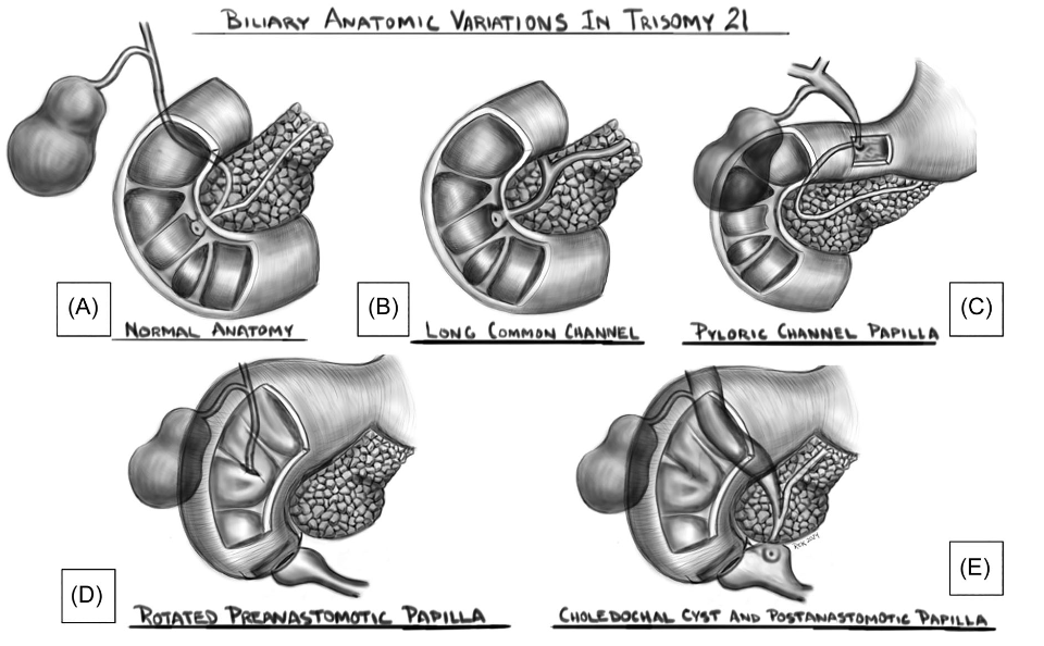
Key findings:

  • Eight patients (66.7%) were found to have an abnormal location of the major papilla. This included three patients’ papillae which were in the blind duodenal pouch created after duodenal atresia surgery (Figure 1B) and two patients had their papilla in the proximal duodenum/bulb (Figure 1D)
  • Two patients (16.7%) had unsuccessful ERCP either due to difficult cannulation or inability to find the major papilla
Figure 1: Biliary anatomic variations in Trisomy 21. Shown are drawings that represent variations of the location of the major papilla.(A) Normal; (B) normal location but with a long common channel; (C) pylorus; (D) postsurgical pre‐anastomotic location; and (E) postanastomotic location with choledochal cyst. Illustrations by Dr. Robert E. Kramer.

My take: In patients with trisomy 21, ERCP may be quite challenging due to anatomic variations and stricturing. In some patients in this cohort, a front-viewing scope was helpful.

Nonspecific Duodenal Histologic Findings Common in Children with Trisomy 21

E Alexander et al. JPGN 2023; 77: 184-190. The Spectrum of Duodenal Histologic Findings in Patients With Trisomy 21: A Multicenter Study

This retrospective study of patients with trisomy 21 (T21) who underwent EGD between 2000-2020. Key findings:

  • Among 836 patients with T21, 419 (50.1%) of whom had duodenal histologic abnormalities.
  • 290 of 419 had villous atrophy (VA) and of those, 172 of 290 met celiac disease (CD) diagnostic criteria, while 118 of 290 did not meet CD criteria (nonspecific VA).
  • Only one of the 118 with nonspecific VA had markedly abnormal celiac serology (TTG IGA >10 times ULN) but had no IELs on biopsy and did not respond to GFD. Four patients with nonspecific VA were later diagnosed with celiac disease over a median of 2.5 years with conversion to abnormal celiac serology.
  • Among the 129 with duodenitis without villous atrophy, 38 (29%) had increased intraepithelial lymphocytes and two patients were diagnosed with CD many years later (development of VA and positive celiac serology.

The authors offer an algorithm (Figure 2) to assist clinical approach based on biopsy results in this population.

  • In those with no villous atrophy, if Marsh score of 0/1, biopsies not consistent with celiac disease. In those with Marsh score of 2 (which is rare), consider as consistent with celiac disease (see next bullet point).
  • In those with villous atrophy consistent with celiac disease, assess response to gluten free diet (GFD). In those without a response, consider RD consult to evaluate exposures and HLA-typing as next steps.
  • In those with villous atrophy NOT consistent with celiac disease, obtain serology (TTG IgA, EMA, IgA, DGP IgG). If serology is negative, consider peptic duodenitis or SIBO treatments and assess/discontinue medications for potential for mucosal damage. If serology is positive, consider HLA-typing, review pathology slides, assess for competing disorders, and could need f/u endoscopy.

My take: Overall, ~20% of patients with T21 undergoing endoscopy, will have pathology consistent with celiac disease; this represents ~40% of those with abnormal histology. In the other 60% with abnormal histology, many have alternative explanations for the histologic findings (like peptic duodenitis). Some will evolve to meeting the diagnostic criteria for CD with time indicating need for ongoing monitoring. This study highlights the diagnostic uncertainty in those with potential for seronegative celiac disease.

Related blog posts:

Down Syndrome -Updated Growth Curves

Full link to Pediatrics article (www.pediatrics.org/cgi/doi/10.1542/peds.2015-1652 DOI: 10.1542/peds.2015-1652): Growth Charts for Children with Down Syndrome in U.S. (Reference from Kipp Ellsworth)

Excerpt:

New DSGS growth charts were developed by using 1520 measurements on
637 participants. DSGS growth charts for children ,36 months of age showed marked improvements in weight compared with older US charts. DSGS charts for 2- to 20-year-olds showed that contemporary males are taller than previous charts showed. Generally, the DSGS growth charts are similar to the UK charts.