Fatigue in Pediatric Inflammatory Bowel Disease

MD Stutvoet et al. J Pediatr Gastroenterol Nutr. 2026;82:699–707. Open Access! Fatigue in pediatric inflammatory bowel disease: Explained by transdiagnostic and disease-focused factors

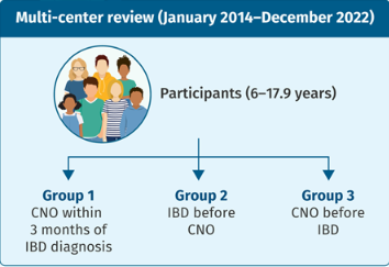
Methods: “The PROactive cohort is a longitudinal study that collects transdiagnostic patient-reported outcomes on fatigue, daily life participation, and psychosocial well-being in children with chronic conditions. It is based at the Wilhelmina Children’s Hospital in the Netherlands.” this study enrolled 127 patients with IBD.

Key findings:

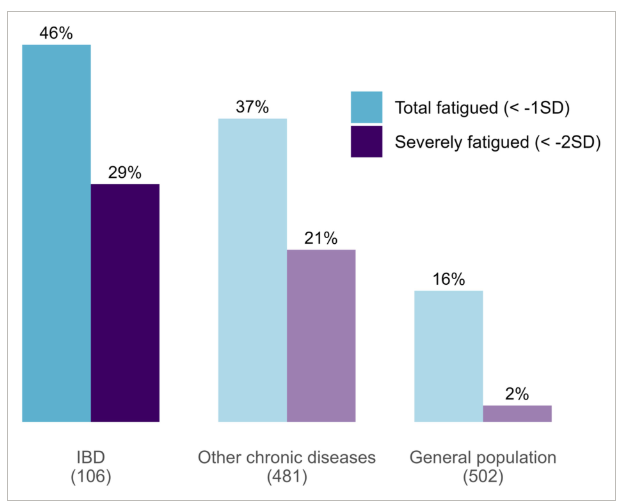
  • Fatigue is highly prevalent in pediatric IBD, with 29% of patients reporting severe fatigue—despite most being in clinical remission
  • 78% of variance in fatigues explained by transdiagnostic lifestyle, psychological and social factors
Fatigue prevalence in pediatric IBD. For reference, fatigue prevalence measured using the same “general fatigue” subscale of the MFS-GCS is also shown for other chronic childhood diseases15 and the general Dutch population.19 

Discussion Points:

  • “Clinical disease activity was significantly associated with fatigue in univariate analysis (p < 0.01). This is consistent with earlier studies showing modest associations between clinical disease activity and fatigue243335 and no association with biochemical markers such as FC, CRP, and anemia.241135 The discrepancy between clinical and biological markers may reflect the inclusion of functional limitations in the clinical disease activity scores, and their weak correlation with measures of biological disease activity as FC.36… Taken together, the association with clinical disease activity shows that fatigue may be closely linked to perceived disease-related limitations rather than inflammatory activity alone.”
  • “Transdiagnostic factors—including depressive symptoms, poor sleep, and social functioning—were strongly associated with fatigue, consistent with previous findings in pediatric IBD cohorts,3433 and other chronic diseases.1416…Our results emphasize the complex interactions among psychosocial factors and support a more integrative approach to fatigue assessment. “

My take (borrowed in part from the authors): “While disease activity may initially precipitate fatigue, other factors—such as poor sleep or school pressure—can perpetuate it, even after inflammation resolves.”

Related blog posts:

2026 Review: Eosinophilic Gastrointestinal Diseases (EGIDs)

P Visaggi, ES Dellon. Gastroenterology 2026;170:476–494. Open Access! Epidemiology, Natural History, and Treatment of Eosinophilic Gastrointestinal Diseases

Diagnosis:

  • EoE is diagnosed in the setting of symptoms of esophageal dysfunction, demonstration of at
    least 15 eosinophils per high-power field (eos/hpf) on esophageal biopsy, and exclusion of competing causes of esophageal eosinophilia. At the time of diagnosis, disease severity can be assessed with the Index of Severity for EoE (I-SEE)
  • Non-EoE EGIDs: the diagnostic process is more challenging and pediatric diagnostic guidelines were only published in 2024…biopsy specimen shows pathologically elevated
    levels of GI tract eosinophils, and other conditions that cause GI eosinophilia are excluded…Symptoms are nonspecific, multiple conditions could explain extraesophageal eosinophilia, inflammation can involve muscular and serosal layers rather than mucosal,

Treatment:

  • EoE: PPIs, Swallowed topical corticosteroids, Monoclonal antibodies and food elimination dets
  • Non-EoE EGIDs: “The management of non-EoE EGIDs is challenging due to lack of established end points, validated outcomes, and of successful RCTs leading to approved treatments. Accordingly, management of non-EoE EGIDs is currently based on low-quality evidence.” Main treatments: corticosteroids (prednisone or budesonide), food elimination diets, and monoclonal antibodies (eg. dupilumab). “The use of PPIs, mast cell stabilizers, leukotriene inhibitors, and immunomodulators for non-EoE EGIDs has been reported,
    with variable results.”

My take: This is a useful review with a number of pointers for epidemiology, natural history, evaluation and management of EoE and Non-EoE EGIDs.

Related blog posts:

What’s Changing For the GI Tract in Patients with Cystic Fibrosis Due to Highly Effective Modulator Therapy

P Saikumar et al. Am J Gastroenterol 2026; 121: 103-111. A Comprehensive Review of Gastrointestinal Manifestations in Cystic Fibrosis in the Era of Highly Effective Modulator Therapy

Thanks to Ben Gold for sharing this article.

Exoocrine Pancreatic Insufficiency (EPI)

  • “Mutlicenter studies, KIWI and ARRIVAL, demonstrated increase in FE-1 (fecal elastse-1) after treatment with ivacaftor, signifying improvement in in pancreatic function”
  • “The PROMISE study, a large 56-center prospective observational study with participants at least 12 years of age…found ETI (Elexacaftor-tezcaftor-ivacaftor) did not result in improvement in pancreatic function…raises the question if age of initiation of CFTR modular therapy has an influence in recovery of pancreatic function.”

Acute Pancreatitis

  • “With the improvement in pancreatic function seen in some patients with CF on HEMT, there have been emerging reports of acute pancreatitis.”

CF Hepatobiliary Involvement

  • “Previously referred to as CF liver disease (CFLD), hepatobiliary complications in CF are now recommended to be classified as CF hepatobiliary involvement (CFHBI) or advanced CFLD”
  • “Vigilant monitoring of transaminase levels and liver function is crucial after CFTR modulator initiation, because drug-induced liver injury (DILI) is a potential concern with elexacaftor/tezacaftor/ivacaftor (ETI) therapy…A disproportionality analysis of Food and Drug Administration adverse event reporting system data from 2019 to 2022 identified 452 reports of DILI associated with ETI use, demonstrating a statistically significant association”
  • “ETI is not recommended for those with Child Pugh Classification C and dose reduction is recommended for Child Pugh classification B in clinically appropriate situation when benefit outweighs the risk”

Nutrition

  • “In addition to macronutrient deficiency, Children with Cystic Fibrosis are at risk of deficiencies in fat-soluble vitamins, minerals, and essential fatty acids (38). With an overt focus on nutrition, PERT, and advancements in CFTR modulators, an opposite trend is now being seen, with a higher incidence of obesity and overweight in this population…[and] associated comorbidities such as coronary heart disease, metabolic dysfunction-associated steatotic liver disease, and metabolic syndrome-including hyperlipidemia and diabetes.”

Eating Disorder

  • “Disordered eating is relatively a new concern in this population. There are controversial data on the prevalence of eating disorder in PwCF” (people with CF).”

GERD

  • “Data indicate that children with CF are 4 times more likely to experience acid gastroesophageal reflux than the general population (59)…RECOVER, a multicenter study of ETI in PwCF displayed decreased GI symptoms, including GERD, after 12 months of treatment (61). Similarly, treatment with IVA was associated with symptomatic relief of GERD at 52 weeks and also showed a decline in extraesophageal reflux in PwCF treated with
  • IVA for 12 months (62).”

Meconium Ileus

  • Recent case reports, “although differing in ETI initiation timing, outcome severity, suggest that prenatal ETI exposure may positively influence MI (69).”

Constipation

  • “There are limited data regarding the effect of ETI on constipation, asthe PROMISE trialshowed only small improvements in Patient Assessment of Constipation-Symptoms score and Patient Assessment of Constipation-Quality of Life”

DIOS

  • “There is paucity of data describing impact of HEMT [highly effective modulator therapry] on DIOS. By correcting the CFTR dysfunction, HEMT may help reset the ion/fluid balance which is considered one of the main factors in pathogenesis of DIOS.”

My take: “CFTR modulators have revolutionized the management of CF by dramatically altering the course of the disease and improving patient outcomes.” They are also altering the clinical GI manifestations.

Related blog posts:

Reduced Dosing of GLP-1 RAs May Work for Maintenance Treatment of Obesity

Roni Rabin, NY Times 3/5/26: Most Patients Keep Weight Off With Fewer GLP-1 Shots, Study Finds

An excerpt:

After 36 weeks of follow-up, most of the patients who spaced out their GLP-1 injections kept the weight off and also maintained health benefits like reduced blood pressure and better blood sugar control…

The study was small, only 34 patients in a relatively homogeneous group — mostly white and privately uninsured… the research, published in February in the journal Obesity, provides a potentially appealing new option for patients who are loath to commit to lifelong weekly injections of a costly medication that may not be covered by insurance and that some fear could have unknown side effects…

The patients reduced the frequency only after achieving their desired weight loss and reaching a weight-loss plateau.

My take: In some patients, less frequent dosing of GLP-1s may be effective as maintenance therapy. Whether this strategy could be applied to the daily oral semaglutide is not clear.

Related blog posts:

Victoria Island, Bariloche Argentina

GLP-1 Use Associated with Lower Risk of Colon Cancer

C Jones. J Clin Oncol 44, 2026: suppl 2; abstract 18 (presented at ASCO 2026). Open Access! GLP-1 receptor agonist vs aspirin for primary prevention of colorectal cancer: Evidence from a real-world head-to-head comparison

Methods: Using de-identified data from TriNetX, which encompasses 150 million patients across 106 health organizations. GLP-1RA users were matched to aspirin users utilizing propensity score matching. The index date was defined as the first documented prescription or administration of either therapy, with follow-up beginning 6 months post-index event. Median follow-up was 2,153 days for GLP-1RA users and 1,743 days for aspirin users. 281,656 patients were analyzed (140,828 per cohort).

Key findings:

  • Colorectal cancer (CRC) incidence was 0.13% (183/140,758) in GLP-1RA users vs 0.176% (247/140,692) in aspirin users
  • GLP-1RA use was associated with a 26% lower risk of CRC [RR: 0.741 (0.612–0.896)].This benefit was consistent across sensitivity analyses at 12 months [RR: 0.738 (0.605–0.900)] and 36 months [RR: 0.779 (0.620–0.979)]
  • Four different GLP-1s were analyzed: HR of 0.436 for liraglutide, 0.471 with dulaglutide, 0.631 with semaglutide, and 0.711 with tirzepatide

The exact mechanism where GLP-1s may exert a protective effect is unclear; perhaps, it is related to a reduction in adverse metabolic parameters and inflammation.

My take: Potential chemopreventive effects are unlikely to be a reason to use GLP-1s in the near future, but may be an added benefit.

Related blog posts:

Bariloche, Argentina

Should You Do a Gut Microbiome Test? No — Here’s Why

J McCreary. MedPage Today; 2/26/26. Open Access! Same Stool Sample, Different Results in Gut Microbiome Tests

An excerpt:

“Direct-to-consumer gut microbiome tests produced markedly different results — even when analyzing the same stool sample, researchers found.

Identical fecal samples sent via 21 home-testing kits to seven anonymized direct-to-consumer testing companies yielded a wide variation in reported bacterial abundance and in the health assessments generated from those data, reported Stephanie L. Servetas, PhD, of the National Institute of Standards and Technology (NIST) in Gaithersburg, Maryland, and colleagues in Communications Biology.

In some cases, there was not even agreement among kits produced by the same company…

When researchers compared 18 commonly reported microbial genera across companies, no single provider aligned with the consensus profile for all 18. Across the full dataset, 1,208 unique taxa were reported, but only three genera appeared in every company’s results…

The authors said the discrepancies likely stem from differences in sample processing, sequencing methods, bioinformatics pipelines, reference databases, reporting thresholds, and quality control standards…

“These tests have become popular, partly because people, I think, are increasingly interested in health and wellness, and partly because the gut microbiome has been linked — at least in the public imagination — to the idea that you can improve a whole range of conditions through diet and lifestyle changes,” said co-author Diane Hoffmann, MS, JD, of the University of Maryland in Baltimore.

“There’s been a lot of hype around that, but the hype doesn’t really match the evidence. These tests often have limited evidence behind them, especially when it comes to informing clinical decisions or even basic dietary recommendations,” she added. “So the marketing can be questionable, and consumers can end up misinterpreting or over-trusting results that aren’t very reliable.”

Related article from Houston Methodist Hospital (2024): Should You Do a Gut Microbiome Test? Key point: “While these tests seem to be effective in mapping the gut microbiome, there is currently no benchmark for what a ‘normal’ gut microbiome looks like. So the question becomes what to do with the results…microbiomes are highly variable — even normal, healthy ones. This makes it incredibly challenging to define the patterns or signatures that suggest a microbiome has become imbalanced. Plus, the at-home steps for correcting microbiome imbalance aren’t established either.”

My take: It is uncomfortable informing families that these gut microbiome tests have little clinical value because there is not a proper way to interpret the results. In addition, this study shows that the tests from one place to another produce wildly different results.

While one’s microbiome is important, we still don’t understand what exactly is a normal microbiome.

Related blog posts:

Highway to El Chalten and Mount Fitz Roy, Argentina

Extent of Eosinophilic Esophagitis and Response to PPI Therapy

DA Hartnett et al. Clin Gastroenterol Hepatol 2026; 24: 375-384. Open Access! Distribution of Esophageal Eosinophilia as a Predictor of Proton Pump Inhibitor Response in Eosinophilic Esophagitis

Methods: This was a retrospective cohort study of newly diagnosed adult patients with EoE — All patients received ≥8-week PPI trial and underwent repeat biopsies to assess response. There were including 66 with isolated distal and 200 with proximal/diffuse disease. 86% of patients received twice daily PPI therapy (73% in those with isolated distal disease and 87% in the diffuse/isolated proximal disease.

Key findings:

PPI response was higher among patients with isolated distal disease:

  • histologic remission [<15 eosinophils/hpf post-PPI]: 63.6% vs 44.5%; P = .01
  • deep remission [<6 eosinophils/hpf]: 54.5% vs 31.0%; P = .001
  • symptom improvement: 92.4% vs 81.0%; P = .03).

The discussion noted that there has been limited studies of EoE distribution and response to treatment. “Godat et al observed that the distribution of esophageal eosinophilia had no impact on clinicohistologic remission rates (defined as ≤2 on a scale of 0–10 for dysphagia/odynophagia in the last 7 days and a peak eosinophil count <5 eos/hpf) in patients treated with budesonide orodispersible tablets.19

“The generally higher PPI response with isolated/predominant distal disease suggests that acid suppression and improved mucosal barrier function likely play a key role in how PPI may lead to EoE remission…prior studies have demonstrated no correlation between findings on ambulatory pH monitoring and PPI response in EoE.25 Therefore, the differential response to PPI based on eosinophil distribution phenotypes may be due to more than comorbid GERD alone.”

My take: While the pathophysiology of how PPIs work for EoE is unclear, it appears that the response to PPIs is better with in those with isolated distal EoE. The difference in response may have been even more pronounced if both groups had a similar percentage of receiving twice daily PPI treatment.

Related blog posts:

Prospective Study: Acute Gastroenteritis Increases Risk of Disorders of Gut-Brain Interaction

A Palorath et al. Am J Gastroenterol 2026; 121: 242-247. Acute Gastroenteritis Is a Risk Factor for the Development of Disorders of Gut-Brain Interaction in Children

Methods:  A prospective, controlled, cohort study. Children (1-15 years) with recent AGE (cases) and siblings (controls) were followed for 6 months. We assessed DGBIs using a validated questionnaire (QPGS IV) per Rome criteria. Patients with underlying GI disorders (eg. Celiac, IBD, recent abdominal surgery) were excluded.

Key findings:

  • Among children without a history of DGBI before the AGE, 6 (12.2%) cases vs 0 control were diagnosed with DGBI (P = 0.01) at follow-up at 3 months
  • At 6 months, 5 cases (1 lost to follow-up) vs 0 control had persistent DGBI (P = 0.03)
  • Severity of AGE was correlated with PI-DGBI. Severe AGE included abdominal pain, ED consullt, and need for IV fluids

My take: It is well-known that previous AGE increases the risk of PI-DGBIs, especially irritable bowel syndrome. However, this is the first prospective cohort study showing this association between AGE and DGBIs in a nonoutbreak setting. In addition, it correlates the risk of DGBI with the severity of AGE.

Related blog posts:

Chronic Nonbacterial Osteomyelitis Associated with Pediatric Inflammatory Bowel Disease

M Matar et al. J Pediatr Gastroenterol Nutr. 2026;82:487–494. Chronic nonbacterial osteomyelitis associated with pediatric inflammatory bowel disease: : A multicenter retrospective study from the Paediatric inflammatory bowel disease Porto Group of ESPGHAN

Methods: Retrospective study with 45 pediatric patients with inflammatory bowel disease (n=32 with Crohn’s disease, n=8 with ulcerative colitis, n=5 with IBD-U)

Key findings:

  • CNO presented in 15 patients (33%) within 3 months of IBD diagnosis, and in additional 20 (44%) patients after IBD diagnosis; in 10 (22%) patients CNO preceded the diagnosis of IBD with a median time 46 (25–248) weeks
  • 11 (24%) subjects displayed at least one additional extra-intestinal manifestations, including arthritis (6, 13%), erythema nodosum (4, 9%), sacro-ileitis (2, 4%), psoriasis (1, 2%), and pyoderma gangrenosum (1, 2%).
  • Complications occurred in six patients and included vertebral collapse, bone fracture, and bone deformity. In eight (18%) subjects vertebral collapse was present already at the time of diagnosis.
  • “While in most patients, diagnosis of CNO was associated with either clinical or laboratory indices of active IBD, especially CD, in some cases, the intestinal disease was quiescent.”
  • “All patients achieved remission of CNO at some point during follow-up, which may support the hypothesis that anti-TNF treatment is unlikely to promote CNO development and does not reinforce the theory of a paradoxical effect that has been suggested by Cordesse et al.22

My take: This is a useful review highlighting the association of CNO with both active disease and quiescent IBD. The authors argue that their data does not support the development of CNO as a paradoxical effect. I disagree with this premise. Many of the extraintestinal manifestations, that anti-TNF agents can treat, rarely can be caused by them. Besides CNO, paradoxical reactions to anti-TNF agents have included the development of rheumatoid arthritis, psoriasis, hidradenitis suppurativa and autoimmune liver disease.

Related blog posts:

QI Project: Increasing H Pylori Eradication

KR Arellanos et al (Senior Author S Bonilla). JPGN Reports. 2026;7:19–27. Standardizing a protocol for Helicobacter pylori gastric biopsy culture: From implementation to sustained practice

Background: “The availability of antibiotic susceptibility data is essential for understanding local and regional resistance patterns and for informing future guidelines on optimal treatment regimens for children.” This quality improvement project report documents how “using a standardized checklist, in combination with educational initiatives for staff physicians and collaborative efforts with endoscopy and laboratory teams, were effective strategies to increase the use of gastric biopsy culture as a diagnostic tool for H. pylori infection and, to a lesser extent, to improve culture yield in patients with positive histology.”

Key steps:

  1. Standardized checklist for biopsy collection used by nurses
  2. Standardized transport protocol
  3. Prompt refridgeration/transportation
  4. Using a single lab for testing (Mayo clinic)

Key findings:

  • There was a consistent increase in culture positivity for H pylori and this was associated with improved eradication rates
  • Overall, antimicrobial resistance was highest for metronidazole (27.5%) and clarithromycin (18.7%), and lower for rifampin (12.2%), levofloxacin (10.1%), and amoxicillin (4.3%). Only one isolate showed resistance to tetracycline

Discussion: “Among the various interventions implemented during the QI project, the one that appears to have contributed most significantly to the dramatic improvement in culture yield among patients with H. pylori-positive histology was the consolidation of specimen processing to a single specialty laboratory [Mayo clinic].” Specific logistics included using a simple sterile container with saline-moistened tissue for collection and sending samples to specialty lab on the same day as collection.

My take: During this project, there was improved use of gastric culture and in culture yield. This resulted in meaningful improvement for patients. However, without clinical leadership to implement these changes, there will continue to be suboptimal eradication rates at other centers.

Related blog posts: