An Esophageal Coin with Moth Eaten Appearance

M Hook et al. JPGN 2025; DOI: 10.1002/jpn3.70221. Esophageal coin with moth eaten appearance: A cause for caution

This case report describes a penny ingestion by a 10 yo with autism that likely happened nine months before attempted retrieval. The coin was not able to be retrieved due to stricturing but was advanced into the stomach (the authors note advancement into duodenum would be better); in addition, there was an esophageal diverticulum that was identified likely secondary to the caustic damage.

In the discussion, the authors note that “the ‘moth eaten’ appearance is a rare finding that indicates prolonged foreign body retention and unforeseen mucosal injury…It is specific to post-1982 pennies, composed of 97.5% zinc.4 Hydrochloric acid exposure dissolves zinc, forming caustic zinc chloride.”

My take: Future generations will not see this radiologic finding. For now, if this finding is seen, it likely indicates a more difficult foreign body removal.

Related blog posts:

Ref: https://www.pbs.org/newshour/nation/u-s-mint-in-philadelphia-to-press-final-penny-as-the-1-cent-coin-gets-canceled

How Often Esophageal Coins Pass Into the Stomach

P Quitadomo et al. Am J Gastroenterol 2025; 120: 1388-1390. “Insert-Coin”: A Prospective Study of Coin Ingestion in Children of Southern Italy

Thanks to Ben Gold for this reference.

This prospective study from Naples, Italy examined children 0-14 yrs of age with a coin ingestion (n=807). Children with coins in the proximal esophagus underwent endoscopic removal within 4 hours whereas those with middle to lower esophageal coins had re-evaluation after 12 hours before removal.

Key findings:

  • 52 of 807 (6.4%) had a coin retained in the esophagus, the remainder were in the stomach or beyond
  • 20 of 52 (38%) were located in the middle to lower esophagus (10 in each)
  • 13 of 20 (65%) coins in the middle to lower esophagus had spontaneous gastric passage
  • The mean age of patients with gastric passage (72 months) was higher than those without passage (48 months)

My take: Only 6% of patients in this study who had a coin ingestion had esophageal retention of the coin. In addition, one-fourth of those with esophageal coins had spontaneous passage into the stomach. This occurred only with the mid-distal esophageal coins; in this subset it occurred in 65%. Thus, in those with mid-distal esophageal coins, watchful waiting for ~12 hrs may be beneficial for patients. The ultimate primary prevention of this problem may occur with more widespread adoption of electronic payments.

Related blog posts:

Agnes Northrup of Louis Tiffany Studio,
Tiffany Garden Landscape Window (1912)
at The Metropolitan Museum of Art
Agnes Northrup of Louis Tiffany Studio,
Autumn Landscape  (1923) at The Metropolitan Museum of Art

Sharp Objects in GI Tract & Good Outcomes

P Quitadamo et al. JPGN 2023; 76: 213-217. Sharp-Pointed Foreign Body Ingestion in Pediatric Age

In this study with 580 children, consecutively recruited from 2016 to 2020, the authors examined outcomes after ingestion of sharp-pointed foreign bodies (FBs).

Key findings:

  • Mean age was 50 months.
  • Sharp/pointed FBs mainly included fragments of metal 270 of 580 (46.55%) and glass 180 of 580 (31%).
  • FBs were endoscopically removed in 79 of 580 (13.6%) children whereas the remaining FBs passed through the gastrointestinal tract over an overall mean time of 29 hours
  • No cases of intestinal perforation nor prolonged retention were observed.
  • The most common metal objects were earrings (n=72). Other objects: screws (n=20), dental works (n=20), nails (n=13), open safety pins (n=7), fish bones (n=65)

My take: I have not had a severe complication from sharp-pointed FBs (in nearly 30 yrs of pediatric GI practice); this article confirms the overall low risk that they pose.

Related blog posts:

Turtle at Chattahoochee Nature Center

Selected Slides from NASPGHAN 2022 Postgraduate Course (Part 1)

Pediatric Foreign Body Ingestions
Esophageal Strictures
Imaging for Acute Pancreatitis
For Acute Liver Failure

Disclaimer: This blog, gutsandgrowth, assumes no responsibility for any use or operation of any method, product, instruction, concept or idea contained in the material herein or for any injury or damage to persons or property (whether products liability, negligence or otherwise) resulting from such use or operation. These blog posts are for educational purposes only. Specific dosing of medications (along with potential adverse effects) should be confirmed by prescribing physician.  Because of rapid advances in the medical sciences, the gutsandgrowth blog cautions that independent verification should be made of diagnosis and drug dosages. The reader is solely responsible for the conduct of any suggested test or procedure.  This content is not a substitute for medical advice, diagnosis or treatment provided by a qualified healthcare provider. Always seek the advice of your physician or other qualified health provider with any questions you may have regarding a condition.

Orbeez Ball Obstruction

Z Patel et al. Gastroenterol 2021: 161. https://doi.org/10.1053/j.gastro.2020.10.051. Full text: An Unusual Case of Small Bowel Obstruction

Case report of a 10 month old who developed a small bowel obstruction due to an “Orbeez” ball. “Orbeez balls are small polymer beads that absorbs water and expands, it can get up to 150 times their original size. When left out of water, the beads shrink back down to its original size. They were originally invented as a way to keep moisture in soil for plants. They are hugely popular with children and often used as toys, flower holders, air fresheners, light jars, and objets d’art in the house.”

Related blog post: Foreign Bodies in Children –Expert Guidance

#NASPGHAN19 Postgraduate Course (Part 1)

Here are some selected slides and notes from this year’s NASPGHAN’s postrgraduate course.  My notes from these lectures may contain errors in omission or transcription.

Link to the full NASPGHAN PG Syllabus 2019

8:00 – 9:00 Module 1 – Endoscopy

11  David Brumbaugh, MD, Children’s Hospital Colorado  Management of foreign bodies

Related blog posts:

22 Petar Mamula, MD, Children’s Hospital of Philadelphia Advanced endoscopic techniques for gastrointestinal bleeding

This talk had some terrific videos (not available in syllabus) and useful practical points.  For example, with cautery, the speaker recommended not just quickly taping the lesion, count for several seconds when applying.  For hemospray, the speaker considers this technically much easier but is using this mainly as a backup option.

Here are two screenshots (not from lecture) which provide information from manufacturer on Hemospray use (link to PDF on Hemospray Manufacturer’s PDF on Hemospray)

Related blog posts:

36 Srinadh Komanduri, MD, Northwestern Medicine  Cancer screening top to bottom

Some of the key points:

  • IBD and colorectal cancer (CRC) screening 8-10 years after disease onset
  • ~10% of CRC in general population occurs between 20-49 years
  • Chromoendoscopy results in higher detection rates of dysplasia

Related blog posts:

Disclaimer: NASPGHAN/gutsandgrowth assumes no responsibility for any use or operation of any method, product, instruction, concept or idea contained in the material herein or for any injury or damage to persons or property (whether products liability, negligence or otherwise) resulting from such use or operation. The discussion, views, and recommendations as to medical procedures, choice of drugs and drug dosages herein are the sole responsibility of the authors. Because of rapid advances in the medical sciences, the Society cautions that independent verification should be made of diagnosis and drug dosages. The reader is solely responsible for the conduct of any suggested test or procedure. Some of the slides reproduced in this syllabus contain animation in the power point version. This cannot be seen in the printed version.

Fidget Spinner Ingestions

A recent study (PT Reeves et al. J Pediatr 2018; 197: 275-9) reviews 3 new cases of fidget spinner ingestion; there had already been 13 cases reported to Consumer Product Safety Risk Management System since 2016.

Key points:

  • “If the object is located within the esophagus, urgent endoscopic retrieval is indicated in some cases, specifically if the object is sharp; has electromagnetic capabilities…)eg. batteries, magnets, light-emitting diodes); or fails to pass into the stomach after 24 hours; or the patient cannot tolerate oral secretions.”
  • The article recommends using the NASPGHAN foreign bodies guidelines (link below)

Related blog posts:

Foreign Bodies in Children -Expert Guidance

The NASPGHAN Endoscopy Committee has published a very helpful “Management of Ingested Foreign Bodies” Report (Kramer RE et al. JPGN 2015; 60: 562-74).  At the current time, one way to access a PDF of the report is the following CME link on the NASPGHAN website (no login required): Management of Ingested Foreign Bodies -Clinical Report

Some key definitiions from the report.

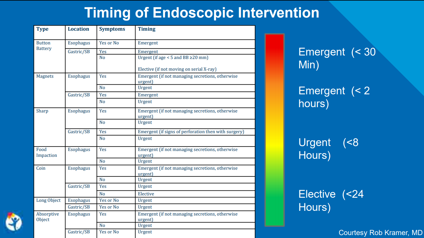
  • Emergent removal <2 hours from presentation regardless of NPO status
  • Urgent removal <24 hours from presentation following usual NPO guidelines

As a general rule, all symptomatic ingestions in the esophagus require emergent removal if feasible.  Most gastric ingestions do not require emergent removal, exceptions include multiple magnets, sharp objects in stomach (possibly with surgery), and possibly absorptive objects (due to obstruction).

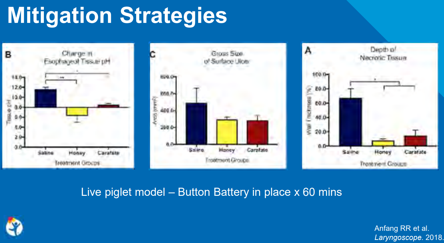
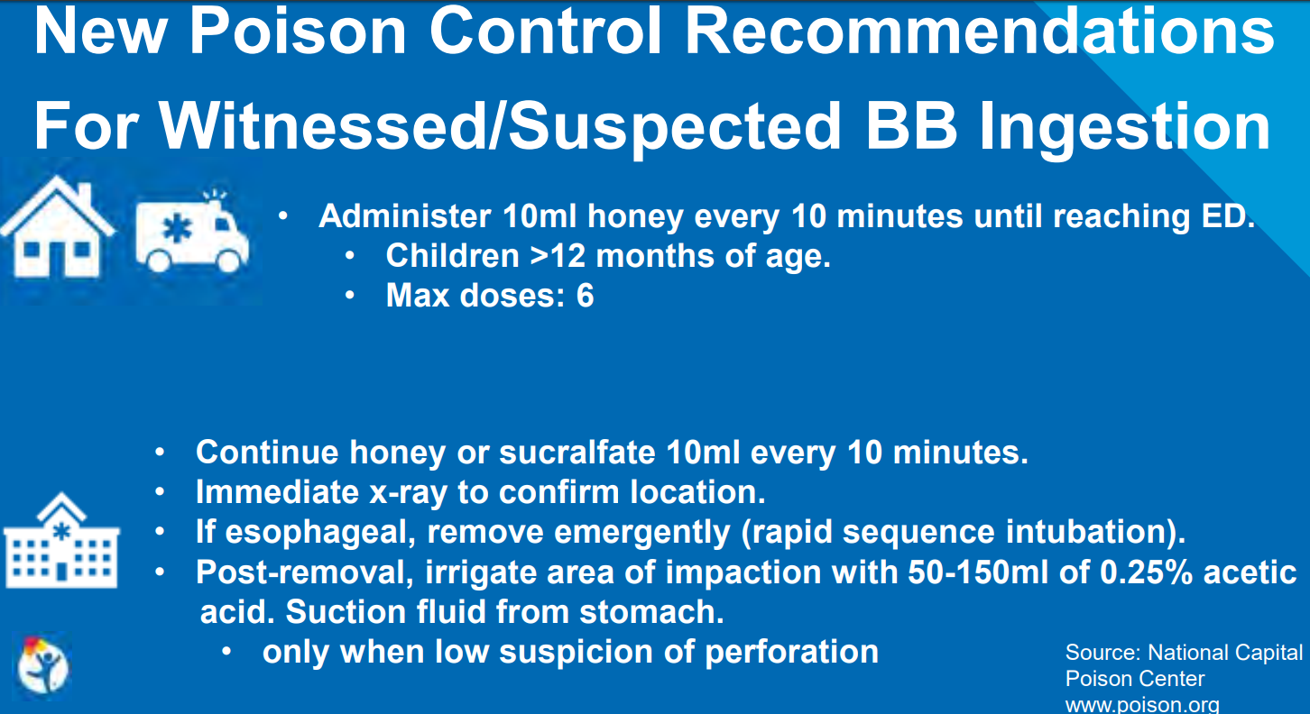
1. Button Batteries:

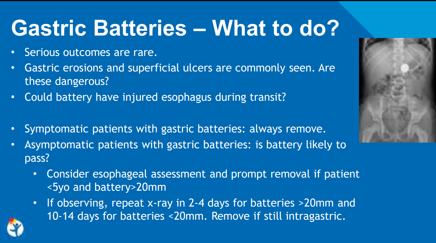
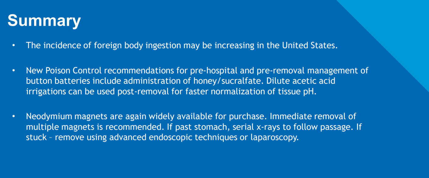
  • Even with “spent” batteries, there is enough residual charge to cause injury and all ingestions (even if asymptomatic) into the esophagus require emergent removal. If these batteries are in the stomach & asymptomatic, urgent removal is recommended if age < 5 years and BB ≥20 mm.

What is different in the proposed algorithm (Figure 1) compared with the Poison Center Guidelines (see:Button Battery Algorithm Link | gutsandgrowth) is more detail regarding concerns about aortoenteric fistula & what to do after endoscopy. Key points:

  • If active bleeding or unstable, endoscopic removal in OR with surgery/cardiovascular surgery is recommended.
  • If any esophageal injury, recommendations include admission, NPO, and IV antibiotics.  Chest imaging (CT angiography &/or MRI of chest) can help decide length of stay.  In those with injury close to aorta, continuation of NPO/antibiotics are recommended along with followup imaging every 5-7 days.  In those with clinical deterioration (eg. hematemesis w/in 21 days of injury), ‘assume aortoenteric fistula and emergently prepare for cardiovascular surgery.’

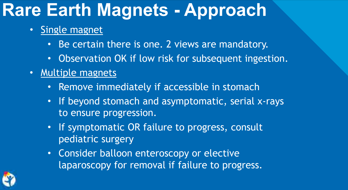
2. Magnets.  Figure 3 provides algorithm for single and multiple magnets (adapted from Hussain SZ et al. JPGN 2012; 55: 239-42).

  • For single magnets, emergent removal from esophagus is recommended (like all other foreign bodies) if difficulty managing secretions, otherwise urgent esophageal removal is suggested.
  • If there are multiple magnets within reach of endoscope, then if symptomatic, emergent removal is recommended, otherwise, urgent removal is suggested.
  • For asymptomatic magnets beyond the reach of an upper endoscopy, potential for colonoscopy, or enteroscopy for removal &/or serial x-rays to follow progression. If there is no progression on X-rays (every 8-12 hrs) &/or development of symptoms, then surgical removal/endoscopic removal is recommended.

3. Sharp objects. Figure 4 provides algorithm.

  • Emergent removal from esophagus/stomach is recommended (like all other foreign bodies) if difficulty managing secretions, otherwise urgent esophageal removal is suggested. For radiolucent objects, if the ingestion was witnessed, urgent removal is suggested; if not witnessed, then further imaging (CT, esophagram, MRI) could be considered.
  • With regard to sharp foreign bodies beyond the reach of an endoscope, “follow clinically with serial x-ray.  Enteroscopy or surgical removal considered if symptoms develop or >3 days without passage.”
  • Despite the low risk of severe morbidity/mortality from sharp objects (beyond esophagus), report recommends urgent “removal of all of the sharp objects within the reach of the endoscope..if possible.”

4. Food impaction in esophagus.  If symptomatic, emergent removal; if asymptomatic, then urgent removal.  Biopsies of the esophagus are recommended with endoscopy (Figure 5).

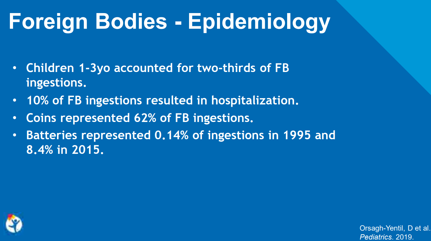
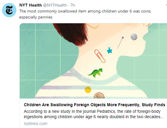
5. Coin ingestions/Blunt objects. Figure 6. “>250,000 ingestions and 20 deaths reported in the United States during a 10-year period.”

  • For esophagus: If symptomatic, emergent removal; if asymptomatic, then urgent removal.  Report recommends check X-ray immediately before sedation. While the report does not address this, a possible alternative to x-ray would be the use of a metal detector.  “Consider glucagon if distal esophageal coin or if endoscopy not readily available.”
  • For stomach: No endoscopy needed. Repeat X-ray at 2 weeks.  Remove if not passed w/in 2-4 weeks. Report recommends check X-ray immediately before sedation. While the report does not address this, a possible alternative to x-ray would be the use of a metal detector.
  • For small bowel: removal (enteroscopy/surgery) if symptomatic.
  • For objects >25 mm width or >6 cm in length –> should be removed from stomach urgently.

6. Superabsorbent objects.  The authors describe ingestions from materials from toys and diapers with polymers that can retain ‘up to 100 times their weight in water.”

  • For esophagus: If symptomatic, emergent removal; if asymptomatic, then urgent removal.
  • Stomach/small intestine: urgent removal is recommended

The authors state these recommendations are based on consensus rather than strong evidence and are “no substitute for clinical judgement.”

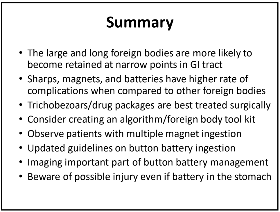
Take-home message: These guidelines are a good starting point to improve the management of children with foreign bodies.

Related blog posts:

Disclaimer: These blog posts are for educational purposes only. Specific dosing of medications/diets (along with potential adverse effects) should be confirmed by prescribing physician/nutritionist.  This content is not a substitute for medical advice, diagnosis or treatment provided by a qualified healthcare provider. Always seek the advice of your physician or other qualified health provider with any questions you may have regarding a condition.

University of Chicago

University of Chicago