Lecture: IBAT Inhibitor for Alagille Syndrome

I recently attended an online lecture which reviewed Alagille Syndrome and the emergence of an IBAT inhibitor for the management of cholestatic pruritus. Selected slides from Mirum Pharmaceutical Lecture: “Updates in the Treatment of Cholestatic Pruritus in Patients With Alagille Syndrome.” *I have no financial disclosures or conflict of interests in this medication or company.

  • The severe itching which is seen in most patients with Alagille is often quite detrimental to quality of life. It impacts sleep, causes irritability, skin damage, and physical distcomfort
  • Typically, the first week of receiving the medication, it is started at1/2 the maintenance dose.
  • Monitoring response can be done with the ItchCheck App, Itch score or Clinical scratch score
  • Monitoring hepatic blood tests and periodic monitoring of fat soluble vitamins is recommended

My take: Though Alagille syndrome is a multisystem disease, improvement in pruritus due to cholestasis with an oral daily medication is an important advance/option. There is little systemic absorption and thus far a reassuring safety profile.

Related blog posts:

FITCH Study of Bezafibrate for Pruritus Due to Cholestatic Liver Disease

In the Fibrates for Itch (FITCH) study, (E de Vries, R Bolier e al. Gastroenterol 2021; 160: 734-743. Full text pdf: Fibrates for Itch (FITCH) in Fibrosing Cholangiopathies:A Double-Blind, Randomized, Placebo-Controlled Trial), the authors study bezafibrate, a broad peroxisome proliferator-activated receptor (PPAR) agonist.

Key findings:

  • 70 patients completed the trial (44 PSC, 24 PBC, 2 SSC) (SSC =secondary sclerosing cholangitis). Treated patients received bezafibrate 400 mg once a day for 21 days.
  • For the primary end point, bezafibrate led in 45% (41% PSC, 55% PBC) and placebo in 11% to >/=50% reduction of severe or moderate pruritus (P ¼ .003).
  • Bezafibrate also reduced serum alkaline phosphatase (35%, P ¼ .03 vs placebo) correlating with improved pruritus (VAS, P ¼ .01) suggesting reduced biliary damage.
  • Serum bile acids and autotaxin activity remained unchanged.

My take: While the majority of patients did not reach the primary end point, bezafibrate merits further investigation and may be a useful agent for pruritus in the setting of cholestatic liver disease. From the associated editorial (pg 649, JK Dyson et al. Bezafibrate for the Treatment of Cholestatic Pruritus: Time for a Change in Management?): “FITCH is an important study and provides novel and important data. It suggests that bezafibrate can be part of the answer to cholestatic pruritus.”

From Editorial:

Current treatment ladder for pruritus and the potential positioning of bezafibrate in the future.

Related blog posts:

Yesterday’s post alluded to alcoholic liver disease. More on that topic from NPR:

Link: Sharp, ‘Off The Charts’ Rise In Alcoholic Liver Disease Among Young Women

Cases of alcoholic liver disease — which includes milder fatty liver and the permanent scarring of cirrhosis, as well as alcoholic hepatitis — are up 30% over the last year at the University of Michigan’s health system, says Dr. Jessica Mellinger, a liver specialist there….

In the U.S., more than 44,000 people died of alcoholic liver disease in 2019. And although liver diseases still affect more men, younger women are driving the increase in deaths, a trend that began several years ago and is now supercharged by the pandemic

NASPGHAN Alagille Syndrome Webinar

​A great and short webinar was recently presented from the ​NASPGHAN Foundation​ with three lectures

Webinar​​: Alagille Syndrome (If this link does not work, the On Demand version of the webinar is now available on LearnOnLine, at https://learnonline.naspghan.org/products/on-demand-advances-in-diagnosis-and-treatment-of-alagille-syndrome.  You can also find it by logging into LearnOnline at https://learnonline.naspghan.org/ and entering the Webinars section.)

The first lecture by Dr. Melissa Gilbert was an excellent overview of the genetics of Alagille Syndrome.

Key points:

  • JAG1 mutations account for ~95% of Alagille syndrome mutations and NOTCH2 about 3%
  • Many mutations identified are due to missense mutations which are often variants of unknown clinical significance (VOUS). In these patients, to determine if it is pathogenic, one has to correlate the clinical picture along with specific amino acid change, location of variant, and frequency of variant in normal population. Dr. Gilbert noted that among the ~97% of cases with genetic abnormalities, about 80% have recognized pathogenic mutations and about 17% have VOUS.
  • There is variability of severity of Alagille syndrome in the same family, likely related to genetic modifiers
  • When using genetic panel, if panel uses only single nucleotide variants, this will miss the deletion/duplication variants which account for ~10% of cases

The second lecture by Binita Kamath was a terrific review and compared the differences between Alagille Syndrome with JAG1 mutations and NOTCH2; the latter are much less likely to have cardiac abnormalities and butterfly vertebrae. The liver phenotype/survival is similar.

Key points:

  • Outcomes of Alagille syndrome by 25 years of age including frequent bone fractures and development of portal hypertension.
  • Severe liver disease is common. 75% in a multi-center cohort (CHILDREN) required liver transplantation by age 18 years and 10% died; in contrast, a large GALA cohort of 911 children, 41% survived with their native liver at 18 years.
  • After transplantation, renal sparing strategies are needed due to frequent renal insufficiency; patients with severe cardiac disease may not be candidates for liver transplantation.
  • There is work on an Alagille Syndrome growth curve.
  • Screening for brain vascular malformations/Moyamoya –Dr. Kamath tends to screen after age 8 years of age at baseline (when child does not need sedation for brain imaging) and then every 4-5 years. Also, an MRI/MRA is done prior to major surgery.
  • Hyperlipidemia in Alagille Syndrome is mainly due to lipoprotein X; this is not a risk factor for cardiac health.

The third (& also excellent) lecture by Saul Karpen (who disclosed his potential conflicts of interest) reviewed current treatments and emerging treatments.

Key points:

  • The current medical therapies have not been carefully tested; rifampin for pruritus may relieve cholestasis in about 50% of patients.
  • IBAT inhibitors interrupt enterohepatic circulation. These agents improve pruritus and decrease serum bile acids.
  • Dr. Karpen reminded the audience to follow fat soluble vitamin levels and if treatment is needed, to provide Vitamin D formulations with TPGS.
On the right hadd panel (above), the orange bar represents those with severe pruritus and the effects of PEBD on pruritus.

Related blog posts:

What Works for Itching?

Two recent articles delve into the issue of pruritus associated with cholestasis:

  • JE Squires et al. JPGN 2017; 64: 425-430.
  • Thebaut et al. JPGN 2017; 64: 431-35.

In the first study, a single-center retrospective review of 8 patients with FIC1 disease who underwent partial exernal biliary diversion (PEBD) showed that all patients had resolution of chronic cholestasis (T bili <2 mg/dL) but 7 of 8 experience episodic cholestatic events. Pruritus improved but did not resolve.  PEBD did not obviate the need for aggressive fat-soluble vitamin supplements.

In the second study, the authors added sertraline to patients who had ongoing pruritus despite ursodeoxycholic acid and rifampin therapy.  Patients had either Alagille syndrome or PFIC (progressive familial intrahepatic cholestasis). Two patients had undergone PEBD. Sertraline was started at 1 mg/kg/day and increased as needed every two weeks to max of 4 mg/kg/day (median daily dose 2.2 mg/kg/day).  6 patients had adverse effects, including agitation (2), skin reactions (2), alopecia (1) and vomiting (1). Key finding:

  • 14 of 20 children had improved “itching score” from 8/10 to 5/10.  This correlated with improved sleep and less skin scratch marks.


Disclaimer: These blog posts are for educational purposes only. Specific dosing of medications/diets (along with potential adverse effects) should be confirmed by prescribing physician/nutritionist.  This content is not a substitute for medical advice, diagnosis or treatment provided by a qualified healthcare provider. Always seek the advice of your physician or other qualified health provider with any questions you may have regarding a condition.

Sawnee Mountain Park

Clinical Features of Byler Disease

A recent article (Morris AL, et al. JPGN 2015; 60: 460-6) provides a detailed analysis of six cases of Byler disease during their first two years of life.  These cases were strictly defined and defined by homozygous c.923G>T mutation of ATP8b1.

Presenting features:

  • 2 with newborn direct hyperbilirubinemia
  • 2 with complications of coagulopathy. “Bleeding diathesis is a particular issue in the Amish community where home delivery is common and vitamin K may not be administered perinatally.”
  • 1 with failure to thrive and rickets
  • 1 was a sibling identified with newborn genetic testing

Key features:

  • Intensive fat-soluble vitamin supplementation was needed. “Vitamin K deficiency can be lethal.”
  • Poor growth was frequent (Figure 2): “growth trajectories were generally at the low end of percentiles and did not reflect parental size.” It was “typically responsive to supplementation with medium-chain triglyceride-based formula. and/or use of 30 cal per ounce formulae.”
  • Elevated serum bile acids and low normal GGT (Υ-glutamyltranspeptidase)
  • Diarrhea was commonly reported
  • Intractable pruritus in 4 of 6 children which developed between 6-12 months of age;  in two patients rifampin therapy was effective.
  • Partial external biliary diversion was used in 4 children during 2nd year of life; there was a “generally favorable response to PEBD.”
  • There were not issues noted with portal hypertension

Bottomline: This report shares some practical experience with this rare disorder.

Related blog posts:

Understanding Cholestatic Pruritus

A recent review (Hepatology 2014; 60: 399-407) sorts out “facts and fiction” with regard to pruritus in cholestasis.

The authors note that for more than 2000 years there has been a search for the potential pruritogen in cholestasis, “when Aretaeus the Cappadocian (2nd century B.C.) stated that ‘pruritus in jaundiced patients is caused by prickly bilious particles.'”

Key points of review:

Pruritus affects a large number of hepatobiliary diseases

  • Hepatocellular cholestasis: intrahepatic cholestasis of pregnancy (ICP), benign recurrent intrahepatic cholestasis (BRIC), progressive familial intrahepatic cholestasis (PFIC1, PFIC2), Hepatitis C
  • Biliary-liver diseases: primary biliary cirrhosis (PBC), primary sclerosing cholangitis (PSC), PFIC3, Alagille, Drug-induced diseases
  • Obstructive cholestasis: gallstones, IgG4-associated cholangitis, biliary atresia, and other causes

Most recently lysophosphatidic acid LPA) and autotaxin (ATX) have been shown to be important pruritogen candidates in cholestasis

  • “ATX is the main source of circulating LPA levels…In cholestasis, serum ATX activity, but not other putative markers of itch such as serum bile salt levels or serum μ-opiod activity, were correlated with itch intensity.”
  • “Rifampicin was found to reduce ATX expression at the transcriptional level.”  This may explain rifampicin’s efficacy for pruritus in cholestasis.
  • “When the enterohepatic circulation is interrupted by nasobiliary drainage, circulating levels of ATX rapidly dropped concomitant with relief of pruritus.”  So, while ATX is not secreted into bile, other substances in bile, like steroid hormones, like play a role in the induction of ATX.

Current therapeutic recommendations (dosing recommendations provided by authors in Table 3)

  • 1st line: Cholestyramine (except in ICP in which ursodeoxycholic acid is considered 1st line)
  • 2nd line: Rifampicin
  • 3rd line: naltrexone
  • 4th line: sertraline
  • Experimental: ondansetron, phenobarbitol, propranolol, lidocaine, dronabinol, butorphanol, phototherapy, nasobiliary drainage, plasmapharesis (and similar treatments), biliary diversion
  • Liver transplantation

Related blog posts:

Disclaimer: These blog posts are for educational purposes only. Specific dosing of medications/diets (along with potential adverse effects) should be confirmed by prescribing physician/nutritionist.  This content is not a substitute for medical advice, diagnosis or treatment provided by a qualified healthcare provider. Always seek the advice of your physician or other qualified health provider with any questions you may have regarding a condition.

Itchy and Scratchy

On TV’s “The Simpsons,” “Itchy” is a sadistic blue mouse and “Scratchy” is a hapless black cat (The Itchy & Scratchy Show – Wikipedia, the free encyclopedia, The Simpsons – Itchy and Scratchy – YouTube).  In real life being real itchy is not a joke; it is a common problem with cholestatic liver disease.  Beneficial treatments for pruritus may be partly due to their effects on serum autotaxin (Hepatology 2012; 56: 1391-1400, editorial 1194).

In this study, blood was drawn from different treatment groups:

  • 17 patients receiving colsevelam (a bile acid sequestrant)
  • 10 patients receiving MARS (molecular absorbance recirculating system)
  • 6 patients receiving rifampicin
  • 5 patients receiving nasobiliary drainage
  • Also, autotaxin levels were drawn from healthy controls, atopic dermatitis, Hodgkin’s disease (with and without pruritus), uremic patients, and cholestatic patients without pruritus

The most common diagnosis in all of the cholestatic groups was primary biliary cirrhosis; though some patients had primary sclerosing cholangitis, progressive familial intrahepatic cholestasis, benign recurrent intrahepatic cholestasis or other liver disorders.

Key findings:

  • Serum autotaxin is increased in pruritic cholestatic liver patients but not in other forms of pruritus.  In addition, autotaxin is normal in cholestatic patients without pruritus.
  • The level of autotaxin correlates closely with the effectiveness of the treatment as gauged by itch intensity

The reasons for pruritus in cholestasis have not been clear.  Several theories have been advanced regarding pathogenesis.  Specifically, retained bile acids and increased opiate activity have been thought to play a role.  More recently, lysophosphatidic acid (LPA) levels have been noted to be elevated in the serum of patients with cholestatic itch.  Injection of LPA into mice results in scratching activity.  LPA activity is a consequence of autotaxin.

Other important points:

  1. Rifampicin has been shown to reduce autotaxin levels in vivo.
  2. MARS and nasobiliary drainage do not directly drain autotaxin; their effects on autotaxin must result by eliminating another factor which stimulates autotaxin production.
  3. Autotaxin is likely to be a useful objective biological marker for pruritus and to evaluate potential novel treatments.
  4. Autotaxin elevation can occur in other noncholestatic inflammatory disease in which pruritus is not a feature; thus, the reasons for pruritus are likely multifactorial and not due to a simple causal role for autotaxin.

Related blog entries:

Challenges with primary sclerosing cholangitis | gutsandgrowth

BRICPFIC, and nasobiliary drainage | gutsandgrowth

Additional references:

  • -J Hepatol 2009; 51: 237-67.  EASL practice guidelines on mgt of cholestatic liver disease
  • -JPGN 2010; 51: 787.  Use of naltrexone in severe pruritus due to cholestasis: 1-2/kg/day. n=4.
  • -JPGN 2008; 46: 241.  Excellent review.  BRIC caused by FIC1 mutations (same as PFIC1).  Nasobiliary drainage of bile may be helpful.
  • -Hepatology 2007; 45: 666.  Use of sertraline (75-100mg; start with 25mg –in adults)
  •  -Clin Gastro & Hep 2007; 5:776 Use of zoloft for pruritus in cholestasis.
  • -Hepatology 2005; 42: 222. summary of cholestasis workshop
  • -JPGN 1999; 29: 442.  Rifampin treatment for cholestasis. 90% response, 40% complete response.  Extrahepatic cases c much better response.  No toxicity observed.
  • -Clin Liver Dis 2006; 10: 27-53.

Gene:                       Disorder (protein)

  1. ABCB11                     PFIC 2, BRIC 2 (BSEP)
  2. ABCB4                       PFIC 3, ICP (MDR3)
  3. CFTR                         CF (CFTR)
  4. ATP8B1                      PFIC1 -Byler’s (FIC1), BRIC, GFC -Greenland Familial
  5. CLDN1                       NISCH (Claudin 1) -neonatal sclerosing cholangitis/icthyosis VPS33B                     ARC syndrome (Vascular protein sorting 33) -arthrogryposis-   renal dysfn-cholestasis, low GGT
  6. AKR1D1                    BAS: Bile acid synthetic defect: neonatal cholestasis with giant cell hepatitis   (5Beta-reductase)
  7. HSD3B7                    BAS (C27-3Beta-HSD)
  8. CYP7BI                     BAS (CYP7BI)
  9. TJP2 (ZO-2)              FHC: Familial hypercholanemia (tight junction protein)
  10. BAAT                        FHC (BAAT)
  11. EPHX1                     FHC (epoxide hydrolase)
  12. JAG1                        Alagille (JAG1) JAG1 is transmembrance cell-surface protein important in   regulating cell fate during embryogenesis
  13. PKHD1                     ARPKD (fibrocystin -important in ciliary function and tubulogenesis)
  14. PRKCSH                  ADPLD (hepatocystin)
  15. ABCC2                     Dubin-Johnson syndrome (MRP2)
  16. CIRH1A                    NAIC -N Amer Indian childhood cirrhosis (Cirhin)

Challenges with primary sclerosing cholangitis

Although primary sclerosing cholangitis (PSC) is an infrequent liver problem in pediatrics, it remains important.  PSC accounts for 2-3% of pediatric liver transplant cases.  A recent report updates some of the challenges for pediatric hepatologists (Shneider BL. Liver Transplantation 2012: 18: 277-81).

First the article examines some of the salient differences between children and adults.  The approach in adults may not apply well to children.  Some inherited diseases and immunologic defects may produce a similar clinical picture.   Specific differences include the following:

  • Defects in the adenosine triphosphate-binding cassette B4 (ABCB4 gene) (multidrug resistance protein 3) may cause a number of small-duct PSC in children
  • Cystic fibrosis can have a similar appearance
  • Overlap syndrome with autoimmune hepatitis is more common in children
  • Children with PSC often have higher alanine aminotransferase enzyme values than adults
  • Cholangiocarcinoma is rare in childhood, with 17 being the youngest reported case

Diagnosis usually requires, in addition to blood tests, cholangiography (eg MRCP), liver biopsy or both.  ERCP may not be needed depending on MRCP results.

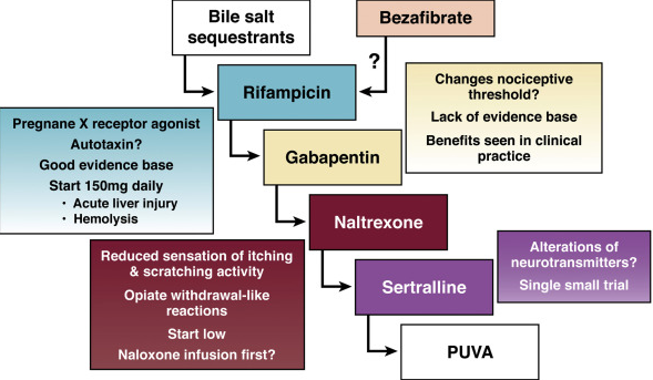
Management: Pruritus may be managed with ursodeoxycholic acid (UDCA), bile acid binding resins, and rifampin.  Bone disease needs to be monitored along with fat soluble vitamin status.

  • While pruritus may improve with UDCA, high-dose UDCA (28-30mg/kg/day) has been associated with increased likelihood of negative endpoints.  It is not clear whether there may be detrimental effects at lower doses.
  • What about immunosuppression?  This (corticosteroids/thiopurines) is accepted in cases with AIH overlap.  Of course, what constitutes overlap is not certain.  So, in many cases, a trial of corticosteroids is considered.
  • Antibiotics?  Oral vancomycin has been tried in a small number with preliminary success.

As noted above, liver transplantation is a needed treatment in some cases. One concern with this approach has been recurrent PSC in about 10% at an average of 18 months after liver transplantation.

Additional references:

  • -Am J Gastro 2011; 106: 1638.  Use of high dose Urso increases risk of colonic neoplasia. n=150.
  • -Hepatology 2011; 54: 1842.  Guidelines on surveillance for PSC.  IF IBD yearly scope.  **Yearly U/S + CA 19-9 or yearly MRI.
  • -Liver Transplantation 201; 17: 925-933.  n=79 pediatric pts.  49% w IBD prior to OLT (46% w UC, 3%) & 9.8% developed IBD p OLT.  1 & 5yr survival for OLT 99% & 87%.  Recurrence in 9.8%.
  • -Clin Gastro & Hepatology 2011; 9: 434.  37% of PSC pts with CA 19-9 >129U/mL do NOT have cholangiocarcinoma.  n=7.
  • -Hepatology 2010; 51: 660-678.  update on dx/mgt.
  • -Gastroenterology 2010; 138: 1102.  genome associations with PSC.
  • -Hepatology 2009; 50: 808, 671 (ed).  Use of urso ~30/kg associated w WORSE outcome @5yrs. n=76 (& 74 controls)
  • -Clin Gastro & Hep 2009; 7:239. n=47 children w PSC.  59% w IBD, 25% w overlap syndrome
  • -JPGN 2008; 47: 61.  Use of oral vancomycin, 50mg/kg/day in patients with PSC/IBD was effective. n=14.
  • -Liver Tx 2008; 14: 735.  Review.  5 yr OLT survival 85%  ReTx rate higher (9.6% vs 4.9%).
  • -Hepatology 2008; 47: 9494-57.  Asymptomatic PSC common in AIH –might be higher than 10%.
  • -Gastroenterology 2008; 134: 975.  Natural hx/o small duct PSC in 83 pts (compared to controls).  Not likely increase in cholangiocarcinoma unless also large duct involvement.
  • -Gastroenterology 2008; 134: 706.  IAC is similar; IAC=IGG4 Associated Cholangitis
  • -Hepatology 2008; 47: 949.  8/79 w AIH also had PSC.
  • -J Pediatr 2007; 151: 255, 230.  association of CFTR mutations in PSC (26%); also present in disease control group with IBD (43%).
  • -Clin Gastro & Hep 2007; 5: 32.  Review vis-a-vis possible cholangiocarcinoma.
  • -Hepatology 2006; 44: 1063.  Review along w secondary causes.
  • -Liver Transplantation 2006; 12: 1813.  Recurrence post Tx 22% AIH, 18% PBC, 11% PSC.  (Review of 43 studies).
  • -Gastroenterology 2005; 129:1464.  High-dose actigall ineffective in 5 yr randomized controlled study.
  • -Gastroenterology 2003; 125: 1364.  Incidence of PSC.
  • -JPGN 2003; 37: 345 (57A). Use of vanco 50/kg/day x 4-6weeks in PSC, n=10, (56A) some pts c normal cholangiograms.
  • -Gastroenterology 2003; 124: 889. Urso prevents colon ca.
  • -Am J Gastro 2001; 96: 1558-62.  High dose UDCA, 25-30/kg/day may help long-term outlook
  • -JPGN 2001; 33:296. PSC-IBD.
  • -NEJM 2002; 346: 271-277.  case c Crohn’s.  PSC assoc c UC, celiac dz, pancreatic insufficiency.
  • -Ann Intern Med 2001; 134: 89-95.  Urso decreases colonic neoplasia in pts c UC & PSC.
    -NEJM 1997; 336: 691. Ursodeoxycolic acid & PSC.
  • -Gregorio et al. Autoimmune hepatitis/sclerosing cholangitis overlap syndrome in childhood: a 16-year prospective study. Hepatology 2001;33:544-553

NASPGHAN 2010 Pointers:
I. -“STOPSC criteria”: 2 of 3 diagnostic criteria needed:
1) elev GGT or alk phos
2) intra and/or extrahepatic bile duct irregularities on cholangiography (i.e. PTC, MRCP, ERCP) c/w PSC
3) liver biopsy abnormalities c/w chronic biliary injury

AND no evidence of a secondary cause of sclerosing cholangitis


II. SUMMARY OF KEY CLINICAL FEATURES:


  • -Sclerosing cholangitis  is associated with IBD, autoimmune and connective tissue disorders, abnormalities of immune function (i.e. immunodeficiencies, GvHD), infiltrative disorders (i.e. Langerhans cell histiocytosis, lymphoma).
  • -PSC may sometimes affect only small intrahepatic bile duct (“small duct PSC”), which may have a better outcome based on adult studies
  • -PSC may have overlapping features of AIH  (“autoimmune sclerosing cholangitis”)
  • -IgG4 –associated cholangitis: patients appear to have PSC with elev IgG4.  They seem to respond very well to steroid therapy in adult studies
  • -No therapy of proven benefit for PSC (ursodiol, antibiotics), some case series have suggested antibiotics may help PSC
  • -5 times higher rate of Colorectal Ca when UC pt has PSC.  Consider yearly endoscopy