Marked Variation with Vibration Controlled Transient Elastography in Pediatric Metabolic Dysfunction Associated Steatotic Liver Disease

Background: “Given the invasiveness of liver biopsy, the development of accurate non-invasive biomarkers of MASLD severity remains an area of active investigation… clinical practice has been shifting towards the use of point-of-care vibration controlled transient elastography (VCTE), based on extensive cross-sectional and longitudinal validation data in adults.11 However, despite increasing clinical use of VCTE to determine MASLD presence and severity in children, this technique lacks well-validated cut-points in this age range and, importantly, there are no data evaluating the implications of longitudinal changes.1215…While a high (>12 kPa) LSM is concerning for progression to advanced fibrosis in adults,11 the optimal cutoff for defining an elevated LSM in pediatric MASLD has yet to be established.1617

Methods: This was a longitudinal retrospective study of youth (n=149, mean age 14 yrs) with MASLD who underwent ≥2 VCTE studies.

Key findings:

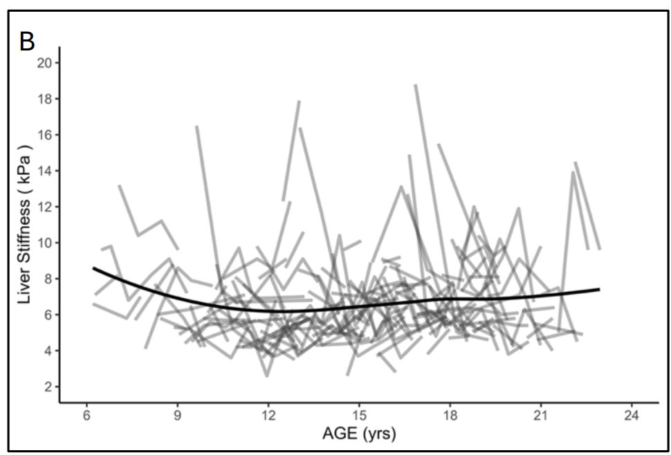
  • Controlled attenuation parameter (CAP) and liver stiffness measurements (LSMs) showed marked intraindividual variability.
  • Changes in CAP and LSM did not predict categorical outcomes of ALT and GGT reduction, however were significantly positively associated with continuous ALT and GGT outcomes, independent of BMI percentile.
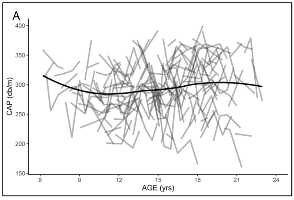
Individual variation in mean controlled attenuation parameter (CAP; panel A) and liver stiffness measures (LSM; panel B) over time. Solid black lines represent Loess lines.

My take (borrowed in part from the authors): “Given the substantial intra-individual variation and the current lack of validated definitions for clinically significant change in VCTE measures (CAP and LSM) in children with MASLD, serial VCTE measurements in individual pediatric patients should be interpreted with caution and not relied upon in isolation for clinical decision-making.”

While transient elastography is not reliable in pediatric MASLD, a related article suggests that it is helpful for pediatric autoimmune hepatitis: W Janczyk et al. J Pediatr Gastroenterol Nutr. 2026;82:1349–1356. Transient elastography for accurate staging of liverfibrosis and predicting complications in children withautoimmune hepatitis

Related blog posts:
Limitations of MRE and TE in Assessing Liver Fibrosis in Pediatric MASLD

N Ravanbakhsh et al J Pediatr Gastroenterol Nutr. 2024;79:1192–1198. Comparing imaging modalities in the assessment of fibrosis in metabolic dysfunction-associated steatotic liver disease

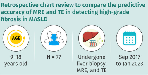
In this retrospective review with 77 patients who had liver biopsy-proven MASLD (2017-2023), the authors examined how well magnetic resonance elastoraphy (MRE) and transient elastography (TE) identified fibrosis.

Key findings:

  • Fibrosis was identified in 90% of liver biopsies
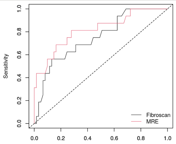
  • The area under the receiver operating characteristic curves (AUROC) of MRE and TE for detection of high-grade fibrosis were 0.817 and 0.750, respectively
  • Only 20% of patients had severe fibrosis on liver biopsy; thus, this is a limitation given the small number
Sensitivity in detecting advanced fibrosis, defined on liver biopsy was defined as Metavir Stage ≥ 3.

Conclusion of authors: “MRE and TE did not accurately predict high-grade fibrosis on liver biopsy. Between the two noninvasive imaging modalities, the correlation of identifying high-grade fibrosis was not statistically different.”

My take: Even MRE is not very accurate at identifying fibrosis. Given the huge numbers of individuals (pediatric and adult) with MASLD, the lack of reliable non-invasive markers is a problematic. As effective treatments become available, being able to determine if they are working is essential.

Related blog posts:

Non-Invasive Studies Often Fail to Detect Advanced Liver Fibrosis in Steatotic Liver Disease

N Ravanbakhsh et al. JGPN 2024; https://doi.org/10.1002/jpn3.12368. Comparing imaging modalities in the assessment of fibrosis in metabolic dysfunction-associated steatotic liver disease

Key findings:

  • TE and MRE did not have high correlation with liver biopsy in the detection of high-grade fibrosis
  • Fibrosis was identified in 90% of liver biopsies with bridging fibrosis in 15 (19%) and cirrhosis in 1 (1%)
  • AUROC curves of MRE and TE for detection of high-grade fibrosis were 0.817 and 0.750, respectively, and not significantly different.

The authors note that previous adults studies suggest that MRE is more accurate in the identification of liver fibrosis than TE (MRE detected ≥ F1 fibrosis with an AUROC of 0.82, while TE detected fibrosis with an AUROC of 0.67).20 

My take: Trying to identify accurate non-invasive testing is crucial to help identify patients most in need of treatment and for limiting costs.

Related blog posts:

Isle of Palms, SC

Defining the Role for Elastography

The ability to determine if a patient has cirrhosis/severe fibrosis with a noninvasive test can help determine appropriate monitoring and treatment for many liver conditions. As such the AGA has provided recommendations for the use of vibration-controlled transient elastography (VCTE).

  • JK Lim et al. Gastroenterol 2017; 152: 1536-43.
  • S Singh et al. Gastroenterol 2017; 152: 1544-77.

Many recommendations are based on the specific unit of measurement, kilopascals (kPa)

Specific recommendations (most with low  or very low quality evidence):

  • “In adults with chronic HCV, we can accurately diagnosis cirrhosis …with VCTE-defined liver stiffness of ≥12.5 (±1) kPa.”  The AGA suggests using VCTE rather than MRE for detection of cirrhosis.
  • “In adults with chronic HCV who have achieved SVR…we can accurately rule out advanced fibrosis (F3 and F4) with post-treatment VCTE-..of ≤9.5 (±1) kPa.” . Even in patients who have had HCV eradicated, if cirrhosis has been identified, careful followup is recommended.
  • “In adults with chronic HBV, we can accurately diagnosis cirrhosis…with VCTE…of ≥11.0 (±1) kPa.”
  • “The AGA makes no recommendation regarding the role of VCTE in the diagnosis of cirrhosis in adults with NAFLD.” For NAFLD, VCTE is not as helpful as with chronic HCV and HBV.  Currently, liver biopsy remains the “gold standard.” However, for noninvasive imaging, “the AGA suggest using MRE, rather than VCTE, for detection of cirrhosis.
  • For adults with suspected compensated cirrhosis, a VCTE of 19.5 or greater can be used “to assess the need for esophagogastroduodenoscopy to identify high risk esophageal varices.”

My take: These elastography recommendations are applicable for adults.  For pediatric patients, these reports suggest that elastography may be helpful in specific circumstances as well.

Related posts:

Omaha Beach

Magnetic Resonance Elastography for Hepatic Fibrosis Assessment

This large case series of 35 children indicates that Magnetic Resonance Elastography (MRE) may be quite useful to assess hepatic fibrosis as well as steatosis (J Pediatr 2014; 164: 186-8).

The study (2011-2012) included 27 patients with nonalcoholic fatty liver disease (NAFLD); 22 of this group had probable or definite nonalcoholic steatohepatitis (NASH).  Other diseases included progressive familial intrahepatic cholestasis (type 2), autoimmune sclerosing hepatitis, Wilson disease, glycogenic hepatopathy (due to type 1 diabetes), and other liver conditions.  All of the patients in the study had undergone liver biopsy as well.

The authors showed that MRE had a high accuracy to detect significant fibrosis and may be better suited for severely obese patients.  At the cutoff they identified, the sensitivity was 88% and the specificity 85% for detecting significant fibrosis.

In severely obese patients, alternative imaging techniques, namely transient elastography and acoustic radiation force imaging have higher technical failure rates.  The authors note that at their institution, more than 100 MRE studies have been completed (including many without liver biopsies); thus far, only two morbidly obese patients failed completion.  In addition, the authors state that this limited study costs about twice that of an ultrasound.

Related posts:

Coming soon –Fibroscan?

Transient elastography as measured by the Fibroscan device is used throughout the world, except notably in the U.S.  That may change soon as an application is under review by the FDA (Gastroenterology & Hepatology 2012; 8: 605-7).

  • Fibroscan works by measuring shear wave velocity with a 50-MHz wave that is “passed into the liver from a small transducer on the end of an ultrasound probe.”
  • “The technology measures the velocity of the sound wave passing through the liver and then converts that measurement into a liver stiffness measurement.”
  • Takes 5-7 minutes to perform
  • Fibroscan is particularly reliable when showing either no fibrosis or advanced fibrosis.  Less accuracy is noted with moderate fibrosis.
  • Technology can be augmented with noninvasive biomarkers of fibrosis
  • Not reliable in several groups: morbidly obese & patients with ascites

Additional references:

  • Am J Gastroenterol 2011; 106: 2121-22. Staging liver fibrosis for HCV
  • Gastroenterol 2005; 128: 343-50.  Comparison of elastography, biomarkers, and liver biopsy for staging fibrosis