Proximal Ileal Crohn’s Disease is Harder to Treat

K Takenaka et al. Clin Gastroenterol Hepatol 2025; 23: 1991-2000. Open Access! Inadequate Efficacy of Biologics for Treating Proximal Ileal Lesions in Crohn’s Disease: A Prospective Multicenter Study

This multicenter prospective study (n=253) examined efficacy of treatment in patients with proximal ileal disease using balloon-assisted enteroscopy (BAE). The recruited patients had a mean disease duration of 4 years. 52% were naive to biologic treatment at baseline.

Key findings:

  • At baseline, 74 patients (29.2%) had proximal ileal ulcerations without terminal ileal ulcerations
  • At week 26, after treatment with anti-TNF therapy (n=103), ustekinumab (n=99) or vedolizumab (n=51), endoscopic remission was achieved in 91 patients (36.0%). Of the patients with complete ulcer healing of the terminal ileum, 28.6% (22/77) had residual ulcers in the proximal ileum
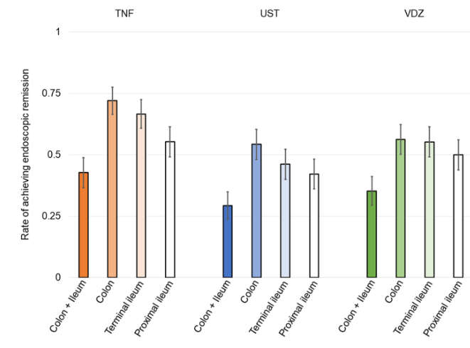
  • The rate of endoscopic remission in the proximal ileum (50.9%) was relatively lower compared with the colon (63.4%) and terminal ileum (56.7%)
  • After a median follow-up of 134 weeks, residual ulcerations in the proximal ileum were associated with a poorer prognosis (P = .0126 for hospitalization and P = .0014 for surgery). In contrast, there was no significant differences in hospitalization and surgery associated with endoscopic activity vs remission in the colon or terminal ileum.

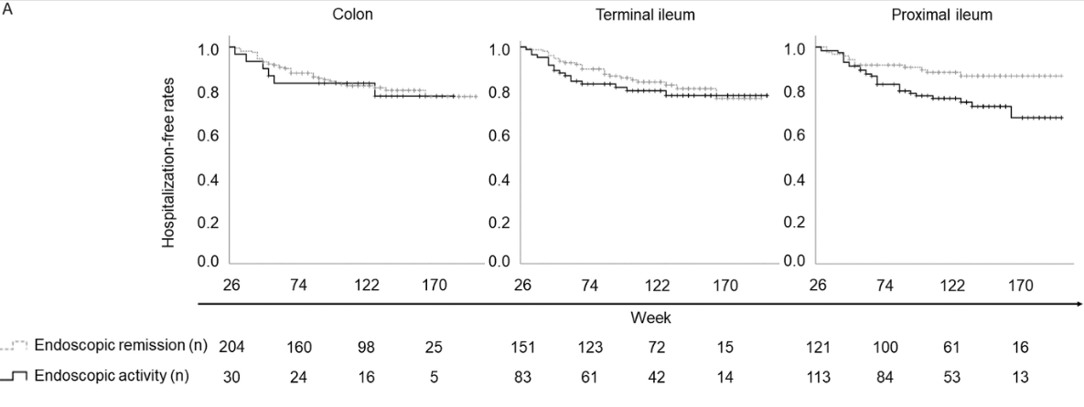
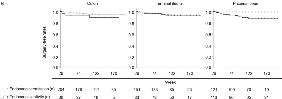
Discussion: Residual “proximal ileal ulcerations … are associated with a poorer prognosis…Additionally, we confirmed that proximal ileal inflammation is less responsive to biologic therapies compared with colonic inflammation. Although the reasons for this disparity remain unclear”

My take: Balloon-assisted enteroscopy is not frequently used in the setting of inflammatory bowel disease, particularly in pediatrics. MRE is typically used to follow proximal small bowel disease, though it has less sensitivity for luminal mucosal disease.

Related blog posts:

Hypercoagulation with Acute Severe Ulcerative Colitis (ASUC) Persists for Months

BJ Griffiths et al. Clin Gastroenterol Hepatol 2025; 23: 1798-1807. Open Access (PDF)! Hypercoagulation After Hospital Discharge in Acute Severe Ulcerative Colitis: A Prospective Study

Methods: In this prospective study, twenty-seven adult patients with ASUC and 25 control patients with quiescent ulcerative colitis were recruited. Thrombin generation (endogenous thrombin potential), rotational thromboelastometry (EXTEM and FIBTEM maximum clot firmness), procoagulant factors, anticoagulant factors, and fibrinolytic markers were assessed for those with ASUC on admission (Day 1), Day 5, 4 weeks, and at 8–12 weeks. These assessments were performed on a single occasion for control patients.

Key findings:

Discussion:

  • “Patients with ASUC had a hypercoagulable profile on initial presentation to the hospital, before receipt of LMWH, which was consistently demonstrated by individual and global assays of coagulation. The most marked elevations of individual factors at presentation were seen in the levels of Clauss fibrinogen, platelets, VWF, and FVIII, alongside heightened
    levels of the inhibitors of fibrinolysis PAI-1 and TAFIa.”
  • “This hypercoagulable state persisted for many weeks after hospital discharge, with levels of FVIII, fibrinogen, VWF, and inhibitors of fibrinolysis (TAFIa) remained significantly elevated at all timepoints up to 12 weeks, compared with the control population. This is despite
    intensive treatment for ASUC in all patients.”
  • “VTE and pulmonary embolism are 1 of the leading causes of morbidity and mortality during IBD flare-ups. The findings from this study reinforce the importance of thromboprophylaxis administration to all patients with ASUC at first presentation to hospital.”

My take: This study is in adults; the risk of VTE is lower in children and guidance on VTE prophylaxis is not clear.

Related blog posts:

Dupilumab for FPIES

M Plassmeyer et al. Journal of Allergy and Clinical Immunology 2025; Dupilumab Opens a Therapeutic Window in Food Protein Induced Enterocolitis Syndrome by un-licensing dendritic cells

Thanks for Ben Enav for this reference.

Methods: This was a two-part study: “(i) a detailed single-patient case of wheat-triggered, endoscopy-confirmed colitic FPIES treated with dupilumab 300 mg subcutaneously every two weeks and (ii) a prospective follow-up of seven additional FPIES patients all of whom initiated dupilumab for approved comorbidities. Serial flow cytometry quantified dendritic-cell OX40L and CD8+ CRTH2+ T-cell subsets before and after treatment; open food challenges assessed clinical tolerance.”

Key Findings:

  • Index case: Within two injections of dupilumab, the wheat sensitive patient tolerated a 50 g wheat protein challenge without gastrointestinal symptoms—this was the first uneventful exposure in 20 years. Discontinuation of dupilumab led to relapse; re-initiation again restored clinical tolerance
  • Cohort: All seven additional patients (ages 2–58 yr; triggers: milk, soy, rice, wheat, shellfish) achieved unrestricted dietary tolerance within three months
  • An important finding in the index case as well as the follow up cohort is the dupilumab induced drop in dendritic cell OX40L. OX40L is a TNF-superfamily co-stimulatory molecule induced on dendritic cells and other antigen-presenting cells.

My take: Dupilumab appears to be a promising medication for FPIES and warrants further study. If confirmed to be effective, it is likely to be targeted to those with approved comorbidities and those with more severe presentations.

Related blog posts:

Amicalola Falls State Park

Kids With Acute Pancreatitis Need Followup

F Ahmed, M Abu-El-Haija. Gastroenterol 2025; 169: 572-584. Open Access! Acute Pancreatitis in Children: It’s Not Just a Simple Attack

This is a really good review of acute pancreatitis covering epidemiology, diagnosis, severity classification, management, microbiome/metabolite derangements, genetics, and complications. Most of these topics have been covered in numerous blog posts (see below).

Selected Key Points:

  • Diagnostic testing -Amylase/Lipase:  “The diagnostic efficacy of amylase for AP, in terms of sensitivity and specificity, is contingent on the selected threshold value. Elevating the cut-off point to 1000 IU/L results in a high specificity of approximately 95%. However, this comes at the cost of reduced sensitivity, which some studies report to be as low as 61%… the activity of serum lipase remains elevated for a longer duration, typically between 8 and 14 days,… Lipase demonstrates superior accuracy with most studies reporting specificities exceeding 95% and sensitivities ranging from 55%–100% at a threshold activity level of 600 IU/L…hese tests have excellent sensitivities, they may have a few limitations such as being poor predictors of severity”
  • Risk Factors in Children (from Figure 1):
  • Incidence and Severity in Children (from Figure 1):

[At a recent lecture, Jay Freeman (How to Upgrade Pancreas Care –Jay Freeman MD (Part 1)) noted that severe pancreatitis is often defined by degree of organ dysfunction (eg. cardiac, pulmonary, renal). A practical definition of severe pancreatitis in children is whether the patient requires admission to an ICU]

  • Diagnostic testing -Imaging: “Imaging techniques are crucial for diagnosing and managing AP in children…NASPGHAN) and the Society for Pediatric Radiology formed consensus guidelines where transabdominal ultrasonography was recommended as the primary imaging technique for pediatric cases with suspected AP…Recent studies in the pediatric population have indicated that US’s sensitivity for AP detection ranges from 47%–52%.25Magnetic resonance cholangiopancreatography (MRCP) is useful for anatomical assessment without radiation but may require sedation”
  • Management: “The cornerstones of therapy are early feeding and intravenous fluids… Allowing patients to eat on admission was feasible and was associated with lower length of stay. Rates of intravenous fluids are recommended at 1.5–2 times maintenance rates,49 and the preferred fluid is Lactated Ringer’s due to limited studies including a recent randomized controlled study that showed that Lactated Ringer’s was associated with a faster discharge rate when administered compared with normal saline.50
  • Genetics:  “A recently conducted study investigated the importance of genetics in pediatric AP patients…use of an extensive panel of 8 genes… PRSS1CFTRSPINK1CPA1, CTRCCLDN2CASR, and SBDS… genetics is a major component in all types of pancreatitis in children, with genetic variants being most prevalent in CP cases at 31%, followed by AP at 19%, and ARP at 6%. A key discovery was that variants in SPINK1CFTR, or PRSS1 genes were associated with faster progression from first episode of AP toward CP.53
  • Complications (from Figure 1): “After the first episode of AP, the QoL is decreased, and it may lead to other disorders such as exocrine dysfunction, endocrine dysfunction and diabetes, nutritional deficiencies, and acute recurrent pancreatitis and CP.”

My take: Even after a single episode of acute pancreatitis, there are risks for long-term complications and patients need to follow-up.

Related blog posts:

Shared Decision-Making in Celiac Disease Diagnostic Approach

Y Sunkoy, S Talathi. Am J Gastroenterol 2025; 2190-2193. Utility of the ESPGHAN Biopsy-Sparing Guidelines for Celiac Disease in Children

Thanks to Ben Gold for this reference.

Methods: Retrospective study of patients (n=2942 children) who had celiac serologies and duodenal biopsies

Key findings:

  • Prevalence of CD in this cohort was 9% (226 of 2942 patients)
  • In those with a high titer (>10xULN), 106 of 107 patients (99%) had celiac disease
  • In this cohort, even in those with with >7XULN, had a Sensitivity of 55.3%, a specificity of 99% and a PPV of 97%

In their discussion, the authors note that “we did not obtain an EMA in a second sample, which is recommended in the ESPGHAN guidelines.”

Associated commentary: Erica Brenner, American Journal of Gastroenterology 120(9):p 1985-1986. The No-Biopsy Approach for Pediatric Celiac Disease: Ready for Prime Time in North America?

  • “Shiha et al (8) found that the PPV ranged from 65% for a 1% CD prevalence to a 99% for a 40% prevalence. As the 9% CD prevalence in the study by Sunkoj et al (4) exceeds the 0.81$-1.4% prevalence in the United States (9), the reported PPV may overestimate reality.” (Related post: No-Biopsy Approach to Celiac Disease Diagnosis and Positive Predictive Value (Based on Population)
  • “Children with type 1 diabetes and trisomy 21 have a higher risk of false-positive serology and therefor may not be appropriated candidates for a no-biopsy approach.”

My take: A larger recent study (Chang et al. Pediatrics. 2025;156(3):e2025070897) found that the no-biopsy approach had a significantly lower PPV in their cohort (94.9% overall, and 95.7% in those without T1DM). Thus, in cohorts with lower prevalence of CD, the no-biopsy approach could lead to 2-4% of children being placed unnecessarily on a gluten free diet. As such, it would be good practice to discuss making a diagnosis via endoscopy vs. the no-biopsy approach as part of shared decision-making.

Related blog posts:

Stercoral Colitis

A Bajer, E Levine. NEJM 2025;393: e23. Stercoral Colitis

This young adult presented to the ED with left-sided abdominal pain and chronic constipation. A CT scan was consistent with a diagnosis of stercoral colitis. “In stercoral colitis, chronically impacted feces distend the colon, resulting in inflammation. In some cases, the fecaloma may lead to focal-pressure necrosis or perforation.”

My take: Most often a CT is not needed in this setting. However, it is important to recognize that a severe impaction can lead to complications.

Related blog posts: