NASPGHAN Postgraduate Course 2017 (Part 3): Biliary Atresia, NAFLD, SMOFlipid, Pancreatic Pain

This blog entry has abbreviated/summarized these presentations. Though not intentional, some important material is likely to have been omitted; in addition, transcription errors are possible as well.

Here is a link to postgraduate course syllabus: NASPGHAN PG Syllabus – 2017

Biliary Atresia: Update on diagnostic and prognostic biomarkers and therapeutic interventions

Cara Mack    Children’s Hospital of Colorado

Key points:

  • 84% of biliary atresia is isolated; 16% are syndromic with other defects
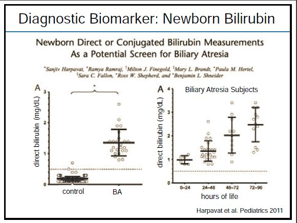
  • Direct bilirubin is (mildly) elevated at birth in patients with biliary atresia
  • Total bilirubin 3 months after Kasai predicts outcome. If <2 mg/dL, then unlikely to need a transplant in the first 2 years of life.
  • Reviewed biomarkers including Th1, Autotaxin, IL-8

Therapeutic interventions:

  • Nutritional support. Better nutrition improves outcomes after liver transplantation.
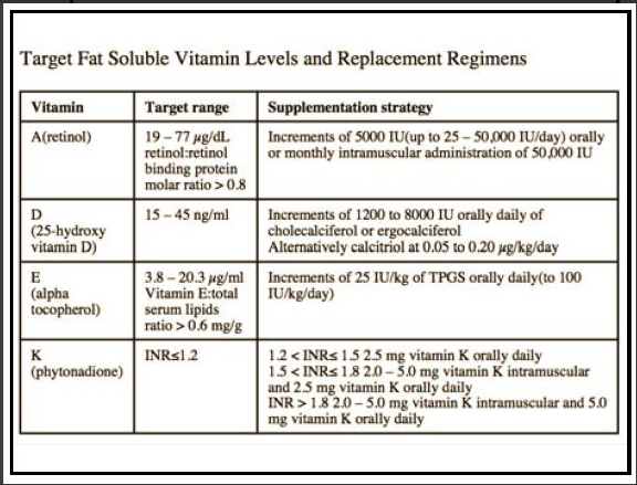
  • Fat soluble vitamin supplementation
  • Cholangitis prevention. Some studies have shown that prophylactic antibiotics may reduce incidence of cholangitis.
  • No therapeutic interventions that delay progression of this disease


CHILDREN Cohort Mgt of Vitamin Supplementation

Steroids are not helpful after Kasai procedure

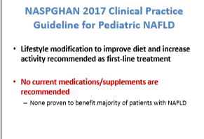
Diagnosis and Management of Pediatric NAFLD 2017

Stavra Xanthokos   Cincinnati Children’s Hospital Medical Center

Key points:

  • NAFLD is #2 cause of liver transplantation in adults and on its way to becoming #1
  • ALT is still the best screening tool; NASPGHAN guidelines recommends screening overweight/obese children 9-11 years of age
  • Ultrasound has poor sensitivity and specificity for NAFLD; it is helpful for detecting gallbladder disease
  • Bariatric surgery has been effective for NAFLD


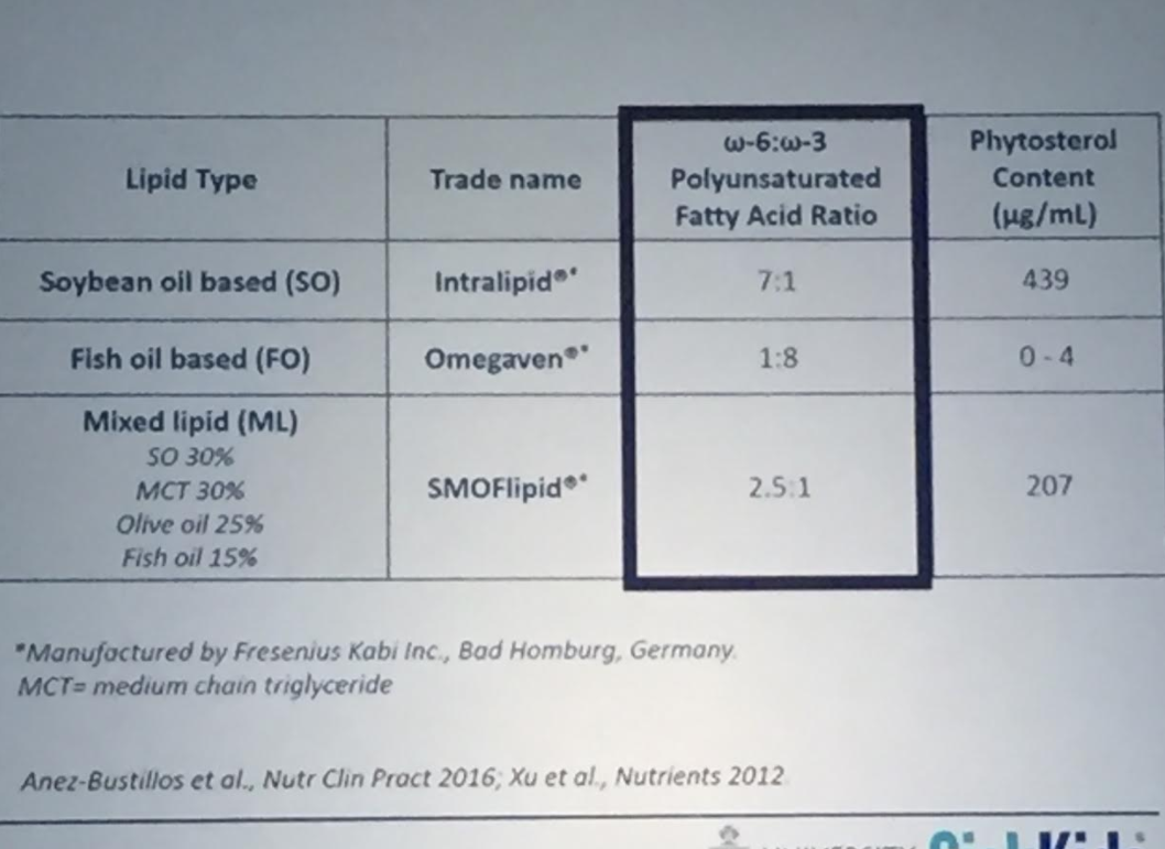
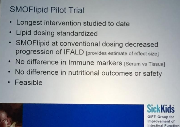
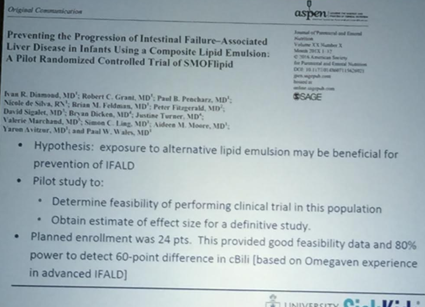
SMOFlipid and the Pediatric Patient

Peter Wales  Hospital for Sick Children (Toronto)

Slides are not available in syllabus

Key points:

  • Improving outcomes noted in the intestinal failure population
  • Dr. Wales reviewed proposed improvements with Omega-3 lipids -less cholestasis, less hepatitis, and less fibrosis
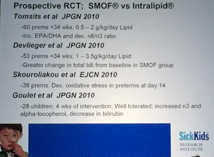
  • Compared improvements with lipid minimization (1 g/kg/day) compared to newer agents: omegaven and SMOFlipid. Additional studies are needed due to limitations of previous studies
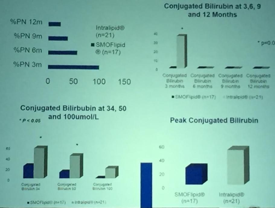
  • Discussed SMOFlipid vs. Intralipid trial at 5 centers in Canada. N=24.
  • At SickKids: SMOFlipid for all preterms at admission & for term infants after 2 weeks of PN. Dosing 2-2.5 g/kg & now accounts for 85% of lipid usage at institution
  • None of the lipid products were designed for preterm infants. Intralipid has a pediatric indication and other products are used off label
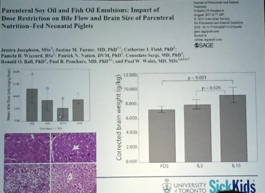
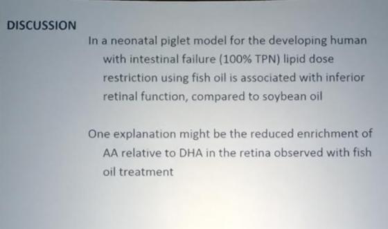
  • Lipid restriction probably affects brain size/development; thus, a lipid agent that allows for higher doses likely will be beneficial for developmental outcomes.  The retina can be used as a biomarker of the brain affects of lipids.

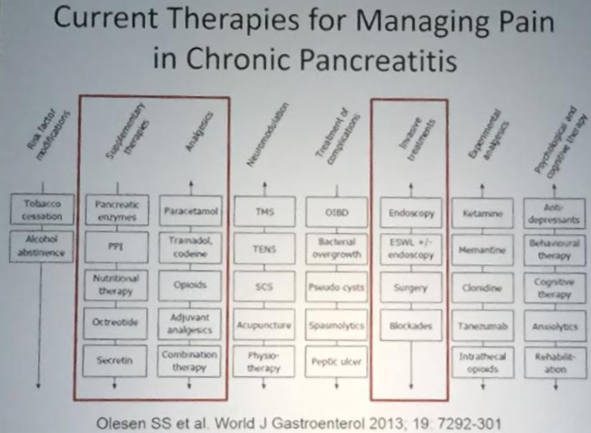
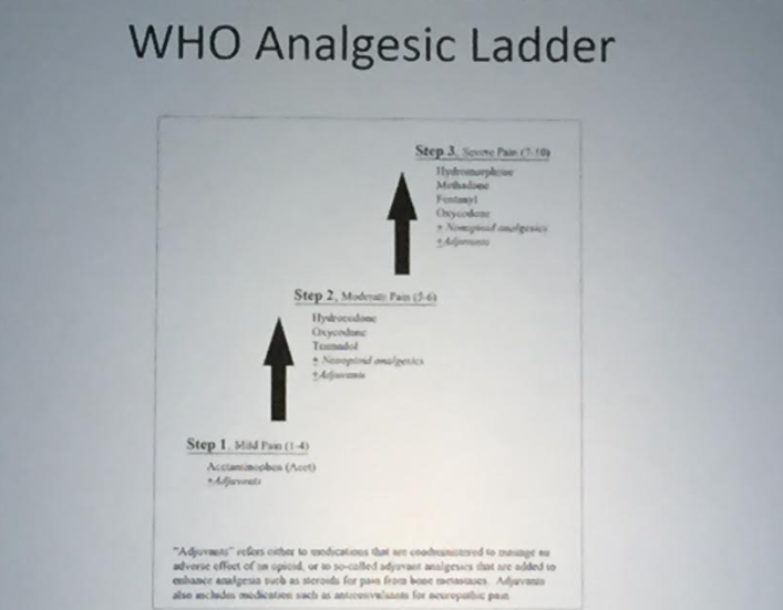
Painful Chronic Pancreatitis: Management/therapeutic interventions

Vikesh Singh  Johns Hopkins University School of Medicine

Slides are not available in syllabus


More on It’s Past Time to Split

In followup to this morning’s post, Pediatric Liver Transplantation: It’s Past Time to Split, one reader pointed out an abstract by Emily R. Perito et al.presented at this year’s AASLD which showed that pediatric deaths would decrease if more livers were split.

ABSTRACT #137

Increasing split liver transplantation in the U.S. could decrease pediatric deaths on the liver transplant waiting list

Emily R. Perito1,3, Garrett Roll2, Jennifer L. Dodge2,

Background: In the United Kingdom, defaulting to split liver transplantation (LT) with suitable deceased donor grafts has virtually eliminated pediatric waitlist (WL) mortality. In the US, only <2% of LTs are split, but 1 in 10 infants die on the WL.

Methods: Using UNOS STAR data, livers for potential split LT were identified from all transplanted, deceased-donor livers 2010-15 who fit strict criteria: age 18-40y, BMI<30, recovered in US after donor brain death, 0-1 vasopressors, a <155meq/L, AST/ALT<100IU/L, bilirubin<3mg/dL, <7d hospitalized, cardiac arrest≤30min, HBV/HCV neg, not CDC high-risk, steatosis≤10% if biopsied, not multi-organ transplant, and no bloodstream infection. Livers allocated to patients high-risk for split LT were also removed: status 1A or MELD/PELD≥40 at WL removal, re-transplant, in the ICU, BMI>34, or >300mi from donor hospital. Pediatric WL deaths included deaths and removals for too sick to transplant, never relisted.

Results: Of 35,461 livers transplanted 2010-15, 6.7% were potentially utilizable for split LT based on donor characteristics. Of these, 95% were transplanted whole (n=2,253). 50% went to recipients deemed possibly high-risk for split LT. This left 1,116 potential livers for split LT (FIGURE); 78% of their primary recipients were listed as willing to accept a segmental liver, and 97% to accept cold ischemia time≥6h (CIT, median 12h). Median donor risk index for this subset was 1.06 (max 1.67). During the same 5y, 261 children died after ≥3d on the WL (median 57d, IQR 15-161)—87% of all pediatric WL deaths. Of these, 56% were <2y of age, 26% 2-12y, 18% 13-18y. Median weight was 9.2kg (IQR 5.9-29.4kg). 36% died at centers that reported doing no pediatric split LTs (15%) or ≤1/year (22%).

Conclusions: Increased utilization of split LT could decrease US pediatric WL mortality—without decreasing LT access for adults. Barriers are significant, but changes to  allocation policy, increasing centers with splitting experience, and splitting on normothermic perfusion could increase access and reduce WL mortality.

Jose Garza, Chelly Dykes, Elvis, and Jay Hochman at Cincinnati Children’s Reception

Pediatric Liver Transplantation: Past Time to Split

A recent study (EK Hsu et al. Gastroentorol 2017; 153: 988-95, editorial 888-89) exposes some deep flaws in organ transplantation in U.S.

The retrospective study examined children on the U.S liver transplant wait-list from 2007-14.  This included 3852 pediatric candidates.  Key findings:

  • Of 27,831 adults who underwent transplantation, 1667 (6%) received livers from pediatric donors (<18 years)
  • Of children who died or were delisted, the centers caring for 173 (55%) had received an offer of 1 or more livers that was subsequently transplanted into another pediatric recipient.  The remaining 45% died or delisted with no offers. High-volume (>15 transplants per year) centers were more likely to accept an organ than a low-volume center (<5 transplants per year).
  • Only 29% of children received a split graft.  When a splittable adult liver graft was allocated to an adult the chance of it being used as a split was 0.6%.

Background:

  • Children have much lower survival rate than adults on waiting list. Of adults who died or delisted, 85% receive at least one transplant offer; whereas, nearly half of all children never even receive an offer.  Children who died/delisted had wait-time of 33 days compared with 92 days for adults who died/delisted.
  • Less than 10% of all liver transplant recipients are pediatric transplants.  Per editorial, “a measure that improves pediatric access by 20% would only reduce adult access by 2%.”
  • There are more than 100 pediatric liver transplant centers in U.S. Certainly, this improves convenience; however, per editorial:  “three-fourths are very low volume centers, performing <5 liver transplantations per year…Death on waiting list” occur 5 times more at low-volume transplantation centers.
  • In this study, only 29% of children received split livers; in comparison, in the UK, >80% receive either a split graft or living donor graft.

The editorial points out that splittable livers that are allocated to adults are virtually never split; this is either due to inconvenience or lack of expertise.  A small increase in liver splitting would dramatically lower the pediatric mortality wait list.  There is no incentive in the current system to split a liver/save a child’s life.

My take: The data from this study points out glaring problems in pediatric liver transplantation.

  1. Children are dying due to lack of prioritization.  Pediatric livers are going to adults.
  2. There is practically no splitting when liver organs are allocated to an adult.  Incentives to increase organ splitting would save many children from dying waiting for an organ.
  3. Large volume pediatric centers are much more likely to accept a liver offer for patients waiting at their centers.  There is an increased wait-list mortality at very low volume centers, perhaps due to lack of expertise and passing up viable organs.  Do hepatologists/surgeons at these centers explain this risk to families at their centers?

Related blog posts:


Assessing Neonatal Jaundice with Smartphone App

A recent study (Taylor JA, et al. Pediatrics 2017; 140 (3) e20170312) reports on the effectiveness of a smartphone app, BiliCam, to detect total serum bilirubin (TSB) in a diverse sample of newborns < 7 days old.  Thanks to Ben Gold for this reference.

BiliCam uses a calibration card which is placed on the infant’s sternum to standardize the color (and jaundice) reading in the photo; the image goes via the internet to a server for analysis.

Key findings:

  • Estimated bilirubin levels using BiliCam were compared with TSB levels in 530 newborns which included 20.8% African American,, 26.3% Hispanic and 21.2% Asian American
  • The overall correlation was 0.91 were similar among all ethnic groups with correlations ranging from 0.88 to 0.92
  •  The sensitivity of Bilicam was 84.6% is for identifying infants with a TSB in the high-risk zone of the Bhutani nomogram. The sensitivity was 100% for identifying TSB > 17 mg/dL. Specificities were 75.1% adn 76.4% respectively.

For more commentary on this article: AAP Journals Blog: Bilirubin phone apps –our future calls!

My take: This article indicates that a digital image with Smartphone app analysis is much more accurate in detecting jaundice that a visual assessment.

Bile Acid Therapy -18 Year Study

JE Heubi et al (JPGN 2017; 65: 321-6) performed a phase 3, open-label, nonrandomized trial on the efficacy and safety of oral cholic acid for patients with Zellweger Spectrum disorders (n=20) and patients with bile acid synthesis disorders (BASD) (n=50). Cholic acid dosing: 10-15 mg/kg/day. Most common BASD were 3β-HSD (n=35), and 5β-reductase (n=10).  Based on this work, cholic acid is an FDA-approved agent.

Key findings:

  • Urine bile acid metabolite scores improved (P<0.0001) with cholic acid
  • Transaminases improved (AST, ALT) (P<0.0001)
  • Growth parameters, improved with weight gain reaching statistical significance
  • “Liver biopsies showed either stable findings or histologic improvement in all parameters except bridging fibrosis”
  • No study drug-related serious adverse events were noted
  • With Zellweger spectrum disorders, it is important to note that “there is no evidence that treatment with cholic acid has any impact on the extrahepatic disease.”

My take: Cholic acid helps the liver in these disorders which is particularly important for BASD. It is unclear if this improves outcomes in patients with Zellweger spectrum disorders as it has not been shown to improve extrahepatic disease.

Related blog post:

Eiffel Tower

Fructose Restriction Improved Fatty Liver Disease in Children

A recent study (J-M Schwarz et al. Gastroenterol 2017; 153: 743-52, editorial MB Vos, IR Goran Gastroenterol 2017; 153: 642-5 ) showed that restriction of fructose quickly improved fatty liver disease.

Several points from the editorial:

  • “The metabolic driver of buildup of fat storage in the liver is de novo lipogenesis (DNL) and fructose is a major substrate of DNL”
  • “In the healthy state, DNL is not expected to be a major contributor to lipid accumulation in the liver….[but] in a fatty liver, it has been estimated that 26% of the fat originates from DNL.”
  • Fructose is “limited in a natural diet…However, it is added to many processed foods and drinks in the form of cane sugar..and other types of sugars, going by ≥57 different names.”
  • Fructose is “commonly used in animal models to induce hepatic steatosis.”

The study is summarized in a recent AGA Journals Blog: Can Restricting Fructose Intake Reduce Fatty Liver Disease in Children?

An excerpt:

Jean-Marc Schwarz et al performed a clinical trial to investigate the effects of reducing fructose intake for 9 days in obese Latino and African American children with habitual high sugar consumption (fructose intake >50 g/day). They measured the effects of isocaloric fructose restriction on de novo lipogenesis, liver fat, visceral fat, subcutaneous fat, and insulin kinetics.

In their study, 41 children, 9−18 years old, had all meals provided for 9 days. The meals had the same energy and macronutrient composition as their standard diet, but with starch substituted for sugar, yielding a final fructose content of 4% of total kilocalories. The authors measured metabolic factors before and after fructose restriction. They measured liver fat, visceral fat, and subcutaneous fat by magnetic resonance spectroscopy and imaging.

Schwarz et al found that on day 10 of the diet, liver fat decreased from a median 7.2% at baseline to 3.8%, and visceral fat decreased from 123 cm3  at baseline to 110 cm3. Liver fat decreased in all but 1 of the 38 participants for whom paired data were available…

De novo lipogenesis decreased significantly after 9 days of fructose restriction; the de novo lipogenesis area under the curve value on day 10 decreased from 68% at baseline to 26% after the diet, in childen with low or high baseline levels of liver fat.

Insulin secretion during fasting and in response to an oral glucose tolerance test decreased significantly in children with low and high baseline levels of liver fat…

In an editorial that accompanies the article, Miriam B. Vos and Michael I. Goran say that it will be important to determine whether the effects of fructose reduction are sustained past 9 days…Vos and Goran state that it is important for physicians, nutritionists, schools, and parents to find ways to reduce fructose in the diets of children and patients with NAFLD.

Related posts:


Moving Away from Liver Biopsies

A recent review (EB Tapper, AS-F Lok. NEJM 2017; 377: 756-68) provides a good review of liver biopsy and liver imaging. My take of this review is that it highlights the emergence of noninvasive tools (imaging & fibrosis markers) which may supplant liver biopsy.  This article does not delve into how more widespread genetic testing may obviate a liver biopsy in many cases as well. The article notes that about 8% of persons in the U.S. have elevated liver enzymes.

Liver biopsy:

  • “A typical liver biopsy samples one fifty-thousandth of the liver.”
  • Limitations of liver biopsy: sampling error is common, biopsy interpretation is subjective, and biopsies can cause complications.  Pain is noted in 30-50% of patients, serious bleeding in 0.6%, injury to other organs (0.08%), and in rare cases, death (up  to 0.1%).
  • Cost: “the average direct cost of a percutaneous liver biopsy is $1448 (in 2016 U.S. dollars).” Transjugular biopsies are much more expensive.  In addition, there are unmeasured indirect costs, due to missing work.

Some prior blogs on liver biopsy

Blood tests:

  • The article details the formulas for biomarker measurements that predict the risk of fibrosis, inlcuding FIB-4, Lok Index, and NAFLD Fibrosis Score.
  • In most liver diseases, aspartate aminotransferase levels “exceed alanine aminotransferase levels when cirrhosis develops.”
  • Thrombocytopenia “is the earliest indicator of cirrhosis among routine blood tests…[due to] diminished liver function (throbopoietin underproduction) and portal hypertension (splenic sequestration).”
  • Proprietary algorithms to assess fibrosis have variable sensitivity, specificity –include FibroTest (aka FibroSure [LabCorp]), FibroMeter, HepaScore (Quest), FIBROSpect, and the Enhanced Liver Fibrosis Score.

Imaging:

  • Elastography with vibration-controlled transient elastography (VCTE) OR magnetic resonance elastography
  • “Elastography offers excellent negative likelihood ratios for advanced fibrosis but much poorer positive likelihood ratios.”
  • Patients with severe obesity are less likely to obtain adequate study with VCTE and could need magnetic resonance elastography to assess fibrosis.

My take: Noninvasive tests have already sharply reduced the need for liver biopsy.

Related posts:

Updated Biliary Atresia Epidemiology

A recent retrospective study (PC Hopkins, N Yazigi, CM Nylund. J Pediatr 2017; 187: 253-7) provides an update on the recent incidence of biliary atresia in the US from 1997-2012. This study relied on coding for biliary atresia or Kasai hepatoportoenterostomy to identify cases using HCUP-KID database.  This database provides a nationally representative sample of pediatric hospitalizations and captures ~96% of pediatric hospitalizations in the US.

Key findings:

  • Incidence of biliary atresia (BA) was 4.47 per 100,000 (1 in 22,371 infants)
  • BA was more common in females (RR 1.43), Asian/Pacific Islanders (RR 1.89), and blacks (RR 1.30)
  • Median age at the time of the Kasai procedure was 63 days with no improvement over the course of the study period.  More than 50% of all children underwent the Kasai procedure after the optimal window of 60 days of life

My take: In my view, at this time, obtaining a blood test for direct bilirubin in the first two weeks of life will need to be adopted broadly if we are going to diagnose biliary atresia at an earlier age.

Related blog posts:

Dry Falls, Highlands NC

Dry Falls, Highlands NC


Recurrent Acute Liver Failure due to NBAS Deficiency

A recent case report (V Cardenas et al. J Pediatr 2017; 186: 179-82) describes recurrent acute liver failure (ALF) in the setting of neuroblastoma amplified sequence deficiency (NBAS).

The case report describes a 2 yo who developed very elevated aminotransferases (ALT >14,000), hypoglycemia, severe coagulopathy (INR 4.5)), lactic acidosis (6.5 mmol/L) and hyperammonemia (282 μmol/L) following a febrile illness.

Genetic testing uncovered 2 variants in the NBAS gene consistent with NBAS deficiency.

Key points:

  • Mutations in NBAS “have been identified as a molecular cause of ALF in children, leading to recurrent episodes of ALF after a febrile illness.”
  • NBAS deficiency should be part of the differential diagnosis of ALF in children
  • In a report of 14 patients with this disorder (J Inherit Metab Dis 2016; 39: 3-16), liver function normalized in between episodes.  Typically, episodes were most severe at younger ages.  ALF “may be prevented through early and effective antipyretic therapy and intravenous application of glucose and lipids.”

My take: NBAS deficiency, along with hemophagocytic lymphohistiocytosis (HLH), infections, and Kawasaki’s disease, needs to be considered in children with severe liver dysfunction associated with fevers.

“Big Improvements for Smallest Recipients” with Bad Liver Disease

A recent study (M Kasahara et al. Liver Transplantation 2017; 23: 1051-7, editorial 977-8) indicates improvement in survival among the smallest liver transplant patients. In this study of 12 patients less than 3 months of age, the cumulative 10-year patient and graft survival for both was 90.9%.

These patients received living donor liver transplantation. Living donors likely contributed to the excellent outcomes both in terms of enhancing the timing of transplantation and also with regard to size.  Whole organs are not likely to fit well in these small abdomens. The size of the patients ranged from 2.8 kg (at 29 days) to 5.5 kg).  11 of 12 had fulminant hepatic failure with 6 of these cases being considered unknown etiology.

Limitation: This was a very small sample size.