Pediatric Drug-Induced Liver Injury

F Monge-Urrea, E Montijo-Barrios. JPGN 2022; 75: 391-395. Drug-induced Liver Injury in Pediatrics

Background: Antibiotics and antiepileptics remain the most frequent causes of DILI. DILI may result in severe outcomes (eg liver transplant) in up to 5% of cases and could result in chronic liver disease in ~20%.

This is a terrific review -Figure 1 is particularly helpful. Figure 1 is an algorithm. Prior to using algorithm, review potential hepatoxcity by searching in NIH Livertox website. Next steps:

  1. Calculate pattern of injury (R score). R= ALT/ULN divided by ALP/ULN (ALP =alkaline phosphatase)
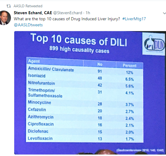
  2. Identify suspect drug. Hepatocellular (R >/=5), Mixed (R=2-5), Cholestatic (R</= 2). Examples of hepatocellular include acetaminophen, NSAIDs, Minocycline. Examples of mixed include azathioprine, and sulfasalazine. Examples of cholestatic include amoxicillin/clavulanate, and TMP/SMX
  3. Exclude alternative causes
  4. Calculate RUCAM Score (detailed in Table 1). This score can also be found at this link (open access): Overview of causality assessment in drug-induced liver injury
  5. Discontinue implicated drug and review specific therapies. For example, N-acetylcysteine for acetaminophen, and carnitine for valproate
  6. Consider liver biopsy only if suspected DILI progresses or fails to resolve on withdrawal of suspect drug (resolution can take 3-4 months)

Drug stop rules are reviewed:

  • ALT or AST values that exceed 8 times the ULN
  • ALT or AST values that exceed 5 times the ULN -hold medication for 2 weeks
  • ALT or AST values >3 times the ULN and Bilirubin >2 times the ULN
  • ALT or AST values that exceed 3 times the ULN with progressive nonspecific symptoms

Related blog posts:

Crow’s Pass Trail, AK

Disclaimer: This blog, gutsandgrowth, assumes no responsibility for any use or operation of any method, product, instruction, concept or idea contained in the material herein or for any injury or damage to persons or property (whether products liability, negligence or otherwise) resulting from such use or operation. These blog posts are for educational purposes only. Specific dosing of medications (along with potential adverse effects) should be confirmed by prescribing physician.  Because of rapid advances in the medical sciences, the gutsandgrowth blog cautions that independent verification should be made of diagnosis and drug dosages. The reader is solely responsible for the conduct of any suggested test or procedure.  This content is not a substitute for medical advice, diagnosis or treatment provided by a qualified healthcare provider. Always seek the advice of your physician or other qualified health provider with any questions you may have regarding a condition.

Liver Shorts -August 2020

V Cardenas et al. JPGN 2020; 71: 197-202.  Incidence and Sequelae of Liver Injury Among Children Treated for Solid Tumors: Analysis of a Large Single-Center Prospective Cohort

  • Of 1136 solid tumor patients, 160 (14%) experienced liver injury, and the overall frequency of DILI was 4%.
  • DILI was the leading identified cause of liver injury (31%), followed by infection (17%), metastatic/malignant biliary disease (13%), and perioperative liver injury (13%).
  • Most DILI cases (>90%) were mild acute hepatocellular injury episodes that did not result in modification to the chemotherapy plan, and all DILI eventually resolved.

N Kapila et al. Hepatology 2020; 72: 32-41. Full Text Link: Hepatitis C Virus NAT‐Positive Solid Organ Allografts Transplanted Into Hepatitis C Virus–Negative Recipients: A Real‐World Experience

Background: As of April 1, 2019, an estimated 103,000 kidney, 13,500 liver, and 3,800 heart transplant (HT) candidates are awaiting transplantation

Key findings:

  • Seventy‐seven patients who were HCV negative underwent solid organ transplantation from a donor who was HCV viremic. Only one has been a HCV-treatment nonresponder (though several have not completed SVR12).
  • “Our study is the largest to describe a real‐world experience of the transplantation of HCV‐viremic organs into recipients who are aviremic. In carefully selected patients, the use of HCV‐viremic grafts in the DAA era appears to be efficacious and well tolerated.”

M Martinello et al. Hepatology 2020; 72: 7-18Short‐Duration Pan‐Genotypic Therapy With Glecaprevir/Pibrentasvir for 6 Weeks Among People With Recent Hepatitis C Viral Infection

  • This was an  open‐label, single‐arm, multicenter, international pilot study; adults with recent HCV (duration of infection < 12 months) received glecaprevir/pibrentasvir 300/120 mg daily for 6 weeks.
  • At baseline, median estimated duration of infection was 29 weeks (range 13, 52) and median HCV RNA was 6.2 log10 IU/mL (range 0.9, 7.7). SVR12 in the intention‐to‐treat and per‐protocol populations was achieved in 90% (27/30) and 96% (27/28), respectively.

H Verkade et al. JPGN 2020; 71: 176-83. Systematic Review and Meta-analysis: Partial External Biliary Diversion in Progressive Familial Intrahepatic Cholestasis

  • With regard to  pruritus improvement, 104/155 (67%) were responders, 14/155 (9%) had partial response, and 37/155 (24%) were nonresponders.

K Patel et al. Hepatology 2020; 72: 58-71. Cilofexor, a Nonsteroidal FXR Agonist, in Patients With Noncirrhotic NASH: A Phase 2 Randomized Controlled Trial

  • “Cilofexor for 24 weeks was well‐tolerated and provided significant reductions in hepatic steatosis, liver biochemistry, and serum bile acids in patients with NASH.”

Liver Shorts August 2019

JB Talcott et al. JPGN 2019; 69: 145-51.  This small study showed an association with prolonged cholestastic liver disease in children and poorest cognitive outcomes despite successful transplantation.  There were 28 participating children in this study, only 12 with chronic liver disease. Acute liver disease was not associated with deficits in cognitive function.  This study “reinforces the need for timely intervention.”

AA Butt et al. Gastroenterol 2019; 156: 987-96.  This study which used a Veterans database for chronic hepatitis C (HCV) infection (n=242,680) found that treatment with direct-acting antiviral therapy (hazard ratio 0.57) was associated with a significant decrease in risk of cardiovascular disease events.

F DiPaola et al JPGN 2019; 69: 152-59.  This study from the drug induced liver injury (DILI) network (2004-2017) with just 57 cases found that antimicrobials (51%) and antiepileptics (21%) were the leading causes of DILI in children. Related blog: Liver toxicity –Where to Look Online

P Huelin et al. Hepatology 2019; 70: 319-33. This study with 320 consecutive cases of acute kidney injury (AKI) in patients hospitalized for cirrhosis found that urinary neutrophil gelatinase-associated lipocalin (NGAL) (best at day 3) helped differentiate acute tubular necrosis from other types of AKI.

Sclerosing Cholangitis-Like Changes Due to Drug Induced Liver Injury

A recent study (J Ahmad et al. Clin Gastroenterol Hepatol 2019; 17: 789-90) reviewed subjects in enrolled in drug-induced liver injury (DILI) prospective cohort to determine the frequency of sclerosing cholangitis (SC)-like changes in this population.  SC-like changes have previously been noted in up to 10% of DILI cases (Dig Liv dis 2015; 47: 502-7). In this study, 233 of 1487 subjects had underwent an MRI.

Key findings:

  • Four of 56 (7%) with adequate quality images had SC-like images (4 with intrahepatic stricture and 1 with a common hepatic duct stricture as well)
  • Patients with SC-like changes had a more severe initial injury noted and were more likely to develop chronic injury as noted by persistent lab abnormalities at 6 months

My take: This study indicates that a severe DILI can result in secondary sclerosing cholangitis.

Related blog posts:


DILI, DILI -Two Studies on Drug-Induced Liver Injury

A Benesic et al. Clin Gastroenterol Hepatol 2018; 16: 1488-94. This prospective study found that monocyte-derived hepatocyte-like (MH) cells isolated from patients could be used to test and identify drugs that triggered acute liver injury.  Among 40 patients, 13 patients had 10 drugs identified which were toxic to MH cells.  Overall, they reported the MH test as having a 92% sensitivity and 100% specificity.

I Medina-Caliz et al. Clin Gastroenterol Hepatol 2018; 16: 1495-1502. Using the Spanish DILI registry (1994-2016), the authors identified 32 of 856 cases of DILI that were due to dietary supplements.  Patients were more often female (63%), and had a mean ALT level 37-fold above ULN.  3 patients (9.4%) progressed to acute liver failure. Many of these supplements were promoted as helpful for weight loss. The authors speculate that reported cases of DILI due to herbal supplements are ‘the tip of the iceberg’ due to under-reporting of cases.

Related blog posts:

Lake Moraine, Banff

Pediatric Liver Transplantation: Past Time to Split

A recent study (EK Hsu et al. Gastroentorol 2017; 153: 988-95, editorial 888-89) exposes some deep flaws in organ transplantation in U.S.

The retrospective study examined children on the U.S liver transplant wait-list from 2007-14.  This included 3852 pediatric candidates.  Key findings:

  • Of 27,831 adults who underwent transplantation, 1667 (6%) received livers from pediatric donors (<18 years)
  • Of children who died or were delisted, the centers caring for 173 (55%) had received an offer of 1 or more livers that was subsequently transplanted into another pediatric recipient.  The remaining 45% died or delisted with no offers. High-volume (>15 transplants per year) centers were more likely to accept an organ than a low-volume center (<5 transplants per year).
  • Only 29% of children received a split graft.  When a splittable adult liver graft was allocated to an adult the chance of it being used as a split was 0.6%.

Background:

  • Children have much lower survival rate than adults on waiting list. Of adults who died or delisted, 85% receive at least one transplant offer; whereas, nearly half of all children never even receive an offer.  Children who died/delisted had wait-time of 33 days compared with 92 days for adults who died/delisted.
  • Less than 10% of all liver transplant recipients are pediatric transplants.  Per editorial, “a measure that improves pediatric access by 20% would only reduce adult access by 2%.”
  • There are more than 100 pediatric liver transplant centers in U.S. Certainly, this improves convenience; however, per editorial:  “three-fourths are very low volume centers, performing <5 liver transplantations per year…Death on waiting list” occur 5 times more at low-volume transplantation centers.
  • In this study, only 29% of children received split livers; in comparison, in the UK, >80% receive either a split graft or living donor graft.

The editorial points out that splittable livers that are allocated to adults are virtually never split; this is either due to inconvenience or lack of expertise.  A small increase in liver splitting would dramatically lower the pediatric mortality wait list.  There is no incentive in the current system to split a liver/save a child’s life.

My take: The data from this study points out glaring problems in pediatric liver transplantation.

  1. Children are dying due to lack of prioritization.  Pediatric livers are going to adults.
  2. There is practically no splitting when liver organs are allocated to an adult.  Incentives to increase organ splitting would save many children from dying waiting for an organ.
  3. Large volume pediatric centers are much more likely to accept a liver offer for patients waiting at their centers.  There is an increased wait-list mortality at very low volume centers, perhaps due to lack of expertise and passing up viable organs.  Do hepatologists/surgeons at these centers explain this risk to families at their centers?

Related blog posts:


Drug-Induced Liver and Skin Reactions

A recent study (H Devarbhavi et al. Hepatology 2016; 63: 993-99 & associated editorial 700–2) provide insight into outcomes and causative agents in patients who had both drug-induced liver injury (DILI) along with severe skin reactions.

With regard to the skin reactions, the authors were specifically focused on Stevens-Johnson syndrome (SJS) and toxic epidermal necrolysis (TEN).  SJS indicates an area of skin detachment of <10% and TEN involves >30%.  SJS/TEN overlap is 10-30%.

The study reviewed a single center DILI registry over 18 years with 748 patients.  There was prospective recruitment during the final 10 years of the study period (1997-2015). 36 (4.8%) had either SJS or TEN (mean age 32 years, 53% females).  9/36 (25%) were <18 years.

Causative agents:

  • Antiepileptics 47%
  • Sulfonamides 18%
  • Nevirapine 16%
  • Multiple agents 61%

Key points:

  • Median duration between drug initiation and onset of rash was 24 days
  • 13/36 (36%) died. 77% of those who died had jaundice.
  • 14/36 (39%) received steroids including 10 survivors and 4 who died.

While a mortality of 36% among those with both DILI and SJS/TEN is high, the discussion notes that the mortality is high even in those without DILI (~18% in ones study).  There were 8/36 in the study with HIV which is associated with a much higher risk of DILI.  There was a lower mortality in the pediatric age group (1 child 11%) and in those with HIV (1 patient 12.5%).

Related blog posts:

Walnut Street Bridge

Walnut Street Bridge

Predicting a Bad End in Drug-Induced Liver Injury

A recent study (VL Re et al. Clin Gastroenterol Hepatol 2015; 13: 2360-68) examined a retrospective cohort of 15,353 patients with presumed drug-induced liver injury (DILI) to formulate a more sensitive model for predicting liver failure.

The authors note that Hy’s Law has good specificity but poor sensitivity.  In their population, Hy’s Law had a specificity of 0.92, negative predictive value of 0.99, sensitivity of 0.68, and a positive predictive value of 0.02.

  • Hy’s law (named for Hyman Zimmerman): AST or ALT > 3 ULN and total bilirubin ≥2 ULN indicate serious hepatotoxicity with >10% mortality rate.

By incorporating data from platelet count and total bilirubin, the authors devised a Drug-Induced Liver Toxicity ALF Score which had a high sensitivity of 0.91 but a lower specificity of 0.76.

  • DrILTox ALF Score = -0.00691292*platelet count + 0.19091500*total bilirubin (per mg/dL)
  • Example: platelet count of 145 & total bilirubin of 3.0 yields a valued of -0.4296 which is above cut off of -1.081 indicating an increased risk of ALF.

Thus, low platelet counts and high bilirubins are strong predictors of acute liver failure (ALF) in the setting of DILI.

My take: Overall, the incidence of ALF due to drugs remains fairly low and determining that a specific drug induced liver injury remains problematic.  This study shows that ALF can occur in those who do not meet Hy’s Law criteria and that more sensitive predictors are needed.

Related blog posts:

Data on Drug-Induced Liver Injury

Two recent studies provide complementary information regarding the causes and consequences of Drug-Induced Liver Injury (DILI).:

  • Chalasani N, et al. Gastroenterol 2015; 148: 1340-52.
  • Goldberg DS, et al. Gastroenterol 2015; 148: 1353-61.

The first study looked at 899 patients with DILI in the DILI Network which is a consortium of several academic institutions funded by the US National Institutes of Health.  Antimicrobials were the most commonly implicated agents (408 cases); however, dietary/herbal supplements were another common cause (145 cases).  Top 10 individual agents:

  • Amoxicillin-clavulanate (Augmentin) (n=91)
  • Isoniazid (n=48)
  • Nitrofurantoin (n=42)
  • Sulfamethoxazole/trimethoprim (n=31)
  • Minocycline (n=28)
  • Cefazolin (n=20)
  • Azithromycin (n=19)
  • Ciprofloxacin (n=16)
  • Levofloxacin (n=13)
  • Diclofenac (n=12)

Key findings:

  • Overall, 10% of patients with DILI died or required liver transplantation.
  • 18% developed chronic injury pattern; this was more common in patients with a cholestatic liver injury.
  • Mortality was high in patients with DILI and concomitant severe skin reactions.  Causative agents of DILI with either Stevens-Johnson Syndrome or Toxic epidermal necrolysis included azithromycin (n=2), lamotrigine (n=3); and one case for each of the following: moxifloxacin, diclofenac, carbamazepine, nitrofurantoin, and possible cephalexin (patient rec’d lamotrigine as well)
  • Preexisting liver disease increased the likelihood of mortality (16% versus 5%)

The second article, a retrospective cohort study using data from >5 million covered individuals over a 7-year span from Kaiser Permanente Northern California, identified 62 inpatients categorized as having definite or possible acute liver failure (ALF).  In this cohort, 32 (52%) had DILI.  Leading agents of DILI-ALF:

  • Acetaminophen n=18
  • Herbal/dietary supplement n=6. Chinese herbals (n=2), pine needle tea, saw palmetto, one unspecified herbal.
  • Antimicrobials n=2

Bottomline: Antibiotics and herbal supplements, both of which are often used without apparent benefit, can lead to liver failure

Related blog posts:

Soapes Creek, Atlanta

Soapes Creek, Atlanta

Liver Disease Associated with Inflammatory Bowel Disease

A useful review on the hepatobiliary manifestations of inflammatory bowel disease (IBD): Inflamm Bowel Dis 2014; 1655-67.

A few topics/comments from review:

Primary sclerosing cholangitis (PSC):

  • “among those with PSC, about 70% to 80% have UC and 15% to 20% have CD.  Those IBD patients with PSC are more likely to develop malignant complications and to require liver transplantation. Conversely, only about 0.4% to 7.5% of patients with IBD will develop PSC.”
  • “Currently, no medical treatment has been proven to decrease the progression of PSC.”
  • “At the time of diagnosis of PSC, IBD must be ruled out and a complete colonoscopy with multiple segmental biopsies of the mucosa needs to be performed.”  Among PSC-IBD patients, “annual surveillance colonoscopy is recommended.”
  • Further surveillance recommendations (eg. annual imaging/CA 19-9 annually) discussed in Table 2.

Cholelithiasis: Gallstones are reported in 13% to 24% of all patients with CD.  In UC, the risk of cholelithiasis “does not seem to be increased.”

Drug-induced liver disease: (see liver tox website)

  • Thiopurines
  • Methotrexate
  • Sulfasalazine/mesalamine
  • Biologic agents

Viral hepatitis in immunosuppressed IBD patients:

Hepatitis B reactivation -algorithm for screening/management of latent hepatitis B provided in Figure 3

Other liver problems seen in IBD patients:

  • Portal Vein Thrombosis
  • Nonalcoholic Fatty Liver Disease
  • Secondary amyloidosis
  • Hepatic abscess

Related blog posts: