NYT: Educate Your Immune System

A recent commentary updates the concept of the hygiene theory and how our lack of exposures to a ‘dirtier’ environment when we are younger can make us more prone to autoimmune diseases, including celiac disease, diabetes, and multiple sclerosis.

Here’s the link: Educate Your Immune

Here’s an excerpt:

People living just over the border in Russian Karelia, as the region is known, have the same prevalence of genes linked to autoimmune disease [as in Finland]. They also live at the same latitude and in the same climate. And yet they have a much lower vulnerability to autoimmune disease. Celiac disease and Type 1 diabetes occur about one-fifth and one-sixth as often, respectively, in Russian Karelia as in Finland. Hay fever and asthma, allergic diseases that also signal a tendency toward immune overreaction, are far less common.

So in a follow-up study, the results of which appeared last month in the journal Cell, Dr. Xavier and his colleagues followed 222 children who were genetically at risk of developing autoimmune diabetes. The newborns were equally divided among Finland, Russia and Estonia, where the prevalence of Type 1 diabetes is on the rise, but still well below Finland’s.

Autoimmune diabetes can be predicted, to some degree, by the appearance of certain antibodies in the bloodstream that attack one’s own tissues. After three years, 16 Finnish children and 14 Estonian children had these antibodies; only four Russian children did. And when the scientists compared the children’s microbiomes in the three countries, they found stark differences. A group of microbes called bacteroides dominated in Finnish and Estonian infants. But in Russia, bifidobacteria and E. coli held sway….

Russian kids have more fecal oral infections, such as hepatitis A, suggesting more sharing not only of pathogens, but of microbes that may benefit health. And previous studies have found that Russian homes harbor a richer and more diverse community of microbes than Finnish ones….

The world today is very different from the one our immune system evolved to anticipate — not just in what we encounter, but in when we first encounter it. Preventing autoimmune disorders may require emulating aspects of that “dirtier” world.

Screen Shot 2016-06-03 at 6.23.18 PM

Best Fecal Marker for Crohn’s Disease: Calprotectin

A recent study (EK Wright et al. Inflamm Bowel Dis 2016; 22: 1086-94) collected data from 135 participants in a prospective, randomized, controlled trial aimed at preventing postoperative Crohn’s disease (CD) recurrence.  As part of this study, serial stool collections enabled comparison of fecal markers: calprotectin (FC), lactoferrin (FL) and S100A12 (FS).

FC was the optimal marker and was superior to CRP and CDAI. Table 4 provides a list of sensitivity, specificity, PPV, and NPV for each of the fecal markers at various cutoffs.

For FC, using the optimal cutoff of 135 mcg/g, the sensitivity was 0.87, specificity was 0.66, PPV was 56%, and NPV 91%.  A lower cutoff (50 mcg/g) improved sensitivity to 0.96 and NPV to 94%; whereas a higher cutoff (200 mcg/g) lowered the sensitivity to 71% but improved the specificity to 0.74 along with raising the PPV% to 59%.

My take: While the yield of a test changes based on the population examined, this report indicates that it is likely that calprotectin would outperform the other fecal inflammatory markers in most settings.

Related blog posts:

Briefly noted: G Gale et al. Inflamm Bowel Dis 2016; 22: 1071-77.  This report describes more extensive disease when there is concomitant orofacial granulomatosis with Crohn’s disease.

Paris from Postcards

Paris from Postcards, Vik Muniz

ParisCaption

Identifying IBD Years Before Symptoms

A while back there was a movie called “Minority Report.”  The movie’s premise was that crimes could be predicted and stopped before they occurred.  A recent study (P Lochhead et al. Clin Gastroenterol Hepatol 2016; 14: 818-24) presents intriguing data suggesting a similar scenario for inflammatory bowel disease (IBD).

The authors used a prospective, nested case control study of participants in the Nurses’ Health Study I and II. Median age of patients with Crohn’s disease (CD) (n=83) and ulcerative colitis (UC) (n=90) was 52.7 years and 50.4 years respectively. Key findings:

  • Median prediagnostic hsCRP levels (mg/L) were 2.3 in CD, 2.2 in UC and 1.5 in controls (n=344).
  • Median prediagnostic IL6 levels (pg/mL) were 1.7 in CD, 1.2 in UC, and 1.0 in controls.
  • Median time interval between blood collection and diagnosis was 6.6 years for CD and 6.8 years for UC.
  • There was increased odds for developing disease even after adjustment for potentially confounding variables like smoking.  This analysis held up even when excluding disease that developed within 2 years of sampling.

Overall, this study suggests that there is a significant population of patients with subclinical IBD which precedes the diagnosis by several years.  This report adds to a number of other studies showing potential “preclinical phase” of many diseases including rheumatoid arthritis and type 1 diabetes.

My take: It is fascinating that bloodwork can be abnormal years before clinical symptoms. However, as in “Minority Report” the problem will be with identifying a crime/disease that might never occur.

Unrelated –Chart Depicting Car Temps:

car temp

IBD School Videos for Patients and Families

While these “IBD School” YouTube videos have been around for several years, I only became aware of them in the past few months.  I think they are good patient education resources.

Here are some links to a few of them:

There are a lot of these videos including the following:

My take: these videos are generally ~4 minutes and a good way to get a lot of information on IBD pretty quickly.

Screen Shot 2016-05-31 at 4.48.09 PM

False-positive Tissue Transglutaminase Antibodies

While tissue transglutaminase IgA antibody is thought to have high specificity for celiac disease, other etiologies need to be considered. As an example, KR Schwartz et al. (NEJM 2016; 374: 1466-76) present a case report which describes a 12 year old who was diagnosed with lymphoma after presenting with anemia, abdominal pain, and fevers. One interesting point was the elevated tissue transglutaminase IgA antibody of 74 (0-15) at presentation; endomysial antibody was negative. The TTG IgA normalized with treatment. The authors note that the presence of TTG IgA antibodies “is not specific for celiac disease but rather is a general phenomenon related to mucosal lesions.”

Related blog posts:

EricCarleCatepillar

Ozanimod for Ulcerative Colitis

The results of a phase 2 trial for Ozanimod have been published: WJ Sandborn et al. NEJM 2016; 374; 1754-62.

Ozanimod (RPC1063) is “an oral agonist of the sphingosine-1-phosphate receptor subtypes 1 and 5 that induces peripheral lymphocyte sequestration, potentially decreasing the number of activated lymphocytes circulating to the gastrointestinal tract.”

From the abstract:

METHODS

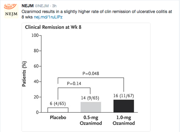
We conducted a double-blind, placebo-controlled phase 2 trial of ozanimod in 197 adults with moderate-to-severe ulcerative colitis. Patients were randomly assigned, in a 1:1:1 ratio, to receive ozanimod at a dose of 0.5 mg or 1 mg or placebo daily for up to 32 weeks. The Mayo Clinic score was used to measure disease activity on a scale from 0 to 12, with higher scores indicating more severe disease; subscores range from 0 to 3, with higher scores indicating more severe disease. The primary outcome was clinical remission (Mayo Clinic score ≤2, with no subscore >1) at 8 weeks.

RESULTS

The primary outcome occurred in 16% of the patients who received 1 mg of ozanimod and in 14% of those who received 0.5 mg of ozanimod, as compared with 6% of those who received placebo (P=0.048 and P=0.14, respectively, for the comparison of the two doses of ozanimod with placebo). Differences in the primary outcome between the group that received 0.5 mg of ozanimod and the placebo group were not significant; therefore, the hierarchical testing plan deemed the analyses of secondary outcomes exploratory. Clinical response (decrease in Mayo Clinic score of ≥3 points and ≥30% and decrease in rectal-bleeding subscore of ≥1 point or a subscore ≤1) at 8 weeks occurred in 57% of those receiving 1 mg of ozanimod and 54% of those receiving 0.5 mg, as compared with 37% of those receiving placebo. At week 32, the rate of clinical remission was 21% in the group that received 1 mg of ozanimod, 26% in the group that received 0.5 mg of ozanimod, and 6% in the group that received placebo; the rate of clinical response was 51%, 35%, and 20%, respectively. At week 8, absolute lymphocyte counts declined 49% from baseline in the group that received 1 mg of ozanimod and 32% from baseline in the group that received 0.5 mg. The most common adverse events overall were anemia and headache.

CONCLUSIONS

In this preliminary trial, ozanimod at a daily dose of 1 mg resulted in a slightly higher rate of clinical remission of ulcerative colitis than placebo. The trial was not large enough or of sufficiently long duration to establish clinical efficacy or assess safety. (Funded by Receptos; TOUCHSTONE ClinicalTrials.gov number, NCT01647516.)

Ozanimod

Related blog post: CCFA Conference Notes 2016 (part 5) -Emerging Therapies …


Endoscopic Surveillance after Esophageal Atresia: Low Yield In Pediatrics

A recent study of patients who had undergone repair of esophageal atresia (Koivusalo et al. JPGN 2016; 62: 562-66) confirms that “routine endoscopic surveillance had limited benefit and seems unnecessary” before 15 years of age.

A retrospective review of pediatric patients with esophageal atresia (EA) from 1980-2014) identified 209 patients with 616 biopsies.  60 patients had undergone antireflux surgery. Key findings:

  • Gr I esophagitis was noted in 37%, Gr II or III in 16%.
  • Gastric metaplasia was found in 17% and reached a prevalence of 15% by age 15 years.
  • Only 9% of patients with gastric metaplasia and 32% of patients with gr II esophagitis were symptomatic.
  • No cases of dysplasia or cancer were identified.

My take: Conditions, including esophageal atresia, that predisposed to chronic reflux esophagitis increase the risk of esophageal malignancy; however, this risk remains very low in childhood.  Therefore, surveillance for asymptomatic children is not needed prior to age 15 years.

Related blog post: Never quite right | gutsandgrowth

Vik Muniz recreation of Mary Cassatt painting

Vik Muniz recreation of Mary Cassatt painting

Up close, one sees this painting is actually a collage of images

Up close, one sees this painting is actually a collage of images

Disclaimer: These blog posts are for educational purposes only. Specific dosing of medications/diets (along with potential adverse effects) should be confirmed by prescribing physician/nutritionist.  This content is not a substitute for medical advice, diagnosis or treatment provided by a qualified healthcare provider. Always seek the advice of your physician or other qualified health provider with any questions you may have regarding a condition.

Extent of Disease: Microscopic or Endoscopic Classification?

Several recent articles highlight the divergence between microscopic and endoscopic classification of disease.

  • Verstraete et al. JPGN 2016; 62: 242-5.
  • Asthon et al. JPGN 2016; 62: 246-51.
  • Pashankar et al. JPGN 2016; 62: 314-16.

The first two references describe histology in comparison to endoscopic extent of disease in pediatric Crohn’s and the 3rd reference provides information on the finding of “duodenitis.”

Verstraete et al selected 60 patients  randomly from their cohort for retrospective review.  Two physicians independently reviewed the patients.  In describing extent of disease, the extent of disease (Paris Classification) was discordant in 34 (56.6%) when comparing  macroscopic disease (imaging and endoscopy) to macroscopic/microscopic combined.  In addition, there was high interobserver variability of the physicians when the physicians reviewed just macroscopic findings (κ= 0.53).

Asthton et al examined data from 172 pediatric patients with inflammatory bowel disease.  They found that histologic disease was more extensive than endoscopic findings.  For example, among those with ileal biopsies, 49% had endoscopic findings compared with 71.3% having histologic disease.

Currently, the Paris classification relies on endoscopic findings; however, together these two studies suggest that the microscopic findings need to be considered as well. How often areas with microscopic disease will eventually develop endoscopic lesions is not clear.

Pashankar et al, reviewed pathology reports over a 5-year periods with 2772 children (mean age 10.6 years).  They identified duodenitis in 352 with a prevalence rate of 12.7%. Gastritis was seen in 64% of children with duodenitis. Interestingly, 63% of the cases with histologic duodenitis had normal endoscopic appearance. Reported reasons for duodenitis:

  • Celiac disease 32%
  • Crohn’s disease 13%
  • Ulcerative colitis 3%
  • Helicobacter pylori infection 6%
  • Functional dyspepsia 7%

The remaining children (36%) were considered to have nonspecific duodenitis.  The authors state: “this finding is similar to the high percentage of nonspecific duodenitis (60%) in adults.”

My take: It is difficult to know how important microscopic findings are in many cases.  With inflammatory bowel disease, whether/how to incorporate microscopic findings in classification is unclear.  With regard to the finding of microscopic duodenitis, when a specific etiology has not been identified, this leads to lots of questions:

  • How important is this finding?
  • How should this be treated?
  • How much additional workup and followup is needed?
  • How helpful is your pathologist –is the threshold for abnormality too low histologically?

Related blog posts:

marriage colonoscopy

What I did not know …a few items

  1. 6-Mercaptopurine (6-MP) often can be used when patients are intolerant of azathioprine. S Hubener et al. Clin Gastroenterol Hepatol 2016; 14: 445-53.  This retrospective study showed that 15 of 20 patients with autoimmune hepatitis and prior azathioprine intolerance responded to 6-MP.  This is somewhat unexpected as azathioprine is metabolized into 6-MP.  However, rather than 6-thiouracil, the “imidazol component of azathioprine, which is cleaved off…might trigger adverse reactions.”  Another artical on thiopurines (Aliment Pharmacol Ther 2016; 43: 863-883) (thanks to Ben God for this reference) provides a thorough review of the pharmacogenetics and pharmocokinetics of these medications.  While this review reinforces the recommendation to check TPMT before treatment, it notes that only a small proportion of thiopurine toxicity is related to deficient TPMT activity.
  2. There is no formal validated or consensus definitions of mild, moderate, or severe IBD. L Peyrin-Biroulet et al. Clin Gastroenterol Hepatol 2016; 14: 348-54.  While the Lemann index measures the cumulative structural bowel disease, the authors propose criteria which involves three areas of severity: impact of the disease on the patient (eg. clinical symptoms), inflammatory burden (eg. biomarkers, mucosal disease, disease extent), and disease course (eg. structural disease, intestinal resection, perianal disease, extraintestinal manifestations)
  3. Fundic gland polyps (often associated with proton pump inhibitor therapy) are not premalignant lesions. There is an “inverse correlation between the FGPs and gastric neoplasia.” S Varghese et al. Gastroenterol Hepatol; 2016; 12: 153-4.
  4. Parents of newborns do not know how to use car seats.  BD Hoffman et al (J Pediatr 2016; 171: 48-54) showed that 95% of car seats were misused (291 families).  Serious misuse was present in 91%.


Related blog posts: Lemann index: Short Takes on IBD Articles | gutsandgrowth

Gibbs Gardens has >20 million daffodils

Gibbs Gardens has >20 million daffodils

Chronic Granulomatous Disease and GI Involvement

Chronic granulomatous disease associated inflammatory bowel disease primarily involves the anus and rectum (SK Khangura et. al. Clin Gastroenterol Hepatol 2016; 14: 395-402).  In this study from the NIH, among 78 patients with CGD-IBD enteric fisula were found in 18% (73% were perianal), colonic stictures were noted in 24% (80% in anorectal area).

Other findings:

  • -EGD showed inflammation in 21%, 74%, and 37% of patients in esophageal, gastric and duodenal biopsies respectively
  • -Small bowel disease was unusual, though two of the four patients with ileostomies had ileal ulcers
  • -” ‘Target-like lesions,’ which have a clear elevated center with surrounding erythema, were characteristic of mild disease
  • -Approximately 40% of CGD patients have GI tract involvement
  • -GI tract malignancy was not observed in this cohort
  • -24 patients had pancolitis

My take: This reference provides comprehensive details of the clinical features of a large cohort with CGD.

Which animals kill the most humans

Which animals kill the most humans