Fidaxomicin Treatment of Clostridioides difficile in Children and Adolescents

MA Conrad et al. The Journal of Pediatrics, Volume 285, 114681. Open Access! Fidaxomicin Treatment of Clostridioides difficile Infections and Recurrences in Children and Adolescents: A Retrospective Multicenter Study

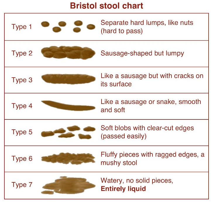
Methods: This was a  a multicenter, retrospective, observational study of fidaxomicin treatment for primary or recurrent CDI in children ages 12 months to 18 years old identified from 2013 to 2021 at 5 centers. Inclusion criteria were active CDI, defined as ≥3 watery stools in 24 hours and a positive laboratory test (toxin enzyme immunoassay positivity and/or polymerase chain reaction [PCR] positivity). Cure was defined as resolution of symptoms.

Patient characteristics:

  • Of the 95 patients included in this study, 84 (88%) were treated with fidaxomicin for a recurrent CDI, and 82 (86%) had at least one medical or surgical comorbidity.  
  • 38 (40%) patients had 4 or more CDI prior to fidaxomicin.
  • 22 (23%) had prior FMT.
  • 29 (31%) had IBD

Key findings:

  • By day 14 (end of treatment): 50 patients (52.6%) had a clinical cure and an additional 29 (30.5%) had improvement of symptoms. Thus, 17% did not respond to treatment.
  • Among 79 patients who responded to fidaxomicin treatment, 17 (21.5%) had a clinical and microbiologically confirmed recurrence of CDI by day 60, likely representing relapse.
  • Patients with inflammatory bowel disease were less likely to achieve clinical cure at day 14 (OR 0.27). 9 of 29 were considered treatment failures.
  • If the patient’s with IBD are excluded (n=66), there were only 7 (11%) treatment failures

Discussion points:

  • “Our clinical experience is that approval for coverage by insurers often is restricted to those with recurrent CDI, and the cost of fidaxomicin may limit availability for use as primary therapy.”
  • “CDI in IBD is a major clinical conundrum as the symptoms of the 2 disorders can overlap, and a positive C. difficile test is not always indicative of its active pathologic role…Therefore, patients who undergo treatment for CDI without response likely have an alternative cause of symptoms…. Current guidelines recommend reassessing symptoms in patients
    with IBD being treated for CDI at day 3 or 4 of the treatment course in order to consider escalation of IBD therapy in those who are not responding clinically to antimicrobial therapy.”

My take (borrowed from the authors): “More extensive studies are necessary to understand how to position fidaxomicin in the treatment algorithm for pediatric CDI.”

Related blog posts:

Chicago Botanic Garden (n Glencoe, IL)

When Celiac Disease Symptoms Continue Despite a Gluten Free Diet

A Kruegger et al. JPGN 2025; 81:596–605. Open Access! The prevalence and predictive factors of overlapping disorders of gut–brain interaction and celiac disease in children

Methods: Single-center, retrospective study of children (4–21 years old, n=191) with biopsy-proven Celiac disease (CeD) who were evaluated for DGBI based on Rome IV criteria. Patients who were adherent to a GFD, demonstrated tissue transglutaminase immunoglobulin A (TTG IgA) decline, and had at least one visit 9–24-months after diagnosis with a pediatric gastroenterologist. For this study, sustained TTG IgA decline required at least two declining TTG IgA values, a 90% decline from baseline, or normalization of TTG IgA.

Key findings:

  • 43% (n = 83) met Rome IV DGBI diagnostic criteria.
  • Functional constipation (27/83, 33%) and functional abdominal pain (24/83, 29%) were the most common DGBI
  • Abdominal pain, constipation, and vomiting at initial presentation as well as comorbid joint hypermobility, headaches, and chronic musculoskeletal pain increased risk of developing DGBI after serological decline

Discussion Points:

  • “The prevalence reported here is similar to a study of adults with CeD who were adherent to a GFD that reported over 50% met criteria for a functional gastrointestinal disorder19 and is higher than previously reported pediatric prevalence rates”
  • “The majority of patients who met DGBI criteria did so through having the persistence of the same gastrointestinal symptoms that were present at CeD diagnosis. This raises the question as to whether the symptoms at presentation were due to CeD, DGBI, or both”
  • “Clinicians could consider discussing that while symptoms related to CeD should improve on a GFD, some symptoms may persist, especially if they have an increased likelihood of having a comorbid DGBI. Such counseling may prevent the misattribution of persistent symptoms to ongoing gluten exposure and mitigate hypervigilance”
  • “Having complete villous blunting on diagnostic biopsy increased the likelihood of having a DGBI. Intuitively, it is possible that complete villous blunting can lead to greater nerve sensitization and subsequently higher rates of DGBI. It is also possible that complete villous blunting is slower to recover”

My take: Given the overlap of DGBI symptoms with CeD, diagnosing DGBI in patients with CeD can be challenging. However, DGBI is much more likely to contribute to lingering symptoms than refractory CeD.

As a practical matter, the high frequency of ongoing GI symptoms despite use of a GFD provides another drawback to relying on a no-biopsy diagnosis. A no-biopsy diagnosis introduces greater uncertainty in the diagnosis and does not allow for a histologic comparison if a subsequent evaluation is needed.

Related blog posts:

Claude Monet, Bridge over a Pond of Water Lilies at The Metropolitan Museum of Art

How Often Esophageal Coins Pass Into the Stomach

P Quitadomo et al. Am J Gastroenterol 2025; 120: 1388-1390. “Insert-Coin”: A Prospective Study of Coin Ingestion in Children of Southern Italy

Thanks to Ben Gold for this reference.

This prospective study from Naples, Italy examined children 0-14 yrs of age with a coin ingestion (n=807). Children with coins in the proximal esophagus underwent endoscopic removal within 4 hours whereas those with middle to lower esophageal coins had re-evaluation after 12 hours before removal.

Key findings:

  • 52 of 807 (6.4%) had a coin retained in the esophagus, the remainder were in the stomach or beyond
  • 20 of 52 (38%) were located in the middle to lower esophagus (10 in each)
  • 13 of 20 (65%) coins in the middle to lower esophagus had spontaneous gastric passage
  • The mean age of patients with gastric passage (72 months) was higher than those without passage (48 months)

My take: Only 6% of patients in this study who had a coin ingestion had esophageal retention of the coin. In addition, one-fourth of those with esophageal coins had spontaneous passage into the stomach. This occurred only with the mid-distal esophageal coins; in this subset it occurred in 65%. Thus, in those with mid-distal esophageal coins, watchful waiting for ~12 hrs may be beneficial for patients. The ultimate primary prevention of this problem may occur with more widespread adoption of electronic payments.

Related blog posts:

Agnes Northrup of Louis Tiffany Studio,
Tiffany Garden Landscape Window (1912)
at The Metropolitan Museum of Art
Agnes Northrup of Louis Tiffany Studio,
Autumn Landscape  (1923) at The Metropolitan Museum of Art

The Best Way to Judge Pediatric Poo

J Orozco et al. Am J Gastroenterol 2025; 120: 1381-1387. Comparison of the Bristol Stool Scale and Modified Version for Children: Use by Providers vs Children

Thanks to Ben Gold for this reference.

Background: The modified Bristol Stool Form Scale for Children (mBSFS-C) removes #3 and #5 from the Bristol Stool Form Scale (BSFS), leaving only one normal image and shortening the options from seven to five.

Methods: Pediatric gastroenterology providers  (21 faculty, 11 fellows, 3 nurse practitioners)  and 200 children/families rated the same 35 stool photographs, reflecting diverse stool forms, using both scales. The order of photograph presentation and scale use were randomized.

Modified Bristol Stool Scale
Bristol Stool Chart

Key findings:

  •  Of 1,225 provider ratings using the mBSFS-C, 90.0% agreed with the provider’s modal ratings vs 77.8% using the BSFS.
  • Of 7,000 child ratings using the mBSFS-C, 84.6% agreed with the children’s modal ratings vs 71.8% using the BSFS.
  • Using providers’ modal ratings as the reference, all mBSFS-C photograph modal ratings matched between children and providers (35/35 photographs) whereas only 86% (30/35 photographs) matched with the BSFS.

Discussion:

  • “Unique and new in this study is the direct head-to-head comparison of the 2 scales (BSFS, mBSFS-C) when used by pediatric gastroenterology providers and children. Both the BSFS and mBSFS-C demonstrated excellent reliability…modal rating agreement was significantly poorer for the BSFS than for the mBSFS-C.”
  • “Almost 20% of the time expert raters using the BSFS (vs. 8% with the mBSFS-C) deemed a stool to be a different clinical delineation than that selected by the majority of their peers.”

Related editorial: Peter Lu, The American Journal of Gastroenterology 120(6):p 1267, June 2025. Is It Time to Scale Down the Bristol?

My take: The modified BSFS is easier and better. This study indicates it should be widely used for children but probably for adults too. As Dr. Lu’s editorial notes, “aren’t adults just big children?”

Related blog posts:

New Data on the Reliability of the No-Biopsy Diagnosis in Pediatric Celiac Disease

Chang D, Wong M, Camila Cardenas M, et al. Pediatrics. 2025;156(3):e2025070897. Positive Predictive Value of Tissue Transglutaminase IgA for Celiac Disease.

Background:For a no-biopsy celiac diagnosis, “the [ESPGHAN] criteria were revised in 2020, to specify that only a highly positive tTG IgA greater than or equal to 10× ULN and a positive EMA on a second blood sample are sufficient to diagnose celiac disease, obviating the need for HLA testing or symptoms.” NASPGHAN “has yet to adopt similar serologic criteria for diagnosing celiac disease. A clinical report in 2016 continued to recommend a confirmatory biopsy in all suspected cases of celiac disease. This recommendation cited concerns of interassay variability.”

Methods: This was a retrospective, multicenter North American cohort study of children (<18 years) with an elevated tTG IgA within 6 months of an esophagogastroduodenoscopy between January 2016 and December 2021. Biopsy-confirmed celiac disease was determined by the presence of intraepithelial lymphocytosis and villous atrophy. The primary outcomes were the PPV of an elevated tTG IgA and tTG IgA greater than or equal to 10 times the upper limit of normal (10× ULN).

The study identified 4019 children with the following characteristics: 63.3% female; 9% type 1 diabetes, and 2% Down syndrome

Key findings:

  • Histologic findings in the entire cohort were consistent with celiac disease for 3321 children (PPV = 82.6%)
  • Among the 1739/4019 (43.2%) children with tTG IgA greater than or equal
    to 10× ULN, 1651 had biopsy-confirmed celiac disease (PPV10× = 94.9%). Five percent (88/1739) of children did not have histologic findings of celiac disease, including 41/1739 (2%) with normal histology
  • EMA only marginally improved specificity as 76% of children without celiac disease and tTG IgA greater than or equal to 10× ULN had a positive EMA, albeit on the same sample. There was variations across providers, practices, and countries, which resulted in EMA not being performed in all cases or tested on the same blood sample as the tTG IgA
  • Assays performed worse in children with type 1 diabetes (PPV10× 89%) than that in those without T1DM (95.7%)
  • Of those with esophageal biopsies, 175/2980 (6%) had at least 15 eos/hpf in at least 1 esophageal biopsy
  • Of those with gastric biopsies, 912/3534 (25.8%) had histologic gastritis, including 1.4% (51/3534) with evidence of Helicobacter infection (n (49 cases of H pylori and 2 cases of H heilmannii)

My take: This study indicates that the no-biopsy diagnostic approach for celiac disease may need to be reconsidered. Though, it is likely that the no-biopsy approach would have had a higher PPV if the guidelines were actually followed (eg. separate EMA confirmatory sample). The values in this study show a much lower PPV than prior studies (96% in those without T1DM). In addition, among the 4% without celiac disease (potential celiac disease), about half of them would likely progress to celiac disease and could potentially benefit from a gluten free diet. Thus, up to 2%of patients, based on this study, would probably be harmed by being placed on a gluten free diet.

Another point to reiterate based on this study, diagnostic confirmation by a specialist prior to dietary changes is recommended. Increasingly, patients are referred after starting a gluten free diet.

Related blog posts:

Brooklyn Botanic Gardens

Pharmacologic Neuromodulation for Bloating Symptoms

Briefy noted: EN Madva et al. Scand J Gastroenterol 2025 Aug 8:1-5. doi: 10.1080/00365521.2025.2544306. Online ahead of print. Pharmacologic neuromodulation for bloating.

This was a small retrospective study of consecutively referred patients with a DGBI (N = 77; ages 18-74, 87% female) to a tertiary neurogastroenterology clinic who were prescribed a neuromodulator for a primary complaint of bloating in 2016-2022.  Duloxetine was the most commonly prescribed neuromodulator (n = 52, 67.5%).

My take: This study shows that neuromodulators are likely beneficial for bloating symptoms. Dr. Garza () previously noted that in patients with bloating “the typical increase in excess gas during bloating symptoms is only 22 mL.” Thus, “A lot of bloating symptoms are due to increased sensitivity and ‘weird gas handling.’ The latter could include compression of diaphragm rather than elevation.”

Related blog posts:

Disclaimer: This blog, gutsandgrowth, assumes no responsibility for any use or operation of any method, product, instruction, concept or idea contained in the material herein or for any injury or damage to persons or property (whether products liability, negligence or otherwise) resulting from such use or operation. These blog posts are for educational purposes only. Specific dosing of medications (along with potential adverse effects) should be confirmed by prescribing physician.  Because of rapid advances in the medical sciences, the gutsandgrowth blog cautions that independent verification should be made of diagnosis and drug dosages. The reader is solely responsible for the conduct of any suggested test or procedure.  This content is not a substitute for medical advice, diagnosis or treatment provided by a qualified healthcare provider. Always seek the advice of your physician or other qualified health provider with any questions you may have regarding a condition.

How to Diagnose Celiac Disease in Patients Already Receiving a Gluten Free Diet

Open Access: Evaluating for Celiac Disease in Patients on a Gluten-Free Diet: A Practical Approach

Algorithm -Figure 2:

Figure 3 lists the content of several common foods -some noted below

While gluten exposure increases the diagnostic yield of currently available tests, there are novel tests being developed “which may aid in the diagnosis of CeD regardless of diet, with a particular focus on immune-based assays. One such innovation involves the use of tetramer-based assays, which enable the direct detection of gluten-specific T cells in the blood. These tetramers, designed to bind to HLA-DQ2 molecules, can help identify T cells that have been activated by gluten exposure. This presents a highly specific immune marker for CeD. Even for those on a GFD, sensitivity (97%) and specificity (95%) have been impressive.”

My take: This article provides practical advice for evaluating whether celiac disease is present in those already consuming a GFD.

Related blog posts:

And news from The Onion 8/26/25: Hummingbird Feels Like Fucking Idiot After Seeing Other Bird Gliding

Immune Dysregulation Can Mimic Celiac Disease

A Turunen et al. JPGN Reports. 2025;6:312–315. Elevated tissue transglutaminase immunoglobulin A: Celiac disease or polytypic plasmacytosis?

Briefly noted: The authors present a case report of an adolescent girl post cardiac transplant with hypergammaglobulinemia and presumed celiac disease (CD), who had a persistently elevated anti‐tissue transglutaminase immunoglobulin A despite a gluten free diet. Ultimately, she was diagnosed with polytypic plasmacytosis from suspected immune dysregulation.

Key point:

  • This case serves as a reminder that when treating those with persistently elevated tTG IgA on a strict GFD, etiologies outside of CD need to be considered.

My take: I had a young girl several years ago who had celiac diagnosis at an outside institution. She had multiple autoimmune disorders. After not responding to treatment, she had a follow-up panendoscopy showing ongoing IELs in duodenum and villous blunting. These findings were noted in the ileum as well. On reviewing her initial biopsies, the pathologist indicated that her findings could be celiac but it was suspected to be an immune dysregulation. Subsequently, she was diagnosed with CTLA4 deficiency and had a marked response to abatacept therapy.

Related blog posts:

Durability of Intensive Feeding Therapy

V Volkert et al. JPGN Reports. 2025;1–7. Long-term outcomes of intensive multidisciplinary intervention for feeding tube dependence and chronic food refusal

Methods: The researchers conducted a 17‐item Qualtrics survey of patients who participated in intensive multidisciplinary intervention treatment to improve the volume and variety of solid food intake and replace enteral feeding due to chronic food refusal an average of 6 years following intervention. 75 met eligibility criteria; 36 caregivers (48%) completed the survey

Key findings:

  • Most patients (80%) who achieved full wean from feeding tube dependence at program discharge maintained their wean at the time of the survey
  • Most caregivers (89%) described their child’s relationship with food as “good” or “neutral”

My take:

  • Our group works closely with the authors and appreciate all of their help
  • Survey studies have a lot of limitations and often conclusions are hampered by low participation
  • It looks like intensive feeding therapy has good durability. It would be interesting if we could know what would have happened to these children without therapy. How many would have gradually improved on their own?

Related blog posts:

On top of Stone Mtn, Georgia.

Dr. Jorge Bezerra: Improving the Care of Children with Biliary Atresia

Recently Dr. Bezerra gave our group a terrific lecture. I have taken some notes and shared some of his slides. There may be inadvertent omissions and mistakes in my notes. In addition, Dr. Bezerra’s presentation included several lengthy animations which helped explain the basic science concepts. These are not included in this summary.

Prior to his presentation to our group, Dr. Bezerra’s accomplishments had been recognized with the Shwachman Award, the highest honor bestowed by our national pediatric GI organization NASPGHAN (NASPGHAN Awards 2025). On a personal note, Jorge was the first person to give me hands-on instruction with an endoscope and I have a great deal of admiration for his humility, thoughtfulness and scientific achievements.

Liver biopsy from patient with BA showing bile duct proliferation
.
Operative cholangiograms from a patient with normal anatomy and one with BA that shows contrast extravasation without patent biliary tract

Key points:

  • Cytomegalovirus (CMV) is frequently associated with biliary atresia (BA). Treatment of CMV when detected may improve outcomes
  • MMP-7 and GGT can help predict the likelihood of BA
Historical controls in red and new protocol in blue
  • The role of the microbiome is being explored as a factor in the predisposition to BA
  • Biliary atresia organoid studies: Biliary atresia organoids had decreased expression of genes related to EGF signaling and FGF2 signaling. When treated with EGF+FGF2, biliary atresia organoids expressed differentiation markers, which restored polarity (Reference: Hepatology 2022 Jan;75(1):89-103. doi: 10.1002/hep.32107. Biliary organoids uncover delayed epithelial development and barrier function in biliary atresia). Thus, these organoids can help understand the underlying pathogenesis and may lead to improved treatments
  • More recently, compared to normal liver organoids, the epithelium of BA organoids was fragmented and peribiliary glands (PBGs) were small, had abnormal intercellular junction (ZO1 expression), and expressed markers of epithelial-mesenchymal transition (EMT), with a prominent expression of TGF-β3. Upon TGF-β inhibition, EMT decreased in the diseased epithelium, the population of PBGs increased, and ZO1 expression improved. In vivo, TGF-β inhibition suppressed the BA phenotype and substantially decreased liver fibrosis in neonatal mice. Thus, the modulation of TGF-β-dependent EMT regulates bile duct epithelial development and influences the susceptibility of neonates to biliary injuries. (Reference: Nat Commun 2025 Jul 17;16:6575. doi: 10.1038/s41467-025-61442-5. Open Access! Cellular crosstalk mediated by TGF-β drives epithelial-mesenchymal transition in patient-derived multi-compartment biliary organoids)
RRV (rhesus rotavirus) can create an experimental model of BA in the mouse. In this mouse model, ILC2 cells (innate lymphoid cells in yellow) play a role in bile duct proliferation, possible attempt at recanalizing damaged bile ducts

Disclaimer: This blog, gutsandgrowth, assumes no responsibility for any use or operation of any method, product, instruction, concept or idea contained in the material herein or for any injury or damage to persons or property (whether products liability, negligence or otherwise) resulting from such use or operation. These blog posts are for educational purposes only. Specific dosing of medications (along with potential adverse effects) should be confirmed by prescribing physician.  Because of rapid advances in the medical sciences, the gutsandgrowth blog cautions that independent verification should be made of diagnosis and drug dosages. The reader is solely responsible for the conduct of any suggested test or procedure.  This content is not a substitute for medical advice, diagnosis or treatment provided by a qualified healthcare provider. Always seek the advice of your physician or other qualified health provider with any questions you may have regarding a condition