Stratifying Risk of Clots in Inflammatory Bowel Disease

T Naito et al. Gastroenterol 2021; 160: 771-780. Full text: Prevalence and Effect of Genetic Risk of Thromboembolic Disease in Inflammatory Bowel Disease

Background: 10% of healthy subjects are genetically at high risk for thromboembolic disease (TED). For adults with inflammatory bowel disease, TED is “largest cause of mortality in
patients”

Key findings:

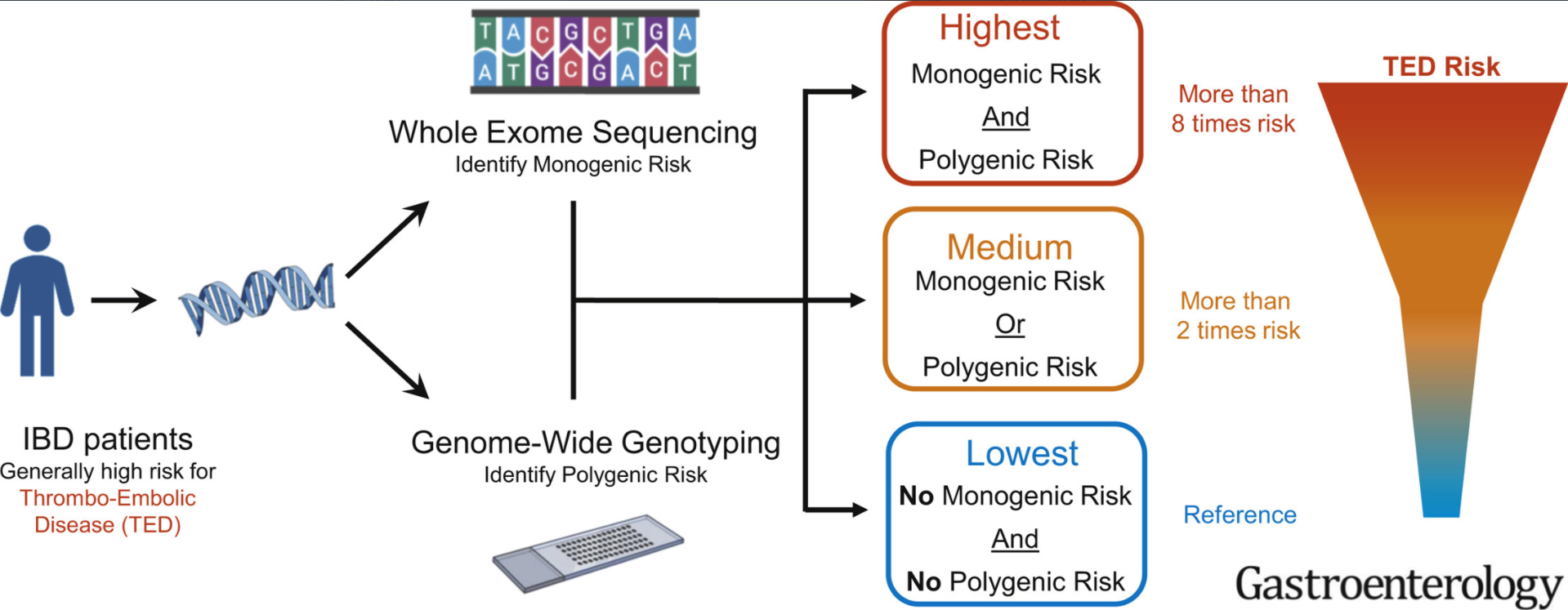
  • In total, this retrospective study had 792 IBD patients who had both whole-exome sequencing and genotyping data to identify thrombophilia pathogenic variants. 122 of 792 IBD patients (15.4%) as genetically high risk for TED.
  • Genetic TED risk was significantly associated with increased TED event (odds ratio,2.5; P ¼ .0036).
  • Patients with high TED genetic risk more frequently had thrombosis at multiple sites (78% vs 42%, odds ratio, 3.96; P ¼ .048)

“Our analyses demonstrate that approximately 1 in 7 patients with IBD have odds 2.5 times higher than nongenetically high-risk patients with IBD for experiencing TED.” The risk of TED in IBD is generally 3- to 4-fold higher than the general population

My take: In children, the risk of clots is much lower than in adults. Thus, the potential to identify those at highest risk would be useful in order to target interventions. Also, patients at higher risk for TED may affect choice of treatment (eg. avoiding JAK inhibitors).

Related blog posts:

From The Onion:

From The Onion

How a Gluten Challenge Could Change

A recent randomized double-blind study (MM Leonard et al. Gastroenterol 2021; 160: 720-733. Full Text: Evaluating Responses to Gluten Challenge: A Randomized, Double-Blind, 2-Dose Gluten Challenge Trial) provide evidence that a gluten challenge could detect evidence of celiac disease in hours, not weeks.

This study consisted of 14 adults with biopsy-proven celiac disease (CD) who were randomized to 3 g or 10 g gluten/day intake for 14 days. Each participant underwent extensive studies to detect histological, visible, and biochemical changes associated with gluten introduction. Data required multiple endosopic duodenal biopsies, VCEs and blood collection.

Key findings:

  • Symptoms and plasma interleukin-2 levels “increased significantly or near significantly at both doses.”
  • Interleukin-2 appeared to be the earliest, most sensitive marker of acute gluten
    exposure. IL-2 increases were observed 4 hours after exposure in patients with CD but not in healthy controls.
  • Intestinal damage is more complex and requires a longer duration and higher dose of gluten exposure. In this study, the higher dose (10 g) of gluten exposure was required for enteropathy within the study time frame.

My take: These study findings need to be confirmed in a broader patient cohort. However, in patients needing a gluten challenge, IL-2 response after a single-dose (measured at 4 hours) could be helpful. Those without IL-2 response are unlikely to have CD. Those with an IL-2 response at 4 hours, could confirm CD by completing a gluten challenge.

Related blog posts:

Polyurethane vs Silcione with Ethanol Locks

Prior recommendations for ethanol locks have favored silicone central lines over polyurethane due to concerns of increased breakage rates when ethanol locks are used with polyurethane catheters. However, a recent small study indicates that this may be incorrect.

K LaRusso et al. JPEN https://doi.org/10.1002/jpen.2056. Prolonged Use of Ethanol Lock Prophylaxis With Polyurethane Catheters in Children With Intestinal Failure: A Single‐Center Experience (Reference from Conrad Cole’s twitter feed)

A 10‐year retrospective study with 10 children comprising 85 CVCs and 13,227 catheter days

Key findings:

  • Breakages were the most common complication: polyurethane 1.46/1000 vs silicone 3.76/1000 catheter days. Silicone catheters had a significantly higher breakage rate (adjusted rate ratio [RR], 2.86; 95% confidence interval [CI], 2.84–2.88; P < .001)
  • Polyurethane catheters had higher rates of occlusion (adjusted RR, 0.14; 95% CI, 0.07–0.28; P < .001) and displacements.
  • There were no differences in the overall catheter replacement rates and any other catheter‐related outcomes.

Related blog post: Central Line Pointers

From The Onion:

from The Onion

1. When You Are Fully Vaccinated 2.Outcomes of Duodenal Atresia and Stenosis

  1. CDC Recommendations: When You’ve Been Fully Vaccinated
  2. GS Bethell et al. JPGN 2021; 72: 239-243. One-year Outcomes of Congenital Duodenal Obstruction: A Population-based Study

This prospective observational study provides helpful outcome data for infants (n=80) born with congenital duodenal obstruction (CDO).

Key findings (also see infographic below):

  • Though there was an 8.4% overall mortality, there were no deaths directly attributed to CDO. 69% had associated anomalies.
  • Median length of stay after repair was 20 days; at 28 days following repair, 76% had been discharged home
  • Failure to achieve full enteral feeds was NOT related to CDO (due instead to other gastrointestinal anomalies). Mean time for full feeds was 13 days post-op; 90% reached full enteral feeds at 28 days.
  • Repair type: 80% had duodenoduodenostomy, 14% had duodenojejunostomy, the others: membrane incision (n=1), membrane resection (n=2), and duodenoplasty (n=2)

My take: This data will inform clinicians of expected outcomes in this population. I hope this cohort is followed long-term to provide more information about long-term outcomes including frequency of pancreatitis.

Must-Read: How to Handle Post-Procedure Fevers

A recent study (JM Boster, M Iwanowski, RE Kramer. JPGN 2021; 72: 250-254. Management of Pediatric Postendoscopy Fever: Reducing Unnecessary Health Care Utilization With a Clinical Care Guideline) provides clear guidance for how to handle post-procedure fevers.

Using a prospective database with 27,100 endoscopies, the authors evaluated a clinical care guideline to reduce unnecessary medical care following endoscopy.

Key findings:

  • Post-endoscopy fever (PEF) occurred in 0.55% (n=150)
  • ONLY 6 of these 150 PEFs (0.4%) were attributed to a procedure complication: 3 had perforations (all with abdominal pain), 2 had aspiration (both had emesis at time of endoscopy) and 1 had a positive blood culture (though had undergone a liver biopsy as well as endoscopy)
  • The authors published their care guideline (Figure 1) which stratifies risk based on whether the procedure was an interventional (high risk) vs diagnostic (low risk), ASA class, duration of fever, concomitant immunosuppression (eg. steroids), and associated symptoms including vomiting, diarrhea, bleeding, new abdominal pain, impact on activities of daily living, and hydration
  • Interestingly, the authors note that their cohort had a total of 23 perforations, but only 3 presented with fever
  • Using the care guideline resulted in a “significant shift in the prevalence of Grade 2 and above (requiring hospital use) to Grade 1 (clinical observation and reassurance) adverse events, dropping ED visits and admissions by 43.6% and 76.4% respectively for the post-endoscopy fever patients.” This shift was not associated with any observed negative patient outcomes or missed diagnoses.

My take: The authors note that fever is often related to release of inflammatory cytokines which can occur with endoscopy in the absence of complications. The authors methodical guideline to post-procedure fever provides a logical approach to this common problem.

Related blog posts:

Disclaimer: This blog, gutsandgrowth, assumes no responsibility for any use or operation of any method, product, instruction, concept or idea contained in the material herein or for any injury or damage to persons or property (whether products liability, negligence or otherwise) resulting from such use or operation. These blog posts are for educational purposes only. Specific dosing of medications (along with potential adverse effects) should be confirmed by prescribing physician.  Because of rapid advances in the medical sciences, the gutsandgrowth blog cautions that independent verification should be made of diagnosis and drug dosages. The reader is solely responsible for the conduct of any suggested test or procedure.  This content is not a substitute for medical advice, diagnosis or treatment provided by a qualified healthcare provider. Always seek the advice of your physician or other qualified health provider with any questions you may have regarding a condition.

Short Bowel Syndrome -YouTube Information

Some good patient educational videos for short bowel syndrome:

Related article: AE Wiskin et al. Clin Nutrition 2021; https://doi.org/10.1016/j.clnesp.2020.12.029. Prevalence of Home Parenteral Nutrition in children Key finding: In 2019, 389 children received HPN (home parenteral nutrition) in the UK; this is nearly double the number last reported in 2012 and is a prevalence of 30 per million children (Thanks to Kipp Ellsworth for this reference)

Other resources:

Related blog posts for Short Bowel Syndrome:

Should We Worry About Osteopenia in Children with Crohn’s Disease?

A recent retrospective study (N Ronel, et al. JPGN 2021; 72: 270-275. Clinical Criteria Can Identify Children With Osteopenia in Newly Diagnosed Crohn Disease) included 116 children (mean age 13 years)

Key finding:

  • In total, 59% of children with BMI z-score <−0.5 had moderate-severe osteopenia and only 18% of those with higher z-scores. 
  • Osteopenia was associated with lower BMI z-score (−0.8 ± 1.2 vs −1.8 ± 1.1, P < 0.001) and higher PCDAI  (33.7 ± 15.2 vs 25.7 ± 16.5; P = 0.009)
  • None of the higher risk patients were receiving long-term corticosteroids 
  • Limitations: retrospective study with relatively small sample size, pubertal stage not recorded, variability in DXA studies, and lack of followup information

My take: The authors have NOT shown that identification of osteopenia at the time of diagnosis improves outcome of Crohn’s disease or bone disease. This is why I disagree with their recommendation to routinely screen children with BMI z-score <−0.5. In those in which finding osteopenia may influence treatment, then a DXA study would be worthwhile.

Related blog posts:

Chicago 2021

Seronegative Villous Atrophy

A recent large retrospective study (R Mandile et al. JPGN 2021; 72: 282-287. Seronegative Villous Atrophy in Children: Clinical and Immunohistochemical Features) provides information about conditions, besides celiac disease (CD) which present with villous atrophy. 64 of 1282 pediatric patients were seronegative with villous atrophy; seronegative was defined as testing negative twice for serology (TTG IgA/EMA or if IgA-deficient, IgG antibody serology).

Key findings:

  • Diagnoses were: inflammatory bowel diseases (IBD) (21/64), food allergy (8/64), infections (7/64, of which 3 HIV infections), immune deficiency (3/64), short bowel syndrome (3/64), congenital diarrhea (2/64), other/inconclusive diagnosis (8/64). In addition, there were 12 with Gastro-Esophageal Reflux Disease (GERD) & the authors speculate that perhaps hyperacidity could play a role in some of these cases.
  • Only one quarter of the seronegative patients had an increased number of intraepithelial lymphocytosis (IELs)
  • Among those with villous atrophy attributed to IBD, this was nearly equally-split between Crohn’s disease and ulcerative colitis, 10 and 11 patients respectively (according to Table 1)
  • The authors note that the ~5% of patients with seronegative villous atrophy with alternative diagnosis than Celiac disease may be an overestimation as more individuals are being diagnosed without biopsy based on serology
  • Despite the large cohort, there are still other rare conditions that were not identified in this study (eg. autoimmune enteropathy, CTLA4B deficiency,drug-induced enteropathy, and tropical sprue)

My take: This article provides a good starting point for patients with villous atrophy and negative serology.

Related article: J Devara et al. JPGN 2021; 72: 288-293. The Significance and Clinical Outcome of Lymphocytic Duodenosis in Children: Mayo Clinic Experience and Systematic Review Background: Lymphocytic duodenosis (LD) defined as increased intraepithelial lymphocytes >25 intraepithelial lymphocytes (IELs) per 100 epithelial cells with normal villous architecture is associated with many gastrointestinal (GI) disorders.

Key findings:

  • During the study period 12,744 children underwent an EGD with biopsies. Of those, we identified 426 children with LD (3%).
  • Among the LD (compared to control group), 5% had celiac disease (vs 0%, P < 0.001), 9% had Crohn disease (3%, P = 0.003) and 3% had Helicobacter pylori gastritis (1%, P = 0.021).

Related blog post: @AmyOxentenkoMD: Celiac Disease and Mimics

Pictographic Constipation Action Plan

A recent study (PT Reeves et al. J Pediatr 2021; 229: 118-126. Full text link: Development and Assessment of a Pictographic Pediatric Constipation Action Plan) highlighted patient education efforts. “This study focused on the design and assessment of a low literacy pictographic CAP for the care of functional constipation in children.”

My take: I agree with the authors that a simple plan like this has “the potential to become an important tool to be used in the care of children with functional constipation, improving both quality-of-care and clinical outcomes.”

Link to PDF: Constipation Action Plan

Related blog posts:

This QR code provides 9 minute explanation of constipation and action plan:

Disclaimer: This blog, gutsandgrowth, assumes no responsibility for any use or operation of any method, product, instruction, concept or idea contained in the material herein or for any injury or damage to persons or property (whether products liability, negligence or otherwise) resulting from such use or operation. These blog posts are for educational purposes only. Specific dosing of medications (along with potential adverse effects) should be confirmed by prescribing physician.  Because of rapid advances in the medical sciences, the gutsandgrowth blog cautions that independent verification should be made of diagnosis and drug dosages. The reader is solely responsible for the conduct of any suggested test or procedure.  This content is not a substitute for medical advice, diagnosis or treatment provided by a qualified healthcare provider. Always seek the advice of your physician or other qualified health provider with any questions you may have regarding a condition

Highly Prevalent Pediatric Feeding Disorders

A recent retrospective study (K Kovacic et al. J Pediatr 2021; 228: 126-131. Pediatric Feeding Disorder: A Nationwide Prevalence Study) provides epidemiologic data for pediatric feeding disorders.

The authors utilized three databases for children aged 2 months to 18 years: Medicaid Databases from Arizona (2009-2017) and Wisconsin (2005-2014) (public insurance databases) and The Truven Health Analytics MarketScan Commercial Claims and Encounters Database (2009-2015) (a nationwide private insurance database).

Key findings:

  • There were 126 002 and 367 256 children 5 years of age or younger with pediatric feeding disorders (PFD) with public and private insurance, respectively
  • In 2014, the annual prevalence of PFD was 1 in 23, 1 in 24, and 1 in 37 in children under 5 years in the publicly insured cohorts in Wisconsin, Arizona, and the privately insured cohort, respectively.
  • The prevalence of PFD in children <5 years (range: 27-44 per 1000 children) exceeds the prevalence of U.S. children with autism spectrum disorder (~17 per 1000 children at age 8 year) and eating disorders like anorexia nervosa and bulimia (8 and 13 per 100,000 persons per year).
  • In an associated editorial (pg 13-14), Rachel Rosen notes that “despite their high prevalence, the lack of studies funded by the National Institutes of Health…is striking.”

My take: This study provides useful data on PFD prevalence. PFD have a wide range of associated diseases, including prematurity, neurologic disorders/developmental delay, congenital heart disease, chronic lung disease, autism, and congenital bowel disorders. In some, PFD are related to poorly-understood feeding aversions.

Related blog posts:

Chicago, IL & Lake Michigan