Slinky for Short Bowel Syndrome?

From GeneralSurgeryNews.com: Open Access: Novel Device Uses Mechanotransduction To Treat Small Bowel Syndrome

An excerpt:

“The basic concept is similar to distraction osteogenesis, which orthopedic surgeons have used for years, applying distraction force to broken bone that will grow up to a millimeter a day,” said Andre Bessette, the CEO and a co-founder of Eclipse Regenesis, Inc

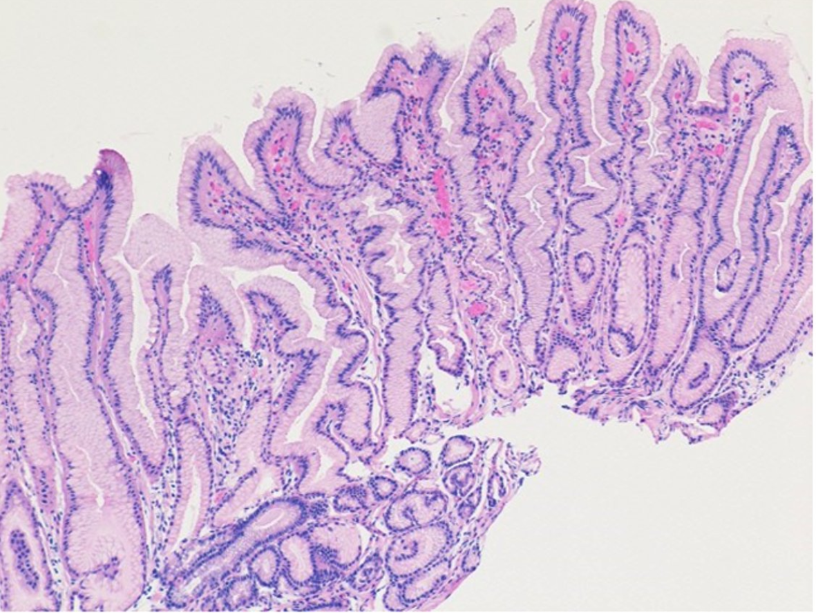
To regenerate small-bowel tissue, a surgeon inserts the device, which looks like a small, compressed coil, inside the small intestine and secures both ends with plication sutures applied to the outside of the intestine. Over two to three weeks, the device slowly expands to its uncompressed state, stimulating new tissue growth—ultimately two to three times the segment’s original length, about 4 cm...[thus]  they’ll need more than one device applied or more than one procedure.

Once this process is complete, the chromic sutures dissolve over about a month, allowing the device to pass through the body to be excreted...

[The researchers] expect to start these [human] trials in the first half of 2022, and they have identified two primary investigator sites: Boston Children’s Hospital and Cincinnati Children’s Hospital

Preclinical studies: Eclipse’s website at www.eclipseregenesis.com/ publications.

The Consequences of Prior Authorizations

Like many clinicians, I would very much like to tell insurance companies how I really feel about their prior authorization policies, and peer-to-peer processes to get approvals needed for treating our patients.

Most of the time I resent the imposition on my time to craft detailed letters explaining my rationale for treatment. Some obstructionist tactics are particularly aggravating. For example, when I am asked to do a peer-to-peer call and find out on the call that the person on the other end is neither a peer (often a pharmacist) and more importantly that this person is not authorized to remedy the situation but only to arrange another call. Another tactic of asking me to write multiple letters at different stages of the authorization process is extremely annoying. All told, these authorization requests are becoming more frequent and further impinging on my free time.

Now it turns out a study has shown the harmful effects of these maneuvers for our patients:

Constant BD, de Zoeten EF, Stahl MG, et al. Delays Related to Prior Authorization in Inflammatory Bowel Disease. Pediatrics. 2022;149(3):e2021052501 (Thanks to Ben Gold for this reference)

In this retrospective study of 190 pediatric patients ((median age 14.5 years) with IBD initiating biologics at a tertiary care hospital, key findings:

  • Prior authorization and complicated prior authorizations (requiring appeal, step therapy, or peer-to-peer review) were associated with 10.2-day (95% confidence interval [CI] 8.2 to 12.3) and 24.6-day (95% CI 16.4 to 32.8) increases in biologic initiation time, respectively.
  • Prior authorizations increased the likelihood of IBD-related healthcare utilization within 180 days by 12.9% (95% CI 2.5 to 23.4) and corticosteroid dependence at 90 days by 14.1% (95% CI 3.3 to 24.8). 

In their discussion, the authors note that “in a recent survey conducted by the American
Medical Association, 94% of physicians reported that prior authorizations delay access to
necessary care, 90% perceived a negative impact on clinical outcomes, and 30% reported that a prior authorization led to a serious adverse event for a patient in their care.”

My take: Prior authorization policies usually delay needed care unnecessarily and lead to complications in children with IBD.

Related blog posts: 

“Do Not Stop Anti-TNF Medications in Children with IBD When They Are Working”

In 2014, one of the posts on this blog addressed stopping anti-TNF therapy: Marriage, Divorce and Separation with Anti-TNF Therapy. My take at that time was “most patients are better off staying married to their anti-TNF therapy.”

Despite changes in therapeutic options, a recent study and editorial come to the same conclusion in 2022:

In the retrospective study, 78 patients with CD and 56 patients with UC underwent endoscopic reassessment. Key findings:

  • Mucosal healing (MH) was achieved by 32 patients with CD (41%) and 30 patients with UC (53.6%); 26 patients with CD (33.3%) and 22 patients with UC (39.3%) achieved histologic healing (HH)
  • Among 45 patients (n=24 CD, n=21 UC) with both MH & HH, anti-TNF therapy was stopped & patients received either an immunomodulatory or mesalamine. 76% of patients with CD had clinical relapse within 3 years and 17% within 1 year. Importantly, objective markers of relapse, including calprotectin and endoscopy were NOT performed; thus, this is certainly an underestimation of relapse rate and time to relapse.

In the commentary, the authors note the high rate of relapse in other studies with anti-TNF withdrawal (eg. STORI trial) and high rate of surgery in patients with perianal CD who stopped therapy. In the STORI trial, “the best outcomes [for infliximab withdrawal] were those with subtherapeutic infliximab trough levels, ie, those for whom infliximab was not responsible for maintaining their remission.”

The data are less certain for UC. The editorial notes that 85% of the 21 patients in the Scarallo study had limited left-sided colitis and only 17 were followed for at least 1 year. In adult studies on anti-TNF discontinuation with UC (Kennedy et al. Aliment Pharm Ther 2016; 43: 910-23 and Molander et al. Inflamm Bowel Dis 2014; 20: 1021-28), 42% and 35% relapsed within 12 months, whereas another small study (Farkas et al. World J Gastroenterol 2014; 20: 2995-3001) found 100% of patients on combination therapy who stopped anti-TNF agent had to restart anti-TNF therapy.

My take (from editorial): “The totality of the currently available evidence suggests that discontinuing anti-TNF medications in children with IBD is associated with a greatly increased risk of disease exacerbation, especially if the anti-TNF trough level was therapeutic.”

Broadwalk; Hollywood, FL

How Effective are Stents for Anastomotic Esophageal Strictures in Patients with Esophageal Atresia

O Baghdadi et al. JPGN 2022; 74: 221-226. Predictors and Outcomes of Fully Covered Stent Treatment for Anastomotic Esophageal Strictures in Esophageal Atresia

This retrospective review of 45 patients (n=92 stents) examine the effectiveness of esophageal stenting in patients with esophageal atresia (EA). All patients had multiple dilatations prior to stenting and/or had a stricture diameter that rapidly narrowed within 2 weeks of dilatation to a diameter that was the same or smaller to predilatation. Patients were observed in the hospital after stent placement and outpatient management was considered if oral nutrition was tolerated for at least 3 days.

Key findings:

  • According to the authors, the stents prevented surgical resection in 41% of patients
  • ΔD (change in diameter) of ≤4 mm (area under the curve = 0.790; 95% confidence interval: 0.655–0.924; P < 0.001) was the optimal cutoff point in differentiating stent success. If the change in diameter decreases by 4 mm or more after stent removal at endoscopic followup, it is likely that stricture contains significant scar tissue and is not amenable to dilatation/stenting.
  • Median stent duration was 11.5 days
  • The most common adverse events were erosions/ulcerations (29%), granulation tissue formation (27%), vomiting/retching (26%) and stent migration (9%). Three stents were complicated by an esophageal leak (treated medically).

My take: Complications were frequent; thus, stenting for refractory strictures requires highly-specialized technical expertise.

Related blog posts:

Azalea trail, near the Chattahoochee River (Atlanta)

Bookmark This Article on Pediatric Acute Liver Failure

JE Squires et al. JPGN 2022 – Volume 74 – Issue 1 – p 138-158. doi: 10.1097/MPG.0000000000003268. Open Access: North American Society for Pediatric Gastroenterology, Hepatology, and Nutrition Position Paper on the Diagnosis and Management of Pediatric Acute Liver Failure

This article provides a terrific summary of the most urgent issues with regard to caring for children with PALF; this article provides helpful information for diagnosis, and management, as well as information on pathophysiology, and associated outcomes.

PDF version: PALF in Children Position Paper

The article makes a number of recommendations for testing/treatment -here are a few of them:

  • While the initial testing does list ferritin, it does not list soluble IL2R as an early test (listed lower in Table 4 under section of hepatic encephalopathy). My colleagues at Emory who specialize in liver transplantation have frequently recommended this test early in the evaluation of severe liver disease/acute liver failure (ALF) as a potential marker of HLH and immune dysregulation.
  • With regard to coagulopathy: “Coagulopathy secondary to vitamin K deficiency should rapidly correct following appropriate repletion. Notably, if coagulopathy persists, efforts to “correct” abnormal coagulation profiles with fresh frozen plasma or other pro-coagulation products should generally be avoided” [in the absence of bleeding or need for invasive procedure].
  • Initial IV Fluids: ” In the absence of the need for volume resuscitation, total intravenous fluids should initially be restricted to around 90% of maintenance fluids to avoid overhydration. Initial fluids should be similar to hypertonic glucose (D10) one-half normal saline and supplemented with 15 mEq of potassium (K+)/L.”
  • Nutritional support: “Patients with PALF are likely catabolic and require more calories than basal needs. Enteral feeding is often preferred over total parenteral nutrition (TPN), and both naso-gastric or naso-jejunal feeds should be considered before TPN.”
  • Neonatal ALF: “GALD results from an intrauterine alloimmune liver injury and is suspected to be the single most common cause of neonatal acute liver failure… Characteristic clinical features of GALD include an ALF presentation usually at birth and almost always in the first days of life. The majority (70–90%) of affected infants are born premature and a history of maternal sibling death is common. Timely exchange transfusion and high-dose intravenous immunoglobulin (IVIG) is the preferred treatment to remove offending antibodies and block their action, including activation of complement (88,91). The mechanism of GALD places subsequent pregnancies at risk, and intrapartum IVIG should be used to prevent recurrences.”
  • Table 8 list common medications implicated in PALF. “Acetaminophen (APAP) …remains the most common cause of DILI, and is the most common identified cause of ALF in children.” 

Related blog posts:

Figure 3: Etiology of acute liver failure in children.
(A) Etiology for 1144 children from the Pediatric Acute Liver Failure Study Group (PALFSG) 1999–2014. (B) Final diagnosis by age (note: figure B includes information on only 985 participants)

Disclaimer: This blog, gutsandgrowth, assumes no responsibility for any use or operation of any method, product, instruction, concept or idea contained in the material herein or for any injury or damage to persons or property (whether products liability, negligence or otherwise) resulting from such use or operation. These blog posts are for educational purposes only. Specific dosing of medications (along with potential adverse effects) should be confirmed by prescribing physician.  Because of rapid advances in the medical sciences, the gutsandgrowth blog cautions that independent verification should be made of diagnosis and drug dosages. The reader is solely responsible for the conduct of any suggested test or procedure.  This content is not a substitute for medical advice, diagnosis or treatment provided by a qualified healthcare provider. Always seek the advice of your physician or other qualified health provider with any questions you may have regarding a condition.

“Show Me the Money:” GI/Liver Expenditures Update

AF Peery et al. Gastroenterol 2022: 162: 621-644. Burden and Cost of Gastrointestinal, Liver, and Pancreatic Diseases in the United States: Update 2021

Using several national surveys (2016-2018), the authors provide data on GI diagnoses and expenditures for adults and children

Key findings:

  • Gastrointestinal health care expenditures totaled $119.6 billion in 2018.
  • Annually, there were more than 36.8 million ambulatory visits for gastrointestinal symptoms and 43.4 million ambulatory visits with a primary gastrointestinal diagnosis
  • A total of 22.2 million gastrointestinal endoscopies were performed, and 284,844 new gastrointestinal cancers were diagnosed
  • Gastrointestinal diseases and cancers caused 255,407 deaths

This article is loaded with statistical details

  • Abdominal pain, reflux, and constipation are the leading GI diagnoses
  • GI bleeding, cholelithiasis, pancreatitis and liver disease are the most common diagnoses associated with admission to the hospital
  • In the pediatric age group, appendicitis, intestinal infection and inflammatory bowel disease are the principal diagnosis associated with GI admissions
  • Trends in GI cancer frequency over the last three decades are shown (including ethnicity)
  • Among non-malignancy, alcohol-associated liver disease followed by cirrhosis and GI bleeding were most common causes of death in GI/Liver categories
  • Data on the frequency of colonoscopies and transplants is provided

Menetrier Disease in a Pediatric Patient

During my training, we reported a case of Menetrier’s disease in a pediatric patient associated with CMV infection (Open Access PDF: Diagnosis of Cytomegalovirus Infection in Pediatric Menetrier’s Disease by In Situ Hybridization).

Recently, JPGN’s twitter feed shared the images below from a recent report (SJ Pathak et al. JPGN January 2022 – Volume 74 – Issue 1 – p e16doi: 10.1097/MPG.0000000000003276. Menetrier Disease in a Pediatric Patient With Ulcerative Colitis and Primary Sclerosing Cholangitis)

From JPGN Twitter Feed

Related blog post: Image Only: Pediatric Menetrier’s Disease

Vedolizumab for Refractory Microscopic Colitis, Plus, Vedolizumab and Serious Infections

LC Shipley et al. Clin Gastroenterol Hepatol 2022; 20: 455-457. Vedolizumab Therapy in Refractory Microscopic Colitis: A Single Center Case Series

In this report, the authors describe nine patients with refractory microscopic colitis (median age 55 years) who were treated with vedolizumab.

Key findings:

  • Clinical response with induction in 9 (100%); time to >50% response ranged from 1 to 7 weeks with 5 patients responding within 2 weeks.
  • Sustained response with maintenance therapy in 6 (67%); duration of follow-up ranged from 1 month to 15 months. The three patients without response had symptom duration of 10 yrs, 12 yrs, and 25 yrs prior to institution of vedolizumab.
  • Only two patients had histologic follow-up. While both had clinical response, the patient with lymphocytic colitis had histologic resolution whereas a patient with collagenous colitis had histologic persistent.

My take: Given vedolizumab’s favorable safety profile, further studies (with endoscopic endpoints) of vedolizumab are needed to define its efficacy for microscopic colitis.

Another study with vedolizumab: J Kirchgesner et al. Clin Gastroenterol Hepatol 2022; 20: 314-324. Risk of Serious Infections With Vedolizumab Versus Tumor Necrosis Factor Antagonists in Patients With Inflammatory Bowel Disease

Key finding: The risk of serious infections was not different between vedolizumab and anti-TNF in the overall IBD cohort (HR, 0.95; 95% CI, 0·79-1.13), while the risk was decreased for vedolizumab users in patients with UC (HR, 0.68; 95% CI, 0.50-0.93), but not CD (HR, 1.10; 95% CI, 0.87-1.38)

Related blog post/related article:

Disclaimer: This blog, gutsandgrowth, assumes no responsibility for any use or operation of any method, product, instruction, concept or idea contained in the material herein or for any injury or damage to persons or property (whether products liability, negligence or otherwise) resulting from such use or operation. These blog posts are for educational purposes only. Specific dosing of medications (along with potential adverse effects) should be confirmed by prescribing physician.  Because of rapid advances in the medical sciences, the gutsandgrowth blog cautions that independent verification should be made of diagnosis and drug dosages. The reader is solely responsible for the conduct of any suggested test or procedure.  This content is not a substitute for medical advice, diagnosis or treatment provided by a qualified healthcare provider. Always seek the advice of your physician or other qualified health provider with any questions you may have regarding a condition.

Food Allergy in Liver Transplant Recipients

F Mori et al. JPGN 2022; 74: 175-179. De Novo Food Allergy in Pediatric Recipients of Liver Transplant

This interesting review has several key points:

  • Reported prevalence of food allergy after liver transplant ranges from 5% to 40%
  • Younger age at transplantation is a risk factor for developing de novo post-transplant food allergy (dnPTFA)
  • Tacrolimus has been implicated as a risk factor for dnPTFA
  • Common dnPTFA are the same as in the general population: milk, egg, wheat, peanouts/nuts, fish, and soy

Management recommendations:

  • If the donor has a food allergy, the recipient should be tested within the first months of transplant for food-specific IgE and “it is advisable to introduce the suspected food in a controlled setting”
  • Elimination diet is mainstay of treatment. Some individuals may need modification of their immunosuppressive medications
  • Provide self-injectable epinephrine in those with food allergy
  • Monitoring of specific IgE/skin prick tests is advised

My take: There are increased allergy issues in kids who have had liver transplantation.

Related blog post: Lots of Allergy & Autoimmunity Issues Following Solid Organ Transplantation

Disclaimer: This blog, gutsandgrowth, assumes no responsibility for any use or operation of any method, product, instruction, concept or idea contained in the material herein or for any injury or damage to persons or property (whether products liability, negligence or otherwise) resulting from such use or operation. These blog posts are for educational purposes only. Specific dosing of medications (along with potential adverse effects) should be confirmed by prescribing physician.  Because of rapid advances in the medical sciences, the gutsandgrowth blog cautions that independent verification should be made of diagnosis and drug dosages. The reader is solely responsible for the conduct of any suggested test or procedure.  This content is not a substitute for medical advice, diagnosis or treatment provided by a qualified healthcare provider. Always seek the advice of your physician or other qualified health provider with any questions you may have regarding a condition.

“Separating Fact from Fiction in the Diagnosis and Management of Food Allergy”

This great article/commentary is available as open access: Separating Fact from Fiction in the Diagnosis and Management of Food Allergy EM Abrams et al. J Pediatr 2022; 241: 221-228.

Key points:

  • “The risk of fatal anaphylaxis is about 1 in 10 million (about equivalent to being struck by lightning)..and fatality on first ingestion of an allergen in infancy has never been described”
  • Food allergy causes considerable morbidity/worsens quality of life (QOL): bullying, meal preparation, stress, social activities, and school attendance (with 10% choosing home schooling because of a food allergy). Some of this detriment on QOL is due to fear “that accidental exposure could result in fatal anaphylaxis”
  • “The impact of peanut allergy on QoL has been noted to be significantly worse than the impact of other chronic childhood diseases such as rheumatologic disease and type 1 diabetes mellitus”
  • Uriticaria does not equal allergy. “Although urticaria is commonly equated with a food allergy, food allergy accounts for less than 10% of the causes of all acute urticaria and is not a cause of chronic urticaria.” Urticaria can be triggered by an acute infection (most common etiology)
  • “Food proteins are digested within several hours; therefore, urticaria associated with a food ingestion will occur within minutes to 2 hours of ingestion, but typically last no more than a few hours (and less with treatment)” (an exception is meat anaphylaxis owing to galactose-α-1,3-galactose allergy)
  • Other common symptoms that are often incorrectly attributed to food allergy include headaches, chronic behavioral symptoms, or chronic nonspecific abdominal pain
  • Allergy testing: “the presence of a food-specific IgE is not itself indicative of an allergy…Although both skin prick testing and food-specific IgE testing are highly sensitive (>90% for skin prick tests, 70%-90% for sIgE), the specificity and positive predictive values of testing are often very low. The rate of false-positive tests is up to 40%.” 
  • Broad “panel testing” (eg, to a panel of “common” foods), or any food allergy diagnostic testing in the absence of a convincing clinical history has high potential to overdiagnose food allergy and result in unnecessary food avoidances
  • Don’t do IgG testing. “There is no evidence that IgG testing marks food sensitivity; in fact, the presence of IgG is both expected and is a potential indicator of ongoing tolerance”
  • For first-line management anaphylaxis, (intramuscular) epinephrine is the “only life-saving intervention available.” Antihistamines are useful only in the treatment of cutaneous symptoms/urticaria.
  • “Although oral corticosteroids are often administered in the setting of anaphylaxis, there is no convincing evidence that their use prevents a biphasic reaction…their routine use is not recommended” (for anaphylaxis)

Related blog posts:

Siesta Beach, FL (2021)