How PPIs Improve Functional Dyspepsia

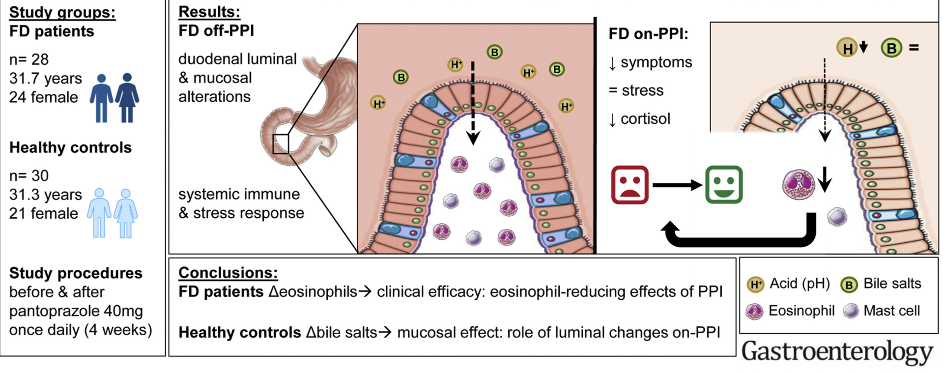
L Wauters et al. Gastroenterol 2021; 160: 1521-1531. Proton Pump Inhibitors Reduce Duodenal Eosinophilia, Mast Cells, and Permeability in Patients With Functional Dyspepsia

In this single-center prospective study, the authors show that pantoprazole (40 mg daily for 4 weeks) improves symptoms and duodenal eosinophilia in adults with functional dyspepsia (FD).

Key finding:

  • Symptoms and duodenal eosinophils, mast cells (all, P < .0001), and paracellular passage (P = .02) were significantly higher in FD-starters (patients new to PPI treatment) vs HVs and reduced with PPI therapy.
  • The authors note that systemic inflammation, subjective stress and salivary cortisol were also higher in patients with FD vs controls (off PPI).

My take: This study indicates that improvement in symptoms in FD related to PPIs is likely often due to improvement in duodenal mucosal inflammation and barrier dysfunction rather than by changing acidity.

Related blog posts:

For the Next Insurance Appeal: Therapeutic Drug Monitoring in Adalimumab Treatment (Pediatrics) & Satire on Prior Authorizations

There is a lot of data supporting the use of therapeutic drug monitoring (TDM) for anti-TNF agents. A recent study (MJ Kim et al. JPGN 2021; 72: 870-876. Therapeutic Drug Monitoring of Adalimumab During Long-term Follow-up in Paediatric Patients With Crohn Disease) adds to this data and supports increased adalimumab (ADL) dosing if below target values.

In this prospective study of 31 pediatric patients with Crohn’s disease, the authors found correlations between ADL values and the endpoints of clinical remission (CR) and mucosal healing (MH). The authors checked TLs at 4 months, 1, 2, and 3 years. Key findings:

  • The median trough levels (TLs) of ADL were higher in patients in CR (7.6 ± 3.5 μg/mL) than in patients with active disease (5.1 ± 2.2 μg/mL).
  • ADL TLs were significantly higher in patients who achieved MH than in those who did not (14.2 ± 7.6 vs 7.8 ± 5.2 μg/mL). 
  • The optimal cut-point for predicting MH at 1 year of ADL treatment was 8.18 μg/mL
  • MH was noted in 42% at 4 months and 55% at 1 yr; CR was noted in 90% at 4 months and 84% at 1 yr. ADL treatment was associated with positive effects on growth indicators as well.

The authors discuss TDM for anti-TNF therapy, noting that for infliximab, the AGA recommends values >5 mcg/mL and the ACG >7.5 mcg/mL. There are fewer studies of ADL TDM -prior studies have indicated goals of >5.8, >7.1, >8, and >8.1; thus, this study is in agreement with these prior studies.

My take: This study further supports the value of TDM; better drug levels correlate with better outcomes.

Related blog posts:

Fort Jefferson, Dry Tortugas. The fort has reportedly 16 million bricks (I didn’t confirm this figure).

More satireOn Prior Authorizations:

Liver Shorts: Stopping immunosuppression after transplant, toxicity of acetaminophen at therapeutic dosing, best imaging of PSC

S Feng et al. Hepatology 2021; 73: 1985-2004. Efficacy and Safety of Immunosuppression Withdrawal in Pediatric Liver Transplant Recipients: Moving Toward Personalized Management. iWITH study found that in highly-selected patients (n=88), immunosuppression could be withdrawn in 38%. Criteria included consistently normal liver biochemistries, >4 years after transplantation, and transplants unrelated to autoimmune etiologies or HBV/HCV.

A Louvet et al. Hepatology 2021; 73: 1945-1955. Acute Liver Injury With Therapeutic Doses of Acetaminophen: A Prospective Study This prospective study (2002-2019) showed that 89 of 400 adult patients with acetaminophen-induced acute liver injury (ALI) had received therapeutic doses of acetaminophen (<6 g). Especially in individuals with underlying alcoholic liver disease or fasting, acetaminophen (more than 2 gm/day) can trigger liver injury.

JE Eaton et al. Hepatology 2021; 73: 1868-1881. Early Cholangiocarcinoma Detection With Magnetic Resonance Imaging Versus Ultrasound in Primary Sclerosing Cholangitis This multicenter retrospective study showed that MRI is superior to ultrasound for the detection of early-stage CCA in patients with PSC. Identification of CCA before the onset of symptoms with MRI is associated with improved outcomes. The authors note that individuals diagnosed with CCA while asymptomatic had a 36% reduction in 5-year mortality and that MRI allowed for a 77% reduction in 5-year mortality among asymptomatic persons. One important limitation of this study would be lead-time bias; that is, those with disease detected at an earlier stage will live longer than those detected at a later stage and thus earlier diagnosis may be ascribed as conferring a greater longevity even with no intervention.

Long-Term Outcomes in Children with Choledochal Malformations

I Hyvarinen et al JPGN 2021; 72: 820-825. Long-term Morbidity of Choledochal Malformations in Children

This single-center retrospective study (n=55 median f/u 6 years) provides data on long-term morbidity of choledochal malformations. Key findings:

  • 21% had long-term complications including cholangitis in 9 (>2 episodes in 5) patients, anastomotic stricture in 2, adhesive volvulus in 1 and hepatocellular carcinoma in 1. 
  • Magnetic resonance cholangiography (MRCP) performed 6.4 (3.6–16) years after hepaticojejunostomy, diameters of both main intrahepatic ducts had decreased significantly to 3.0 (2.5–3.5) mm (P = 0.0001) but a distal cyst stump was remaining in 30% with a length of 6.0 (4.0–20) mm

My take: Despite surgical management (hepaticojejunostomy), biliary tract malignancy is still possible in patients with choledochal cysts. Regular CA 19-9 testing is probably worthwhile, especially in teens and older. The authors note that in patients with type 1 choledochal malformations, some have recommended annual liver biochemistries and ultrasonography following successful surgery (J Gastroenterol Hepatol 2019; 34: 966-974).

Figure from JPGNonline twitter feed:

Identifying BAD (bile acid diarrhea) in IBS-D

BC Beinvogl et al. JPGN 2021; 72: 859-865. Markers of Bile Acid Metabolism in Pediatric Diarrhea Predominant Irritable Bowel Syndrome and Healthy Controls

Background: Up to ~30% of adults with IBS-D may have bile acid diarrhea (BAD); however, identification has been hampered by cumbersome testing. In the U.S., the most reliable test has been a 48-hr fecal bile acid (FBA) level of >2337 micromol/48 h. Alternatively, blood tests have been used:

  1. 7alpha-hydroxy-4-cholesten-3-one (C4)–a direct measure of BA production
  2. Fibroblast growth factor-19 (FGF-19)–an indirect measure of ileal BA resorption

This prospective cross-sectional study of adolescents (n=26 and 56 healthy controls) examined these blood tests and 48-h FBA . Key findings:

  • 20% of IBS-D patients had elevated C4 levels based on 90% of serum C4 in healthy controls (HC). Mean value in HC was 12 and mean value in IBS-D was 16; 90th% was 22 in HC.
  • 28% had decreased fasting serum FGF-19 based on 10% of HC. Mean value in HC was 128 pg/mL compared with 93 in IBS-D; 10th% was 45 in HC.
  • There was good correlation between C4 and 48-h FBA and there was an inverse relationship between serum C4 and FGF-19. Mean value for 48-h FBA in HC was 490 micromol/48 h compared with 824 in IBS-D; 90th% was 972 in HC.

The authors argue that a definitive diagnosis of BAD is beneficial compared to empiric use of bile acid sequestrants. They point to studies showing that treatment is more effective in those with known BAD, up to 75% response rate. In addition, the use of empiric treatment “has not been validated as a diagnostic test for BAD.” Furthermore, definitive diagnosis would help with adherence to long-term treatment and avoid drug interactions/side effects in those who are unlikely to respond to treatment.

My take: This study shows that C4 could help identify BAD in IBS-D in adolescents and is in agreement with studies in adults (Mayo Clinic labs does run this test: Mayo Clinic: 7AC4, Bile Acid Synthesis, Serum).

Related blog posts:

Below is a sign from the broadwalk in Hollywood, FL. Watch out if you are eating something!

Is the Rectal Exam Obsolete?

A recent review (S Rajindrajith et al. JPGN 2021; 72: 794-801. Functional Fecal Incontinence in Children: Epidemiology, Pathophysiology, Evaluation, and Management) provides a quick review of fecal incontinence in children but one part left me unsettled.

Under the section titled, “Clinical Evaluation,” the authors state the following:

Digital examination of the rectum is not needed as general history and examination reveal the diagnosis in most cases; however, in case of diagnostic uncertainty, digital examination should be performed and can provide information about the integrity of the spincter complex…the presence of a large fecal mass helps to differentiate between constipation-associated FI [fecal incontinence] and FNRFI [functional nonretentive fecal incontinence].”

Of course, there are many situations in which a rectal exam should be deferred. But I think it is a big mistake to state in a leading pediatric GI journal that the default position is that a rectal exam is unnecessary. Here’s why:

  1. A rectal exam is the best way to determine if a patient needs a ‘cleanout’ prior to a routine management plan.
  2. A rectal exam can help avoid unnecessary hospitalizations. I have been made aware of patients in the inpatient setting who have been subjected to cleanouts when they did not need this. Unnecessary cleanouts for outpatients also happen. This can occur in children with irritable bowel syndrome who are having regular bowel movements and are told by practitioners that they are backed up due to flimsy evidence (like a normal abdominal xray).
  3. A rectal exam does not add any additional costs to the evaluation. Later in this same review the authors describe many potential expensive investigations including colonic transit studies, anorectal or colonic manometry, and MRI of lower spine. Is it really a good idea to order any of these tests without completing a rectal exam first?
  4. The article also reviews potential treatments beyond fiber and pharmacology including psychological interventions, transanal irrigation, botulinum toxin injection, antegrade continence enemas, bowel resection and neuromodulation. In my view, none of these should be undertaken prior to a rectal exam.

The review does state that guidelines recommend against using plain abdominal X-ray for evaluation for defecation disorders, noting that “the sensitivity and specificity are not sufficient to provide the required diagnostic accuracy.”

My take: I fundamentally disagree with the premise that a rectal exam is NOT part of the routine evaluation of children with defecation disorders.

Related blog posts:

Picture below from “Robert Is Here” Fruitstand (Homestead, FL)

What is the likelihood of an alternative liver etiology in children with obesity/overweight?

T Yodoshi et al. Pediatrics: 2021, 147 (4) e2020009829; DOI: https://doi.org/10.1542/peds.2020-009829. Alternative Etiologies of Liver Disease in Children With Suspected NAFLD

METHODS: Multicenter, retrospective cohort study of patients aged ≤18 years with overweight and obesity and evidence of elevated serum aminotransferases and/or hepatic steatosis on imaging, referred for suspected NAFLD to Cincinnati Children’s Hospital Medical Center (2009–2017) or Yale New Haven Children’s Hospital (2012–2017). Testing was performed to exclude the following: autoimmune hepatitis (AIH), Wilson disease, viral hepatitis (B and C), thyroid dysfunction, celiac disease, α-1 antitrypsin deficiency, and hemochromatosis

RESULTS: A total of 900 children with overweight and obesity (63% boys, 26% Hispanic ethnicity) were referred, with a median age of 13 years (range: 2–18). Most had severe obesity (n = 666; 76%) with a median BMI z score of 2.45 (interquartile range [IQR]: 2.2–2.7). Median alanine aminotransferase level at presentation was 64 U/L (IQR: 42–95). A clinically indicated liver biopsy was performed in 358 children (40%) at a median of 6 months (IQR: 1–14) post initial visit; of those, 46% had confirmed nonalcoholic steatohepatitis. Positive autoantibodies were observed in 13% of the cohort, but none met criteria for AIH. Only 19 (2%) were found to have other causes of liver disease, with no cases of viral hepatitis or Wilson disease detected.

Specific diseases included thyroid dysfunction in 11 (1.2%), celiac disease in 3 (0.4%), A1AT deficiency in 3 (0.4%), and non-Hodgkin’s lymphoma in 1. A prior study had indicated that AIH was the second-most common etiology (Aliment Pharmacol Ther 2013; 38: 1267-77); this study indicated a much higher rate of alternative diagnosis (24%) in children undergoing a liver biopsy for suspected NAFLD (see related blogs: Screening for NAFLD and Concise Review: Fatty Liver in Pediatrics).

My take: The yield of extensive testing in children with suspected fatty liver disease is very low. I suspect that a cost-effective analysis would indicate a much more limited role for further liver evaluations.

Related blog posts:

Sisyphus -Rock Art

New Guidelines from AGA for Crohn’s Disease

Summary from AGA website: New AGA guidelines on the medical management of moderate-to-severe Crohn’s disease (see below for main takeaways). These guidelines are for adults.

Fulltext Guideline: JD Feuerstein et al. AGA Clinical Practice Guidelines on the Medical Management of Moderate to Severe Luminal and Perianal Fistulizing Crohn’s Disease. DOI:https://doi.org/10.1053/j.gastro.2021.04.022

Resources:

Mediterranean Diet vs Specific Carbohydrate Diet for Crohn’s Disease

It appears that the Mediterranean diet works as well as the specific carbohydrate diet for adults with Crohn’s disease.

A Randomized Trial Comparing the Specific Carbohydrate Diet to a Mediterranean Diet in Adults with Crohn’s Disease (in press -thanks to Kipp Ellsworth for this reference) JD Lewis et al. Gastroenterol 2021; https://doi.org/10.1053/j.gastro.2021.05.047

Abstract:

Background & Aims

This study compared the effectiveness of the Specific Carbohydrate Diet (SCD) to the Mediterranean Diet (MD) as treatment for Crohn’s disease (CD) with mild to moderate symptoms.

Methods

Adult patients with CD and with mild-moderate symptoms were randomly assigned 1:1 to consume the MD or SCD for 12 weeks. For the first 6-weeks, participants received prepared meals and snacks according to their assigned diet. After 6-weeks, participants were instructed to follow the diet independently. The primary outcome was symptomatic remission at week 6. Key secondary outcomes at week 6 included: fecal calprotectin (FC) response (FC <250 μg/g and reduction by >50% among those with baseline FC >250 μg/g) and C-Reactive Protein (CRP) response (high-sensitivity CRP (hsCRP) <5 mg/L and >50% reduction from baseline among those with hsCRP >5mg/L).

Results

194 patients were randomized, and 191 were included in the efficacy analyses. The percentage of participants who achieved symptomatic remission at week 6 was not superior with SCD (SCD 46.5%, MD 43.5%; P = .77). FC response was achieved in 8/23 participants (34.8%) with SCD and 4/13 participants (30.8%) with MD (P = .83). CRP response was achieved in 2/37 participants (5.4%) with SCD and 1/28 participant (3.6%) with MD (P = .68).

Conclusions

SCD was not superior to MD to achieve symptomatic remission, FC response and CRP response. CRP response was uncommon. Given these results, the greater ease of following the MD, and other health benefits associated with MD, the MD may be preferred to the SCD for most patients with CD with mild to moderate symptoms.

Related blog post: