The Rise of Eosinophilic GI Diseases –Not Likely Connected to Helicobacter Pylori

A recent prospective case-control study (J Molina-Infante et al. Am J Gastroenterol 2018; 113: 972-9 -thanks to Ben Gold for this reference) examined the potential connection between Helicobacter pylori and eosinophilic GI diseases.  They examined 808 individuals (404 cases of eosinophilic esophagitis [EoE], 404 controls). Key findings:

  • H pylori prevalence was not different between cases and controls (37% vs. 40%, odds ratio 0.97).  The authors conclude that H pylori which has declined in prevalence globally is not inversely associated with EoE as had been suggested in some previous reports

In an associated editorial, (pg 941-4), the authors note that there has been a dramatic increase in atopic diseases over the past 30 years.  One hypothesis has suggested that these epidemiologic changes are related to a changing microbiome.  This in turn may be related to frequent antibiotic usage.  An example of the proliferation of antibiotics: “20-25% of Swedish adults receive an antibiotic prescription annually.”

While H pylori may be a biomarker associated with poor hygiene/less antimicrobial exposure, it does not appear to be directly related to EoE.  The authors indicate that until we have a better understanding, “in the meantime attention to healthier diets and minimizing antibiotic exposure may optimize public health in terms of atopic disease risk.”

My take: Since our genetics do not change quickly, the dramatic changes in disease frequency of conditions like EoE and Crohn’s disease must be influenced by environmental exposures.  How to lower the risk of these conditions remains uncertain.

Related blog posts:

Not a joke –WIFI hotspot at Sunshine Meadows, Banff National Park

Budesonide Looks Better for Eosinophilic Esophagitis

A recent retrospective study (JM Fable et al. JPGN 2018; 66: 26-32) found that patients with eosinophilic esophagitis (EoE) who were treated with oral viscous budesonide (OVB) had more favorable outcomes than those treated with fluticasone propionate (FP).  This single center study included 68 pediatric patients (mean age 10.6 years) with 20 receiving FP and 48 OVB.

Dosing in study:

  • FP 110 mcg/actuation 2 puffs twice a day if 1-10 years, and 220 mcg/actuation 2 puffs twice a day if >10 years
  • OVB: 0.5 mg twice a day if 1-10 years, and 1 mg twice a day if >10 years
  • The authors noted that Duocal (which contains cornstarch and coconut oil) was a suitable alternative to Splenda.  They note that Neocate Nutra is effective too (limited by cost/coverage) as is pasteurized honey and maple syrup.

Key findings:

  • Histologic response (<15 eos/hpf) was noted in 75% (36/48) of OVB group and 40% (8/20) of FP group
  • Mean post-treatment peak eos/hpf was 12 ± 16 in OVB group and 20 ± 29 in the FP group (P=0.002)
  • Histologic remission (<5 eos/hpf) was noted in 54% OVB group and 35% FP group
  • In OVB-treated patients, those without asthma were more likely to achieve a histologic response (P=0.031)

Since this is a retrospective study, there are several potential limitations, including possible selection bias.  In addition, higher doses of topical agents have been shown to have higher response rates.

My take: Budesonide is probably better than fluticasone for EoE and its high first-pass metabolism indicates that it is probably safer as well.

Related blog posts:

Cytosponge for Eosinophilic Esophagitis

A recent prospective study (DA Katzka et al. Am J Gastroenterol 2017; 112: 1538-44 -thanks to Ben Gold for this reference) provided more information regarding the potential utility of the cytosponge for eosinophilic esophagitis (EoE); the cytosponge has been studied for Barrett’s esophagus.

Background: 86 adult patients were recruited; 6 could not swallow sponge.  In the remainder, 105 procedures were performed comparing the cytosponge to standard endoscopic biopsies. The cytosponge technique can be completed in ~5 minutes without sedation. “All that is required is centrifuging the cytosponge specimen in its preservative to create a pellet followed by routine paraffin embedding and processing.”

Key findings:

  • Cytosponge was considered to have adequate specimen in 102 of 105 cases, compared with 104 of 105 with endoscopic sampling
  • Using a cutoff of <15 eos/hpf for inactive disease, the authors found that the cytosponge had a sensitivity of 75% and a specificity of 86%.
  • Six patients had active EoE on cytosponge with negative endoscopic biopsies.
  • 14 patients with active EoE with endoscopic biopsies had <15 eos/hpf with cytosponge
  • No complications were noted with cytosponge.

The sensitivity of 75% is likely due to inadequate contact between cytosponge and esophageal wall which could be related to technique, especially in those with a normal caliber esophagus.

My take: The cytosponge appears to identify active EoE in the majority of adult patients.  In those with abnormal cytosponge, the likelihood of active EoE would be very high; as such, it could be a useful biomarker.  It is still probable that many with normal cytosponge result would need endoscopy due to suboptimal sensitivity.

Related blog posts:

NEJM 2017; 377: e22. In this patient with lingual papillomas, hx/o melenoma, and both hyperplastic and adenomatous polyps, a genetic mutation identifying Cowden syndrome was identified.

#NASPGHAN17 Selected Abstracts

Some of the abstracts that were presented at this year’s meeting –see below.  For a listing of the titles/authors presented, use this link: NASPGHAN Annual Mtg 2017

For complete abstracts: NASPGHAN 2017 Scientific Abstracts

Using a standardized approach along with a protocol for oral cleanouts and saline enemas if needed, the authors showed a marked decline in admissions for fecal impaction:

In this study, the authors found that low risk patients had a 91% likelihood of a negative scope.  However, on closer inspection, this rate OVERESTIMATES the likelihood of finding anything significant.  Most findings in the low risk group had questionable benefit from being identified on endoscopy including “acute colitis,” and H pylori.

The following abstract showed that in patients with EoE and not PPI-REE that topical steroids alone were as effective as PPI with topical steroids.

The following slides indicate the development of A4250, a bile acid transporter, which reduces pruritus. The presenter stated that this drug essentially is a chemical diversion which could replace biliary diversion for pruritic conditions like PFIC and Alagille syndrome.

Changing the Dietary Approach with Eosinophilic Esophagitis

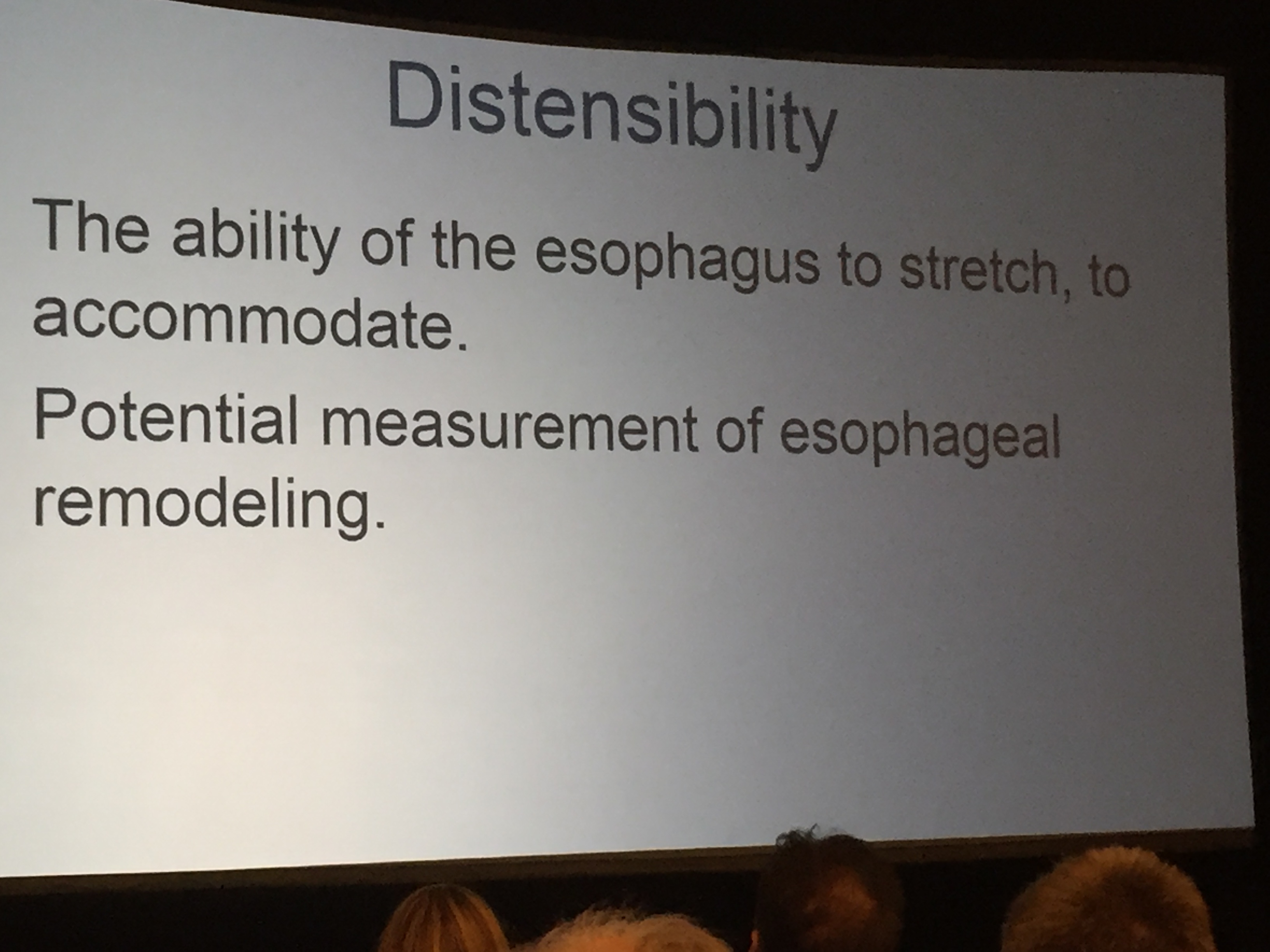
A recent study (AF Kagalwalla, JB Wechsler, K Amsen, S Schwartz, M Makhija, A Olive, CM Davis, M Manuel-Rubio, Seth Marcus et al.  Clin Gastroenterol Hepaot, 2017; 1698-1701) is going to help shake up the dietary management of eosinophilic esophagitis (EoE).  Our group GI Care for Kids, led by Seth Marcus, was one of the four centers which participated in this study of a four food elimination diet (FFED or 4-FED).

This prospective study enrolled 78 patients. IN those who did not respond to twice daily proton pump inhibitor therapy, subjects were instructed to restrict cow’s milk, wheat, egg, and soy. Patients underwent serial challenges (8 week for a challenge) with followup upper endoscopy to each food to determine response; 25 patients completed the challenge to all four foods. Key findings:

  • 64% (n=50) had remission (Eos <15/hpf) with the FFED
  • Symptom scores decreased in 91% of the histologic responders
  • Among those who completed additional challenges: 22/26 (85%) had reactions to cow’s milk, 10/30 (33%) had reactions to wheat, 14/40 (35%) had reactions to eggs, and 8/43 (19%) had reactions to soy
  • Among those (n=25) who completed challenges to all four foods, reactions to food: 84% milk, 28% wheat, 8% eggs, 8% wheat
  • Among those (n=25) who completed challenges to all four foods, reactions occurred to a single food-groups in 64%, two food-groups in 20%, three-food groups in 8%, and four-food groups in 8%.  Thus 36% had more than a single-food group reaction.

This study shows that the FFED provides similar efficacy to the six-food elimination diet (SFED) and is less restrictive.  It offers the prospect of less time to complete food reintroduction and fewer upper endoscopies

Limitations: nonrandomization, absence of control group, selection bias.  The fact that 24 patients dropped our before completing reintroduction highlights the difficulty of maintaining these diets in clinical practice.

In the associated editorial (S Eluri, ES Dellon, pages: 1668-9), the authors reiterate that the SFED (or 6-FED), which includes nuts and seafood, had histologic response rates typically between 69%-72% and that this study shows a similar response. They note that dairy elimination alone has had response rates ranging from 43% to 65%.

In addition, they discuss a 2-food elimination diet (milk and wheat).  In a recent study (J Molina-Infante, et al. Gastroenterol 2017; 152: S207), the authors started with a 2-FED with histologic response of 43% and if not responding would advance to 4-FED and 6-FED.  This approach was more efficient and further limited endoscopies.

My take: In those patients who are treated with a dietary approach, the 4-FED is a sensible initial therapeutic approach and an improvement from the 6-FED.  Though there is slightly higher initial response to 6-FED, the 4-FED allows more efficiency at identifying the trigger foods and lessened patient burdens with regard to endoscopy, diet complexity, and cost.

Related study: EA Erwin et al. JPGN 2017; 65: 520-5. This study showed that patients multiple IgE antibodies (≥ 0.1 IU/mL) to foods correlated with the likelihood of identifying eosionphilic esophagitis (≥ 15 eos/hpf):

  • In this cohort, among males, positive IgE antibodies to zero foods had a positive predictive value of 22%, 1-3 foods 52%, and 4-5 foods of 77%
  • In this cohort, among females, positive IgE antibodies to zero foods had a positive predictive value of 10%, 1-3 foods 29%, and 4-5 foods of 56%

Related blog posts:

Disclaimer: These blog posts are for educational purposes only. Specific dosing of medications (along with potential adverse effects) should be confirmed by prescribing physician.  This content is not a substitute for medical advice, diagnosis or treatment provided by a qualified healthcare provider. Always seek the advice of your physician or other qualified health provider with any questions you may have regarding a condition.


#NASPGHAN17 Eosinophilic Esophagitis Session

This blog entry has abbreviated/summarized these presentations. Though not intentional, some important material is likely to have been omitted; in addition, transcription errors are possible as well.

This is a long post –highlighting four separate talks on eosinophilic esophagitis.

PPI Use in Esophageal Eosinophilia: Recommendations from the recent AGREE conference

Glenn Furuta  Children’s Hospital of Colorado

Key points:

The term PPI-REE (proton pump inhibitor-responsive eosinophilic esophagitis) may not be needed.  PPI-REE is quite similar to eosinophilic esophagitis based on molecular and clinical features.  The main difference being that this subset responds to PPI therapy.


Characterization of CYP2C19*17 Polymporphisms Among Children with PPI Responsive EoE and EoE

James Franciosis et al.  Nemours Children’s Hospital Orlando

My take: This cool presentation offered a potential explanation of why some patients respond to PPIs (so called “PPI-REE”) from those with EoE that does not respond to PPIs.  This is pertinent because on a molecular basis the disease appears to be the same.  The difference in PPI-REE from EoE may be how the patient metabolizes PPI.  Those EoE patients who metabolize PPIs “extensively” are much less likely to respond to this therapy.

Eosinophilic esophagitis: Now an “Oldie” -But with increased interest and new research, a “Goodie”

Chris Liacouras  Children’s Hospital of Philadelphia

This lecture covered an enormous amount of material.  Here are a few slides.

Final Lecture (from November 3rd presentation):

Key points:

  • Endoflip is a new tool that helps determine esophageal distensibility.  Improved distensibility indicates less fibrostenotic disease which is one long-term goal.
  • Response to treatment has been correlated in improvement in Endoflip measurements.
  • There are no FDA approved medications at this point for EoE, though topical steroids may be approved soon.

Drug Development for Eosinophilic Esophagitis

Full text link: White Paper AGA: Drug Development for Eosinophilic Esophagitis (EoE)

I Hirano et al. Clin Gastroenterol Hepatol 2017; 15: 1173-83. This article reviews diagnostic criteria for EoE, clinical endpoints, and current/emerging treatments.

From AGA website-an excerpt:

Four important points from the white paper:

1. There is a complex inter-relationship between EoE, gastroesophageal reflux disease (GERD) and proton pump inhibitor-responsive esophageal eosinophilia (PPI-REE). A substantial proportion of patients with esophageal eosinophilia will improve with PPI treatment.

2. The clinical features and presentation of EoE differ between children and adults. Poor symptom specificity among children has limited the ability to identify appropriate candidates for enrollment into clinical trials and has impeded the development of pediatric patient-reported outcome instruments.

3. Aside from pharmacologic approaches, EoE can be addressed with dietary modifications and/or endoscopic dilation.

4. We should remember that the diagnosis of EoE carries a potentially major financial burden on patients’ families, making identifying new, effective and affordable treatment options a priority.

A Better Budesonide for Eosinophilic Esophagitis

A recent study (S Olivia et al. JPGN 2017; 64: 218-24) examines a preprepared viscous budesonide (PVB) for eosinophilic esophagitis (EoE).

The authors used higher doses than in previous studies: 1 mg twice a day if height <150 cm and 2 mg twice a day if height >150 cm.  Treatment period was 12 weeks.

Key findings:

  • 32 of 36 (89%) showed macroscopic remission at 12 weeks and median eosinophils count in histology dropped from 42.2 to 2.9 cells/hpf.  46.7% maintained remission (off therapy) at 36 weeks.
  • 89% achieved eosinophil count <20 cells/hpf at 12 weeks.
  • In this short study, the authors did not identify any changes in cortisol levels.

My take: A reliable composition from a manufacturer, if not too expensive, would be a big improvement for many kids with EoE. Higher doses of budesonide may be warranted in some cases of EoE.

Related article: “How I Approach the Management of Eosinophilic Esophagitis in Adults” I Hirano. Am J Gastroenterol 2017; 112: 197-99. (Thanks to Seth Marcus for this reference). The author states that he prefers to perform a baseline assessment prior to PPI initiation.  After diagnosis, he will use PPI and if no response, advance to either a dietary approach or topical steroids (he prefers fluticasone using the diskus formulations). His goals for therapy include: elimination of esophageal eosinophilia (<5-15 eos/hpf), resolution of dysphagia, and maintenance of esophageal diameter ≥16 mm. He does advocate annual testing for adrenal insufficiency for those taking long-term topical steroids.

Related blog posts:

screenshot-162

Eosinophilic Disease in Children with Intestinal Failure

Last week, this blog posted an abstract regarding the use of “real foods” for short gut kids.  This post looks into whether certain foods may provoke an allergic response.

A large (n=105) single center retrospective study (C Duggan et al. JPGN 2016; 63: 336-39) examined the histology from 208 endoscopic procedures to determine the frequency of eosinophilic disease in children with intestinal failure.

Key findings:

  • 37% of patients had evidence of eosinophilic inflammation in at least one section of the GI tract.
  • Most common sites for eosinophilic disease: colon/rectosigmoid 18/68 (26%), esophagus 17/83 (20%), ileum 9/54 (17%) and duodenum 4/83 (5%)
  • Both peripheral eosinophilia and hematochezia correlated with eosinophilic colitis
  • The authors state that “a strict elemental diet for 3 months before endoscopy was not associated with a decreased frequency of eosinophilic inflammation.”

While a strict elemental diet was not shown to be effective in this study, the limitations of the study design (eg. retrospective, small number on amino acid diet) preclude a definitive answer about the utility of these diets.  Other confounders, including ongoing parenteral nutrition support, also ‘muddy’ the picture.  A prospective study would be able to determine more conclusively how effective elemental diets are at minimizing eosinophilic inflammation and to allow for a more uniform definition of abnormal tissue eosinophilia.

Given the frequency of elemental diets early in life along with prior GI insults, the propensity to eosinophilic disease may have its origins well before this study period.  In healthy children, the LEAP, LEAP-ON, and EAT studies indicated that earlier exposure to allergens reduces the risk of allergic disease.

My take: This study shows a high prevalence of GI eosinophilic inflammation among children with intestinal failure.  Thus, in children with hematochezia and intestinal failure, eosinophilic colitis needs to be considered.

Related blog posts:

Grinnell Glacier, Glacier Natl Park

Glacier Natl Park