Unknown's avatar

About gutsandgrowth

I am a pediatric gastroenterologist at GI Care for Kids (previously called CCDHC) in Atlanta, Georgia. The goal of my blog is to share some of my reading in my field more broadly. In addition, I wanted to provide my voice to a wide range of topics that often have inaccurate or incomplete information. Before starting this blog in 2011, I would tear out articles from journals and/or keep notes in a palm pilot. This blog helps provide an updated source of information that is easy to access and search, along with links to useful multimedia sources. I was born and raised in Chattanooga. After graduating from the University of Virginia, I attended Baylor College of Medicine. I completed residency and fellowship training at the University of Cincinnati at the Children’s Hospital Medical Center. I received funding from the National Institutes of Health for molecular biology research of the gastrointestinal tract. During my fellowship, I had the opportunity to work with some of the most amazing pediatric gastroenterologists and mentors. Some of these individuals included Mitchell Cohen, William Balistreri, James Heubi, Jorge Bezerra, Colin Rudolph, John Bucuvalas, and Michael Farrell. I am grateful for their teaching and their friendship. During my training with their help, I received a nationwide award for the best research by a GI fellow. I have authored numerous publications/presentations including original research, case reports, review articles, and textbook chapters on various pediatric gastrointestinal problems. In addition, I have been recognized by Atlanta Magazine as a "Top Doctor" in my field multiple times. Currently, I am the vice chair of the section of nutrition for the Georgia Chapter of the American Academy of Pediatrics. In addition, I am an adjunct Associate Clinical Professor of Pediatrics at Emory University School of Medicine. Other society memberships have included the North American Society for Pediatric Gastroenterology Hepatology and Nutrition (NASPGHAN), American Academy of Pediatrics, the Food Allergy Network, the American Gastroenterology Association, the American Association for the Study of Liver Diseases, and the Crohn’s and Colitis Foundation. As part of a national pediatric GI organization called NASPGHAN (and its affiliated website GIKids), I have helped develop educational materials on a wide-range of gastrointestinal and liver diseases which are used across the country. Also, I have been an invited speaker for national campaigns to improve the evaluation and treatment of gastroesophageal reflux disease, celiac disease, eosinophilic esophagitis, hepatitis C, and inflammatory bowel disease (IBD). Some information on these topics has been posted at my work website, www.gicareforkids.com, which has links to multiple other useful resources. I am fortunate to work at GI Care For Kids. Our group has 17 terrific physicians with a wide range of subspecialization, including liver diseases, feeding disorders, eosinophilic diseases, inflammatory bowel disease, cystic fibrosis, DiGeorge/22q, celiac disease, and motility disorders. Many of our physicians are recognized nationally for their achievements. Our group of physicians have worked closely together for many years. None of the physicians in our group have ever left to join other groups. I have also worked with the same nurse (Bernadette) since I moved to Atlanta in 1997. For many families, more practical matters about our office include the following: – 14 office/satellite locations – physicians who speak Spanish – cutting edge research – on-site nutritionists – on-site psychology support for abdominal pain and feeding disorders – participation in ImproveCareNow to better the outcomes for children with inflammatory bowel disease – office endoscopy suite (lower costs and easier scheduling) – office infusion center (lower costs and easier for families) – easy access to nursing advice (each physician has at least one nurse) I am married and have two sons (both adults). I like to read, walk/hike, bike, swim, and play tennis with my free time. I do not have any financial relationships with pharmaceutical companies or other financial relationships to disclose. I have helped enroll patients in industry-sponsored research studies.

Hypercoagulation with Acute Severe Ulcerative Colitis (ASUC) Persists for Months

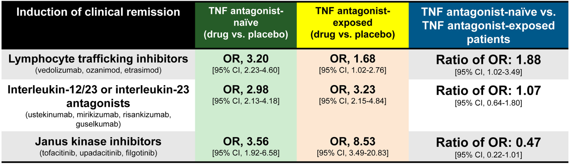
BJ Griffiths et al. Clin Gastroenterol Hepatol 2025; 23: 1798-1807. Open Access (PDF)! Hypercoagulation After Hospital Discharge in Acute Severe Ulcerative Colitis: A Prospective Study

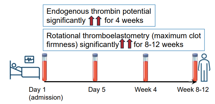
Methods: In this prospective study, twenty-seven adult patients with ASUC and 25 control patients with quiescent ulcerative colitis were recruited. Thrombin generation (endogenous thrombin potential), rotational thromboelastometry (EXTEM and FIBTEM maximum clot firmness), procoagulant factors, anticoagulant factors, and fibrinolytic markers were assessed for those with ASUC on admission (Day 1), Day 5, 4 weeks, and at 8–12 weeks. These assessments were performed on a single occasion for control patients.

Key findings:

Discussion:

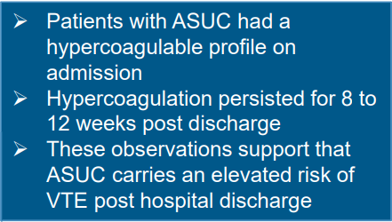
  • “Patients with ASUC had a hypercoagulable profile on initial presentation to the hospital, before receipt of LMWH, which was consistently demonstrated by individual and global assays of coagulation. The most marked elevations of individual factors at presentation were seen in the levels of Clauss fibrinogen, platelets, VWF, and FVIII, alongside heightened
    levels of the inhibitors of fibrinolysis PAI-1 and TAFIa.”
  • “This hypercoagulable state persisted for many weeks after hospital discharge, with levels of FVIII, fibrinogen, VWF, and inhibitors of fibrinolysis (TAFIa) remained significantly elevated at all timepoints up to 12 weeks, compared with the control population. This is despite
    intensive treatment for ASUC in all patients.”
  • “VTE and pulmonary embolism are 1 of the leading causes of morbidity and mortality during IBD flare-ups. The findings from this study reinforce the importance of thromboprophylaxis administration to all patients with ASUC at first presentation to hospital.”

My take: This study is in adults; the risk of VTE is lower in children and guidance on VTE prophylaxis is not clear.

Related blog posts:

Dupilumab for FPIES

M Plassmeyer et al. Journal of Allergy and Clinical Immunology 2025; Dupilumab Opens a Therapeutic Window in Food Protein Induced Enterocolitis Syndrome by un-licensing dendritic cells

Thanks for Ben Enav for this reference.

Methods: This was a two-part study: “(i) a detailed single-patient case of wheat-triggered, endoscopy-confirmed colitic FPIES treated with dupilumab 300 mg subcutaneously every two weeks and (ii) a prospective follow-up of seven additional FPIES patients all of whom initiated dupilumab for approved comorbidities. Serial flow cytometry quantified dendritic-cell OX40L and CD8+ CRTH2+ T-cell subsets before and after treatment; open food challenges assessed clinical tolerance.”

Key Findings:

  • Index case: Within two injections of dupilumab, the wheat sensitive patient tolerated a 50 g wheat protein challenge without gastrointestinal symptoms—this was the first uneventful exposure in 20 years. Discontinuation of dupilumab led to relapse; re-initiation again restored clinical tolerance
  • Cohort: All seven additional patients (ages 2–58 yr; triggers: milk, soy, rice, wheat, shellfish) achieved unrestricted dietary tolerance within three months
  • An important finding in the index case as well as the follow up cohort is the dupilumab induced drop in dendritic cell OX40L. OX40L is a TNF-superfamily co-stimulatory molecule induced on dendritic cells and other antigen-presenting cells.

My take: Dupilumab appears to be a promising medication for FPIES and warrants further study. If confirmed to be effective, it is likely to be targeted to those with approved comorbidities and those with more severe presentations.

Related blog posts:

Amicalola Falls State Park

Kids With Acute Pancreatitis Need Followup

F Ahmed, M Abu-El-Haija. Gastroenterol 2025; 169: 572-584. Open Access! Acute Pancreatitis in Children: It’s Not Just a Simple Attack

This is a really good review of acute pancreatitis covering epidemiology, diagnosis, severity classification, management, microbiome/metabolite derangements, genetics, and complications. Most of these topics have been covered in numerous blog posts (see below).

Selected Key Points:

  • Diagnostic testing -Amylase/Lipase:  “The diagnostic efficacy of amylase for AP, in terms of sensitivity and specificity, is contingent on the selected threshold value. Elevating the cut-off point to 1000 IU/L results in a high specificity of approximately 95%. However, this comes at the cost of reduced sensitivity, which some studies report to be as low as 61%… the activity of serum lipase remains elevated for a longer duration, typically between 8 and 14 days,… Lipase demonstrates superior accuracy with most studies reporting specificities exceeding 95% and sensitivities ranging from 55%–100% at a threshold activity level of 600 IU/L…hese tests have excellent sensitivities, they may have a few limitations such as being poor predictors of severity”
  • Risk Factors in Children (from Figure 1):
  • Incidence and Severity in Children (from Figure 1):

[At a recent lecture, Jay Freeman (How to Upgrade Pancreas Care –Jay Freeman MD (Part 1)) noted that severe pancreatitis is often defined by degree of organ dysfunction (eg. cardiac, pulmonary, renal). A practical definition of severe pancreatitis in children is whether the patient requires admission to an ICU]

  • Diagnostic testing -Imaging: “Imaging techniques are crucial for diagnosing and managing AP in children…NASPGHAN) and the Society for Pediatric Radiology formed consensus guidelines where transabdominal ultrasonography was recommended as the primary imaging technique for pediatric cases with suspected AP…Recent studies in the pediatric population have indicated that US’s sensitivity for AP detection ranges from 47%–52%.25Magnetic resonance cholangiopancreatography (MRCP) is useful for anatomical assessment without radiation but may require sedation”
  • Management: “The cornerstones of therapy are early feeding and intravenous fluids… Allowing patients to eat on admission was feasible and was associated with lower length of stay. Rates of intravenous fluids are recommended at 1.5–2 times maintenance rates,49 and the preferred fluid is Lactated Ringer’s due to limited studies including a recent randomized controlled study that showed that Lactated Ringer’s was associated with a faster discharge rate when administered compared with normal saline.50
  • Genetics:  “A recently conducted study investigated the importance of genetics in pediatric AP patients…use of an extensive panel of 8 genes… PRSS1CFTRSPINK1CPA1, CTRCCLDN2CASR, and SBDS… genetics is a major component in all types of pancreatitis in children, with genetic variants being most prevalent in CP cases at 31%, followed by AP at 19%, and ARP at 6%. A key discovery was that variants in SPINK1CFTR, or PRSS1 genes were associated with faster progression from first episode of AP toward CP.53
  • Complications (from Figure 1): “After the first episode of AP, the QoL is decreased, and it may lead to other disorders such as exocrine dysfunction, endocrine dysfunction and diabetes, nutritional deficiencies, and acute recurrent pancreatitis and CP.”

My take: Even after a single episode of acute pancreatitis, there are risks for long-term complications and patients need to follow-up.

Related blog posts:

Shared Decision-Making in Celiac Disease Diagnostic Approach

Y Sunkoy, S Talathi. Am J Gastroenterol 2025; 2190-2193. Utility of the ESPGHAN Biopsy-Sparing Guidelines for Celiac Disease in Children

Thanks to Ben Gold for this reference.

Methods: Retrospective study of patients (n=2942 children) who had celiac serologies and duodenal biopsies

Key findings:

  • Prevalence of CD in this cohort was 9% (226 of 2942 patients)
  • In those with a high titer (>10xULN), 106 of 107 patients (99%) had celiac disease
  • In this cohort, even in those with with >7XULN, had a Sensitivity of 55.3%, a specificity of 99% and a PPV of 97%

In their discussion, the authors note that “we did not obtain an EMA in a second sample, which is recommended in the ESPGHAN guidelines.”

Associated commentary: Erica Brenner, American Journal of Gastroenterology 120(9):p 1985-1986. The No-Biopsy Approach for Pediatric Celiac Disease: Ready for Prime Time in North America?

  • “Shiha et al (8) found that the PPV ranged from 65% for a 1% CD prevalence to a 99% for a 40% prevalence. As the 9% CD prevalence in the study by Sunkoj et al (4) exceeds the 0.81$-1.4% prevalence in the United States (9), the reported PPV may overestimate reality.” (Related post: No-Biopsy Approach to Celiac Disease Diagnosis and Positive Predictive Value (Based on Population)
  • “Children with type 1 diabetes and trisomy 21 have a higher risk of false-positive serology and therefor may not be appropriated candidates for a no-biopsy approach.”

My take: A larger recent study (Chang et al. Pediatrics. 2025;156(3):e2025070897) found that the no-biopsy approach had a significantly lower PPV in their cohort (94.9% overall, and 95.7% in those without T1DM). Thus, in cohorts with lower prevalence of CD, the no-biopsy approach could lead to 2-4% of children being placed unnecessarily on a gluten free diet. As such, it would be good practice to discuss making a diagnosis via endoscopy vs. the no-biopsy approach as part of shared decision-making.

Related blog posts:

Stercoral Colitis

A Bajer, E Levine. NEJM 2025;393: e23. Stercoral Colitis

This young adult presented to the ED with left-sided abdominal pain and chronic constipation. A CT scan was consistent with a diagnosis of stercoral colitis. “In stercoral colitis, chronically impacted feces distend the colon, resulting in inflammation. In some cases, the fecaloma may lead to focal-pressure necrosis or perforation.”

My take: Most often a CT is not needed in this setting. However, it is important to recognize that a severe impaction can lead to complications.

Related blog posts:

Selective Acid Suppression for Esophageal Atresia Patients

This year’s masterpiece!

Link from AAP HealthyChildren.org: Halloween Fun & Safety Tips for Kids of All Ages


S Zeneddin et al. J Pediatr Gastroenterol Nutr. 2025;81:960–966. Acid suppression after esophageal atresia repair: Some infants do benefit

Methods: The authors performed a retrospective study using the Pediatric Health Information System for infants undergoing EA/TEF repair between 2010 and 2022 (n=1445 infants). Acid suppression was defined as receipt of an H2 blocker or proton pump inhibitor on the day of discharge or longer than 30 inpatient days. Complex EA/TEF repair was defined as delayed repair (>7 days), G-tube placement before repair (likely a sign of a long gap or type A anomaly), prolonged hospitalization (>60 days), or multiple inpatient fluoroscopies. The authors defined stricture solely if it required intervention.

Key findings:

  • 257 (17.8%) required dilation by 1 year. Of the 688 (47.6%) infants who met criteria for complex EA/TEF, 126 (18.6%) required a dilation.
  • At 1 year, stricture rate was similar in infants with simple EA/TEF, with or without acid suppression (17.5% vs. 17.0%, p = 0.90)
  • In infants with complex EA/TEF, stricture rates were lower among those who received acid suppression compared to those who did not (15.3% vs. 26.0%, p = 0.001).

The associated editorial (D George, DK Robie. J Pediatr Gastroenterol Nutr. 2025;81:911–912) reviews some of the limitations of the study but does not provide clear recommendations on utilization of acid suppression therapy: the decision should be “should be individualized, weighing the potential benefits against the risks.”

My take: It is not surprising that more complex EA would have higher stricture rates. In my training (in the 1990s!), it was routine practice to use indefinite acid suppression. This article indicates that patients with low risk EA likely do not need acid suppression. In high risk patients, the algorithm by Yasuda et al (see post below J Am Coll Surg 2024; 238: 831-843) provides their approach to weaning acid suppression.

Related blog posts:

Disclaimer: This blog, gutsandgrowth, assumes no responsibility for any use or operation of any method, product, instruction, concept or idea contained in the material herein or for any injury or damage to persons or property (whether products liability, negligence or otherwise) resulting from such use or operation. These blog posts are for educational purposes only. Specific dosing of medications (along with potential adverse effects) should be confirmed by prescribing physician.  Because of rapid advances in the medical sciences, the gutsandgrowth blog cautions that independent verification should be made of diagnosis and drug dosages. The reader is solely responsible for the conduct of any suggested test or procedure.  This content is not a substitute for medical advice, diagnosis or treatment provided by a qualified healthcare provider. Always seek the advice of your physician or other qualified health provider with any questions you may have regarding a condition.

Prior Exposure to TNF Antagonists May Increase Response to JAK Inhibitors in Patients with Ulcerative Colitis

HH Lee et al. Clinical Gastroenterology and Hepatology 2025; 23, 2102 – 2114. Open Access! Differential Efficacy of Advanced Therapies in Inducing Remission in Ulcerative Colitis Based on Prior Exposure to TNF Antagonists

Methods: Meta-analysis of 17 randomized controlled trials in 8871 adults with moderate-severe UC. The authors calculated the ratio of odds ratio of achieving remission with active drug vs placebo, in TNF antagonist–naïve vs TNF antagonist–exposed patients.

Key findings:

  • JAK inhibitors: Less efficacious in TNF antagonist–naïve vs exposed patients (6 trials; ratio of OR, 0.47)
  • IL-23 antagonists: No significant difference was observed in efficacy of selective interleukin-23 antagonists vs placebo in TNF antagonist–naïve vs exposed patients (6 trials; ratio of OR, 1.07)
  • Lymphocyte trafficking inhibitors: More efficacious in TNF antagonist–naïve vs exposed patients (5 trials; odds ratio [OR], 1.88)

Discussion:

  • This study “confirmed prior observations that exposure to TNF antagonists significantly reduces the efficacy of lymphocyte trafficking inhibitors in inducing remission, including both vedolizumab and S1P receptor modulators, by approximately 50%.In contrast, prior exposure to TNF antagonists was associated with a significant increase in the efficacy of JAK inhibitors in inducing remission, with 2-fold higher efficacy in TNF antagonist–exposed vs TNF antagonist–naïve patients”
  • In the SELECT-COMPARE trial in patients with rheumatoid arthritis, there was also an improved response to upadacitinib in patients with prior adalimumab.
  • “The current findings raise the intriguing possibility that exposure to TNF antagonists could result in lasting effects on the immune system that differentially alter responsiveness to therapies with distinct mechanisms of action”

My take: This study suggests that JAK inhibitors are a good choice for secondary therapy after anti-TNF agents. Other factors, besides efficacy, including safety, extraintestinal manifestations, and cost, have to be considered as well.

Related blog posts:

Disclaimer: This blog, gutsandgrowth, assumes no responsibility for any use or operation of any method, product, instruction, concept or idea contained in the material herein or for any injury or damage to persons or property (whether products liability, negligence or otherwise) resulting from such use or operation. These blog posts are for educational purposes only. Specific dosing of medications (along with potential adverse effects) should be confirmed by prescribing physician.  Because of rapid advances in the medical sciences, the gutsandgrowth blog cautions that independent verification should be made of diagnosis and drug dosages. The reader is solely responsible for the conduct of any suggested test or procedure.  This content is not a substitute for medical advice, diagnosis or treatment provided by a qualified healthcare provider. Always seek the advice of your physician or other qualified health provider with any questions you may have regarding a condition.

Comprehensive ACG Clinical Guidelines for Crohn’s Disease (2025)

GR Lichenstein et al. The American Journal of Gastroenterology 120(6):p 1225-1264, June 2025. Open Access!!  ACG Clinical Guideline: Management of Crohn’s Disease in Adults

Yesterday and Today I am highlighting two adult clinical guidelines both of which are equivalent to up-to-date textbook chapters with specific recommendations; both are open access. In addition, the articles have accompanying author podcasts. Thanks to Ben Gold for these references.

Selected Management Recommendations:

  • Table 1, #3: We suggest against requiring failure of conventional therapy before initiation of advanced therapy for the management of CD
  • Table 1, #13: We recommend combination therapy of intravenous infliximab with immunomodulators (thiopurines) as compared with treatment with either immunomodulators alone or intravenous infliximab alone in patients with CD who are naive to those agents
  • Table 1, #33: In patients with high-risk CD, we recommend anti-TNF therapy to prevent postoperative endoscopic recurrence

Key Concepts:

  • Table 2, #9: Symptoms of CD do not correlate well with the presence of active inflammation and therefore should not be the sole guide for therapy. Objective evaluation by endoscopic or cross-sectional imaging should be undertaken periodically to avoid errors of under- or over-treatment.
  • Table 2, #14: The 10-year cumulative risk of major abdominal surgery in CD is 40%–55%, although recent studies performed in the biologic era suggest that the 10-year risk may have decreased to 30%. The 10-year risk of a second resection after the first is 35%, although again more recent studies suggest that this may have dropped to closer to 30%.
  • Table 2, #15: In CD, the 5-year rate of symptomatic postoperative recurrence is ∼50%.
  • Table 2, #29: Small bowel imaging should be performed as part of the initial diagnostic workup for patients with suspected CD.
  • Table 2, #31: Because of the absence of radiation exposure, magnetic resonance enterography should be used preferentially in young patients (younger than 35 years) and in patients in whom it is likely that serial exams will need to be performed.
  • Table 2, #38: Mucosal healing as determined by endoscopy is a goal of therapy. Scoring systems are available to measure the endoscopic disease activity and may be used to monitor response to therapy.
  • Table 2, #41: Antibiotics are not an effective treatment for luminal inflammatory CD and should not be used as a primary therapy.

My take: Given the rapid changes in available therapies, it would be optimal to make these collaborative guidelines (AGA, ACG, NASPGHAN) available online with frequent updates (similar to HCVguidelines.org).

Related blog posts:

Comprehensive ACG Clinical Guidelines for Ulcerative Coliits (2025)

D Rubin et al. The American Journal of Gastroenterology 120(6):p 1187-1224, June 2025. Open Access! ACG Clinical Guideline Update: Ulcerative Colitis in Adults

Today and tomorrow I am highlighting two adult clinical guidelines both of which are equivalent to up-to-date textbook chapters with specific recommendations; both are open access. In addition, the articles have accompanying author podcasts. Thanks to Ben Gold for these references.

Table 2 in the UC guideline makes 54 recommendations and Table 3 provides 57 key concepts.

Selected Management Recommendations:

  • Table 2, #4: We recommend treating patients with UC to achieve endoscopic improvement (Mayo score 0 or 1) to increase the likelihood of sustained steroid-free remission and to prevent hospitalization and surgery
  • Table 2, #5: We recommend the use of FC (fecal calprotectin) in UC to assess response to therapy, to evaluate suspected relapse, and during maintenance
  • Table 2, #33: When infliximab is used as induction therapy for patients with moderately to severely active UC, we recommend combination therapy with a thiopurine
  • Table 2, #43: Recommend continuing tofacitinib or upadacitinib as compared with no treatment for maintenance of remission in patients with prior moderately to severely active UC now in remission after induction with tofacitinib or upadacitinib. **The authors recommend continuing each biologic that achieved remission with induction therapy (#38-#43)
  • Table 2, #51: In patients with ASUC failing to adequately respond to intravenous corticosteroids (IVCS) by 3 days, we recommend medical rescue therapy with infliximab or cyclosporine (Strong recommendation, moderate quality of evidence).

Key concepts:

  • Table 3, #29: Patients who are primary nonresponders to an anti-TNF (defined as lack of therapeutic benefit after induction and despite sufficient serum drug concentrations) should be evaluated and considered for alternative mechanisms of disease control (e.g., in a different class of therapy) rather than cycling to another drug within the anti-TNF class.
  • Table 3, #31:  Subcutaneous infliximab and vedolizumab are considered equivalent to the standard intravenous maintenance dosing of these agents. The equivalence of the subcutaneous formulations for induction or as substitution for escalated doses of these therapies has not been robustly established.
  • Table 3, #41: Patients with UC should have available all medical options as recommended by their doctor and healthcare team. Third-party payers and requirements for step therapy should not come between the patient and their healthcare team in making decisions about treatment for UC.
  • Table 3, #48: All patients with ASUC should undergo a flexible sigmoidoscopy within 72 hours and preferably within 24 hours of admission. This should be used to assess endoscopic severity of inflammation and to obtain biopsies to evaluate for cytomegalovirus (CMV) colitis.
  • Table 3, #51: Nonsteroidal anti-inflammatory drugs (NSAIDs), narcotics, and medications with anticholinergic side effects should be avoided in ASUC.
  • Table 3, #57: In patients with ASUC initiating infliximab, dose intensification should be considered for those patients with low serum albumin (<2.5 g/dL).

My take: This article does an excellent job of summarizing current available evidence and good practice. Many of the recommendations may be helpful in garnering approval from third party payers.

Related blog posts:

Medical Imaging of Children/Adolescents and Risk of Cancer (2025)

R Smith-Bindman et al. NEJM 2025; 393: 1269-1278. Medical Imaging and Pediatric and
Adolescent Hematologic Cancer Risk

Methods: This was a retrospective cohort of 3,724,623 children born between 1996 and
2016 in six U.S. health care systems and Ontario, Canada, until the earliest of cancer
or benign-tumor diagnosis, death, end of health care coverage, an age of 21 years, or December 31, 2017.

Key findings:

  • During 35,715,325 person-years of follow-up (mean, 10.1 years per person), 2961 hematologic cancers were diagnosed, primarily lymphoid cancers (2349 [79.3%]), myeloid cancers or acute leukemia (460 [15.5%]), and histiocytic- or dendritic-cell cancers
    (129 [4.4%]).
  • The excess cumulative incidence of hematologic cancers by 21 years of age among children exposed to at least 30 mGy (mean, 57 mGy) was 25.6 per 10,000
  • The authors estimated that 10.1% of hematologic cancers may have been attributable to radiation exposure from medical imaging, with higher risks from the higher-dose medical-imaging tests such as CT
Cumulative Incidence of Hematologic Cancer According to Attained Age and
Radiation Dose to Bone Marrow among Children without Down’s Syndrome

Discussion Points:

  • “A 15-to-30-mGy exposure equivalent to one to two CT scans of the head was associated with an increased risk by a factor of 1.8”
  • “Although CT and other radiation-based imaging techniques may be lifesaving, our
    findings underscore the importance of carefully considering and minimizing radiation exposure during pediatric imaging to protect children’s long-term health”
  • “Research on Japanese atomic-bombing survivors showed that leukemia rates peaked 6 to 8 years after exposure, with excess risk lasting for more than five decades, particularly for acute myeloid leukemia”
  • This study tried to avoid concerns about reverse causation — in which imaging is performed because of existing cancer symptoms –by lagged exposures by 6 and 24 months
  • “The increasing use of low-value imaging in children and excessive radiation doses in CT are well documented…In many cases, reducing the imaging dose or substituting magnetic resonance imaging or ultrasonography may be more feasible than avoiding imaging altogether”

While the risks in aggregate appear quite substantial, the editorial (L Morton. NEJM 2025; 393; 1337-1339.Studying Cancer Risks Associated with Diagnostic Procedures –Interpret Wisely) makes the point that the risks for the individual are very small. “Fewer than 1% of youths in this study accumulated doses of 30 mGy or more from medical imaging and even at this exposure level, the excess cumulative incidence of hematologic cancers was low (25.6 per 10,000)…we need to ensure that all involved in medical imaging…wisely interpret the results…to understand the balance of the very small risks and the notable benefits of necessary imaging examinations to provide optimal patient care.”

My take: This study is a reminder to carefully evaluate the benefits, risks and alternatives when using ionizing radiation studies.

Related blog posts: