Deconstructing PPI-Associated Risks with Nearly 8 Billion Data Points and More on COVID-19 GI Symptoms (Video)

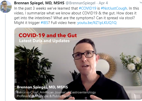
Link: 22 minute video —COVID-19 and the GI Tract -What We Know Right Now

———————

A recent study (C Ma et al. Gastroenterol 2020; 158: 780-82) used cross-sectional data from the National Ambulatory Medical Care Survey (NAMCS) (2006-2015) with a total 7,872,115,883 weighted observations.  They used this data to evaluate medication exposures and outcomes.

Key findings:

  • There was no association between PPI use and dementia, pneumonia, or intestinal infections.  There was a trend towards intestinal infections (AOR 1.48, CI 0.80-2.71) but this did not reach statistical significance. “Sensitivity analysis showed an association between PPI use and C difficile.”
  • There was an association with chronic kidney disease (CKD) (AOR 1.26); however, this was seen with a multitude of drug classes including statins, calcium channel blockers, and beta-blockers.

Discussion:

  • This study notes that a recent large randomized controlled trial found no statistically significant differences between those receiving PPIs and those receiving placebo except for intestinal infections.
  • With regard to CKD, “it is extremely unlikely that all of these medications increase the risk of CKD, and therefore, it is likely that these findings are due to residual confounding.”

My take: With the exception of C difficile/intestinal infections, this study provides further evidence of the safety of PPIs and a lack of association between these medications and purported PPI-related adverse events.  That said, it is still a good idea to limit use for appropriate indications.

Related blog posts:


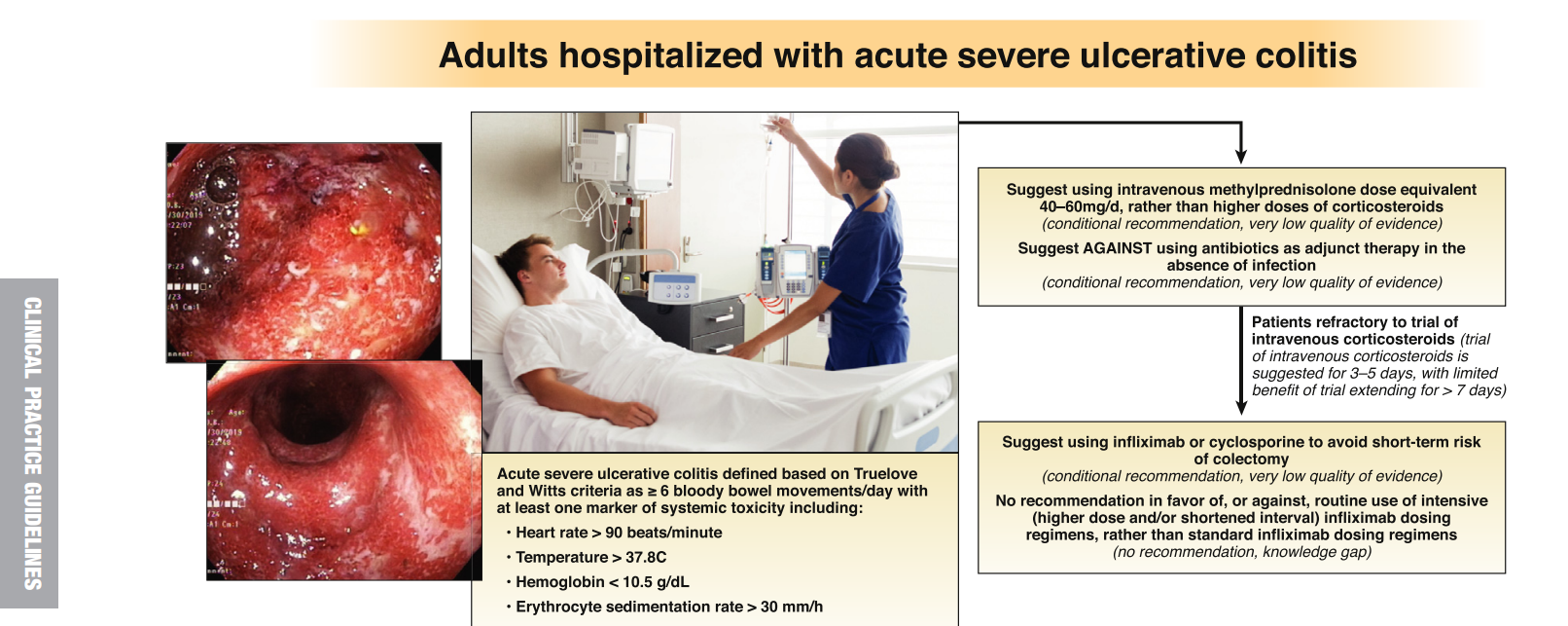
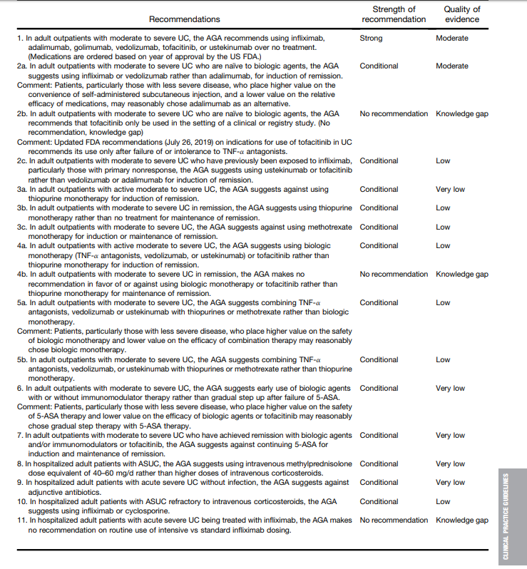
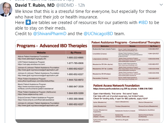
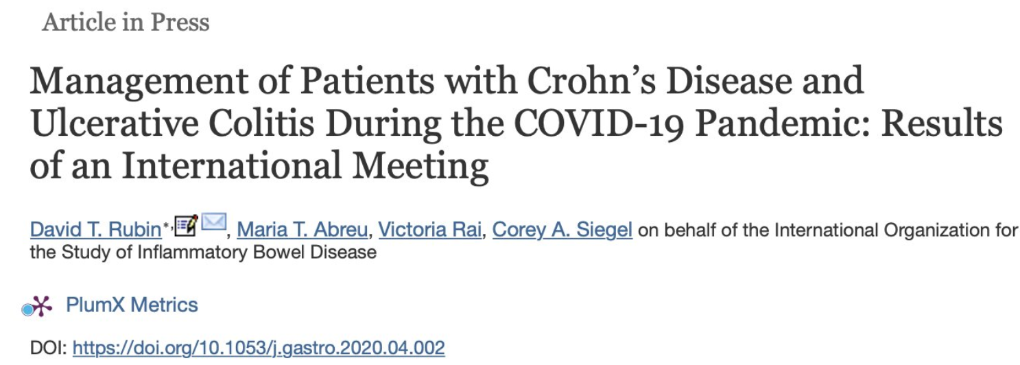
Also, IOIBD recommendations for IBD patients and COVID-19 have been published.

Here is link as well:

IOIBD (International Organization for the Study of Inflammatory Bowel Disease) Recommendations (#76) for IBD Patients with Regard to COVID-19:

Full link: IOIBD Update on COVID19 for Patients with Crohn’s Disease and Ulcerative Colitis (3/26/20)


What is the Current Standard of Care for PPE and Endoscopy Cases?

CC Thompson et al. Gastointestinal Endoscopy (EPUB), in a letter to the editor, respond to two recent studies on SARS-CoV-2 virus/COVID-19 and provide recommendations for PPE use in this era of COVID-19.

Here’s a link to manuscript: COVID-19 in Endoscopy: Time to do more?

Key points:

  • Reduce non-urgent cases. “We have cut our daily endoscopy volume by over 80% and closed our ambulatory endoscopy practice.”
  • Increase the use of telemedicine. “At present, telemedicine or virtual visits make up 91% of our upcoming clinic appointments.”
  • Physical distancing as advocated recently by WHO throughout a patient’s time in the endoscopy unit is stressed in the papers, with a 6-foot minimum between individuals.
  • Suggests “the need for a separate toilet as part of the isolation to minimize spread of infection due to bioaerosols from the toilet plume”
  • Our hospital system has recently changed policy to mandate that all employees wear surgical masks at all times while in the hospital and attest to their wellness online before reporting to work.
  • We suggest labeling each computer so the same provider uses that computer and chair for the entire day, and separating by at least 6 feet.
  • All endoscopic procedures (upper endoscopy, colonoscopy, EUS, ERCP) are aerosol-generating, referencing studies that show contamination of the endoscopist’s face during routine procedures. This makes all endoscopic procedures high risk from an infectious standpoint, and appropriate PPE is
    recommended… It makes little sense for healthcare providers to perform
    aerosolizing procedures, with patients coughing or passing gas on them, while not wearing an N95 mask or better
  • “It is important to use full PPE for all endoscopic procedures while in a pandemic such as this especially in areas with community spread, because no one is truly low risk given our ongoing difficulties with testing.”
  • “The mask can be reused as long as it is functional, not soiled, and not used in a suspected or COVIDpositive patient. It is important to cover the N95 to prevent soiling.”
  • “A study from China showed that no medical staff working in high-risk departments who wore N95s and practiced strict hand hygiene regardless of patient’s infection status became infected.”
  • “Testing all patients before high-risk procedures such as endoscopy is likely the best approach; however, this will depend on significant expansion of testing capabilities. Hopefully, the development of point-of-care testing with rapid results and increasing testing availability will make this a reality soon”

My take (in part from authors): “We are living through an unprecedented time and are all trying our best to protect our patients and ourselves under suboptimal conditions of limited PPE, limited testing, and limited data. ”  The recommendations in this article are based mainly on expert opinion and may need modifications based on new data and circumstances.

—————————-

IOIBD (International Organization for the Study of Iinflammatory Bowel Disease) Recommendations (#76) for IBD Patients with Regard to COVID-19:

Full link: IOIBD Update on COVID19 for Patients with Crohn’s Disease and Ulcerative Colitis (3/26/20)


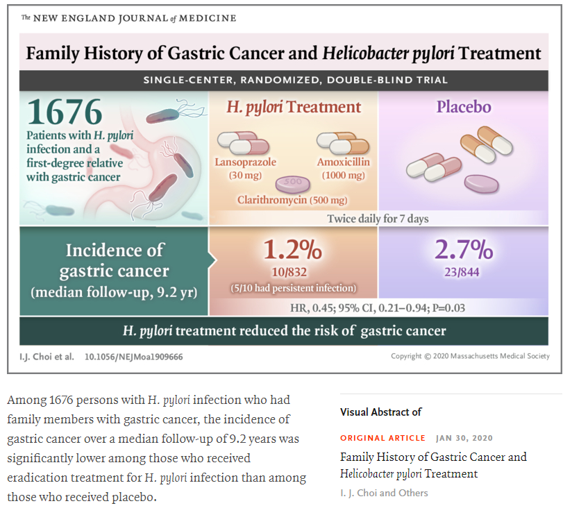
Treating Helicobacter Pylori Lowers The Risk of Gastric Cancer

Related blog posts:

Clinical Practice Advice: Pancreatic Necrosis

Recently the AGA published expert practice advice for pancreatic necrosis: TH Baron et al. Gastroenterol 2020; 158: 67-75.

Link to full-text PDF:  American Gastroenterological Association Clinical Practice Update: Management of Pancreatic Necrosis.  The link includes 5 figures which provide algorithms based on their recommendations.

I’ve copied their 15 best practice advice below and highlighted the most useful.  Early in the course of pancreatic necrosis, it can be difficult to discern if an infection is present due to a robust inflammatory response; some findings suggestive of infection include gas in the collection, bacteremia, sepsis, or clinical deterioration.  Generally, surgical, endoscopic or radiologic intervention is more optimal when there is a walled-off pancreatic necrosis (WON) which typically takes 4 weeks or more.

Best Practice Advice 1

Pancreatic necrosis is associated with substantial morbidity and mortality and optimal management requires a multidisciplinary approach, including gastroenterologists, surgeons, interventional radiologists, and specialists in critical care medicine, infectious disease, and nutrition. In situations where clinical expertise may be limited, consideration should be given to transferring patients with significant pancreatic necrosis to an appropriate tertiary-care center.

Best Practice Advice 2

Antimicrobial therapy is best indicated for culture-proven infection in pancreatic necrosis or when infection is strongly suspected (ie, gas in the collection, bacteremia, sepsis, or clinical deterioration). Routine use of prophylactic antibiotics to prevent infection of sterile necrosis is not recommended.

Best Practice Advice 3

When infected necrosis is suspected, broad-spectrum intravenous antibiotics with ability to penetrate pancreatic necrosis should be favored (eg, carbapenems, quinolones, and metronidazole). Routine use of antifungal agents is not recommended. Computed tomography–guided fine-needle aspiration for Gram stain and cultures is unnecessary in the majority of cases.

Best Practice Advice 4

In patients with pancreatic necrosis, enteral feeding should be initiated early to decrease the risk of infected necrosis. A trial of oral nutrition is recommended immediately in patients in whom there is absence of nausea and vomiting and no signs of severe ileus or gastrointestinal luminal obstruction. When oral nutrition is not feasible, enteral nutrition by either nasogastric/duodenal or nasojejunal tube should be initiated as soon as possible. Total parenteral nutrition should be considered only in cases where oral or enteral feeds are not feasible or tolerated.

Best Practice Advice 5

Drainage and/or debridement of pancreatic necrosis is indicated in patients with infected necrosis. Drainage and/or debridement may be required in patients with sterile pancreatic necrosis and persistent unwellness marked by abdominal pain, nausea, vomiting, and nutritional failure or with associated complications, including gastrointestinal luminal obstruction; biliary obstruction; recurrent acute pancreatitis; fistulas; or persistent systemic inflammatory response syndrome.

Best Practice Advice 6

Pancreatic debridement should be avoided in the early, acute period (first 2 weeks), as it has been associated with increased morbidity and mortality. Debridement should be optimally delayed for 4 weeks and performed earlier only when there is an organized collection and a strong indication.

Best Practice Advice 7

Percutaneous drainage and transmural endoscopic drainage are both appropriate first-line, nonsurgical approaches in managing patients with walled-off pancreatic necrosis (WON). Endoscopic therapy through transmural drainage of WON may be preferred, as it avoids the risk of forming a pancreatocutaneous fistula.

Best Practice Advice 8

Percutaneous drainage of pancreatic necrosis should be considered in patients with infected or symptomatic necrotic collections in the early, acute period (<2 weeks), and in those with WON who are too ill to undergo endoscopic or surgical intervention. Percutaneous drainage should be strongly considered as an adjunct to endoscopic drainage for WON with deep extension into the paracolic gutters and pelvis or for salvage therapy after endoscopic or surgical debridement with residual necrosis burden.

Best Practice Advice 9

Self-expanding metal stents in the form of lumen-apposing metal stents appear to be superior to plastic stents for endoscopic transmural drainage of necrosis.

Best Practice Advice 10

The use of direct endoscopic necrosectomy should be reserved for those patients with limited necrosis who do not adequately respond to endoscopic transmural drainage using large-bore, self-expanding metal stents/lumen-apposing metal stents alone or plastic stents combined with irrigation. Direct endoscopic necrosectomy is a therapeutic option in patients with large amounts of infected necrosis, but should be performed at referral centers with the necessary endoscopic expertise and interventional radiology and surgical backup.

Best Practice Advice 11

Minimally invasive operative approaches to the debridement of acute necrotizing pancreatitis are preferred to open surgical necrosectomy when possible, given lower morbidity.

Best Practice Advice 12

Multiple minimally invasive surgical techniques are feasible and effective, including videoscopic-assisted retroperitoneal debridement, laparoscopic transgastric debridement, and open transgastric debridement. Selection of approach is best determined by pattern of disease, physiology of the patient, experience and expertise of the multidisciplinary team, and available resources.

Best Practice Advice 13

Open operative debridement maintains a role in the modern management of acute necrotizing pancreatitis in cases not amenable to less invasive endoscopic and/or surgical procedures.

Best Practice Advice 14

For patients with disconnected left pancreatic remnant after acute necrotizing mid-body necrosis, definitive surgical management with distal pancreatectomy should be undertaken in patients with reasonable operative candidacy. Insufficient evidence exists to support the management of the disconnected left pancreatic remnant with long-term transenteric endoscopic stenting.

Best Practice Advice 15

A step-up approach consisting of percutaneous drainage or endoscopic transmural drainage using either plastic stents and irrigation or self-expanding metal stents/lumen-apposing metal stents alone, followed by direct endoscopic necrosectomy, and then surgical debridement is reasonable, although approaches may vary based on the available clinical expertise.

Related blog post: NASPGHAN 2017 Postgraduate Course Part 1 -includes slides on pancreatic fluid collection management


ACG Guideline for Small Intestinal Bacterial Overgrowth

Link to full PDF: ACG Clinical Guideline: Small Intestinal Bacterial Overgrowth

M Pimentel et al. Am J Gastroenterol 2020;00:1–14. https://doi.org/10.14309/ ajg.0000000000000501; published online January 8, 2020

One key point is that the authors acknowledge that almost all of their recommendations are based on a very low level of evidence.  In fact, only one of their 6 recommendations in Table 1 is considered to have more evidence and it is rated as low level of evidence.

The article provides an in-depth review of small intestinal bacterial overgrowth including underlying homeostasis mechanisms/pathophysiology (Tables 3 & 4) and treatments.  For those needing treatment, the authors list options in Table 5 including rifaximin, amoxicillin-clavulanic acid, ciprofloxacin, doxycycline, metronidazole, neomycin, norfloxacin, tetracycline, and trimethoprim-sulfamethoxazole.

Image Available from Lizzie Aby Feed

Related blog posts:

Disclaimer: This blog, gutsandgrowth, assumes no responsibility for any use or operation of any method, product, instruction, concept or idea contained in the material herein or for any injury or damage to persons or property (whether products liability, negligence or otherwise) resulting from such use or operation. These blog posts are for educational purposes only. Specific dosing of medications (along with potential adverse effects) should be confirmed by prescribing physician.  Because of rapid advances in the medical sciences, the gutsandgrowth blog cautions that independent verification should be made of diagnosis and drug dosages. The reader is solely responsible for the conduct of any suggested test or procedure.  This content is not a substitute for medical advice, diagnosis or treatment provided by a qualified healthcare provider. Always seek the advice of your physician or other qualified health provider with any questions you may have regarding a condition.


New Therapy for Eosinophilic Esophagitis

In a report (I Hirano, ES Dellon et al. Gastroenterol 2020; 158: 111-22) on a phase 2 trial with 47 adults, the authors show that weekly subcutaneous injections of dupilumab (300 mg) was associated with improvement in eosionophilic esophagitis.

Dupilumab, a fully human monoclonal antibody, antagonizes the interleukin-4 receptor-alpha component of the type 2 receptor, thereby inhibiting signaling of IL-4 and IL-13. Dupilumab has shown “efficacy in pediatric and adult atopic dermatitis, asthma, and chronic sinusitis with nasal polyposis” and is FDA-approved for use in these disorders.

Key findings:

  • Dupilumab was associated with improvement in dysphagia as measured by the Straumann Dysphagia Instrument with a mean reduction of 3.0 compared to 1.3 in the placebo group at week 10 (P=.0304)
  • Dupilumab was associated with improvement in histology, at week 12, the peak eosinophil count had dropped by a mean of 86.8 (P<.0001 vs. placebo)
  • Dupilumab was associated with improvement in endoscopic parameters as assessed by endoscopic reference score which dropped by 1.6 (P -.0006 vs placebo)
  • No serious adverse effects were evident.  Nasopharyngitis was noted in 17% in the dupilumab group compared to 4% of placebo-treated patients.  Injection site erythema was noted in 35% (vs. 8% in placebo group

Of note, the study was sponsored by pharmaceutical companies and many of the authors (including both lead authors) were either affiliated with these companies.

My take: This study indicates that Dupilumab has at least short term efficacy and safety for eosinophilic esophagitis.

Related blog posts:

Disclaimer: This blog, gutsandgrowth, assumes no responsibility for any use or operation of any method, product, instruction, concept or idea contained in the material herein or for any injury or damage to persons or property (whether products liability, negligence or otherwise) resulting from such use or operation. These blog posts are for educational purposes only. Specific dosing of medications (along with potential adverse effects) should be confirmed by prescribing physician.  Because of rapid advances in the medical sciences, the gutsandgrowth blog cautions that independent verification should be made of diagnosis and drug dosages. The reader is solely responsible for the conduct of any suggested test or procedure.  This content is not a substitute for medical advice, diagnosis or treatment provided by a qualified healthcare provider. Always seek the advice of your physician or other qualified health provider with any questions you may have regarding a condition

Neurologic Toll of Celiac Disease

A recent prospective cohort study (M Hadjivassiliou et al. Clin Gastroenterol Hepatol 2019; 17: 2678-86) shows an alarmingly-high level of neurologic deficits in 100 consecutive adults (mean age 43 years) with a new diagnosis of celiac disease.

Key findings:

  • Gait instability in 24%
  • Persistent sensory symptoms in 12%; peripheral neuropathy was identified in 2%
  • Frequent headaches in 42%
  • Abnormal results from Brain MRI in 60%; 25% had brain white matter lesions beyond expectation for age group and 46% had abnormal MR spectroscopy of the cerebellum
  • Anti-TG6 antibodies were detected in 40% of patients and this subgroup had significant atrophy of subcortical brain regions compared to patients who were Anti-TG6 antibody-negative

Some neurologic findings improve on a gluten-free diet (GFD).  In previous studies of patients with CD and headaches, 75-80% improved or subsided after a year of strict adherence to a GFD.

My take: This study indicates that early diagnosis of celiac disease along with strict adherence to a gluten-free diet is likely to prevent permanent neurologic disability.

Related blog posts:

How Often Do Adults Develop Celiac Disease After Negative Testing?

RS Choung et al. Gastroenterol 2020; 158: 151-9.  

Full abstract link: (which has link to full text): “Community-Based Study of Celiac Disease Autoimmunity Progression in Adults”

Methods: In this prospective cohort study, waste blood samples from residents of a community were tested for CeD autoimmunity at 2 time points. We analyzed waste blood samples from 15,551 adults for tTGA and, if titer results were above 2 U/mL, for endomysial antibody. The median interval between the two time points was 8.8 years.

Results:

  • Of the serum samples collected at the first time point, 15,398 had negative results for tTGA, and 153 had positive results for tTGA (>4 U/mL). Based on medical records, 6 individuals received a diagnosis of celiac disease, for a cumulative incidence of celiac disease diagnosis of 0.06% over 10 years.
  • Forty-nine (0.32%) individuals with a negative result from the first serologic test for tTGA had a positive result from the second test
  • Among the 153 adults who were tTGA positive at the first time point, 31 (20%) had a subsequent diagnosis of celiac disease, 81 (53%) remained positive for tTGA without a clinical diagnosis of celiac disease, and 41 (27%) had negative test results for tTGA at the second time point.

Highlights in IBD from Two 2019 Meetings: American College of Gastroenterology and United European Gastroenterology Week

Gastroenterology & Hepatology. December 2019 – Volume 15, Issue 12, Supplement 5

Excerpts from William Sandborn Commentary which are at the end of this supplement along with references:

Vedolizumab

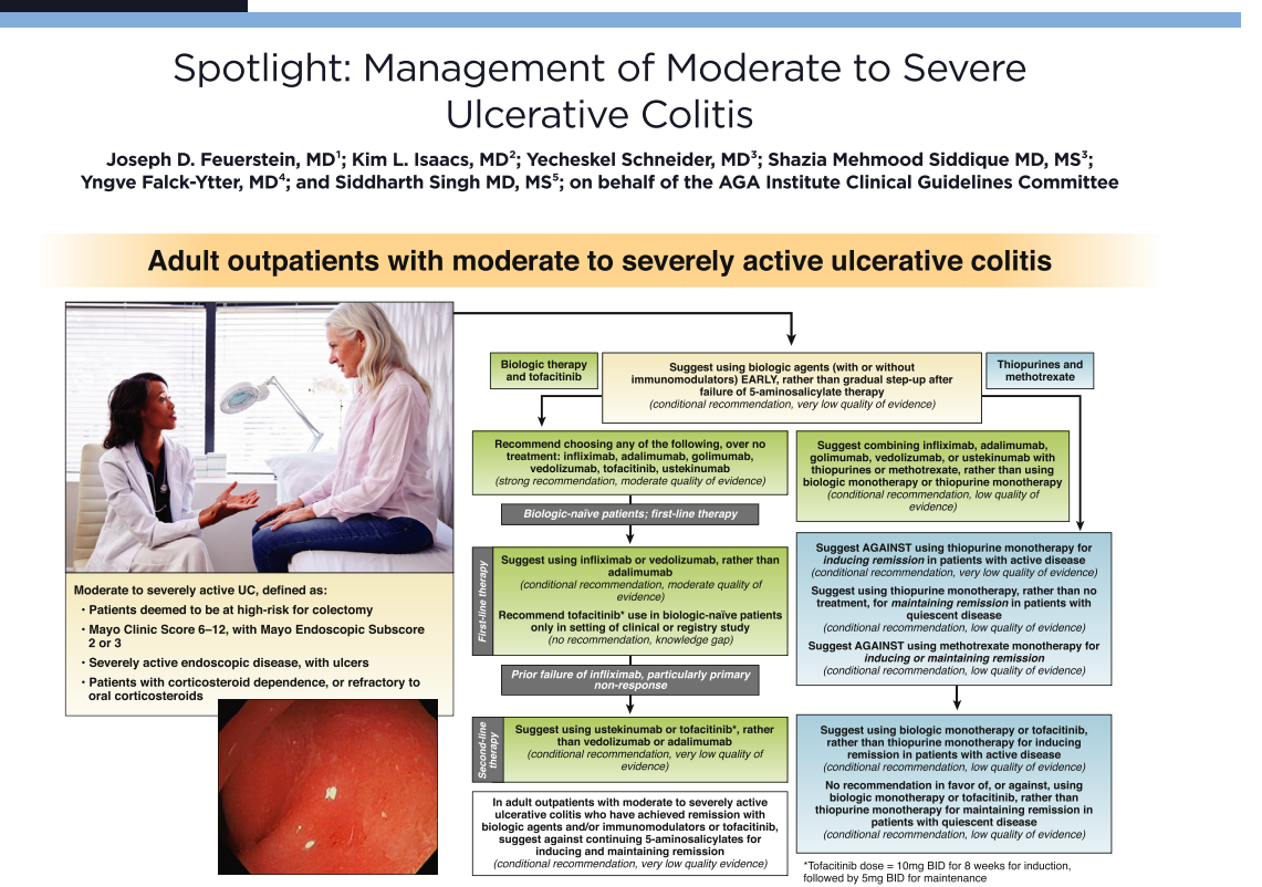
In the VARSITY study (An Efficacy and Safety Study of Vedolizumab Intravenous [IV] Compared to Adalimumab Subcutaneous [SC] in Participants With Ulcerative Colitis), 769 patients with ulcerative colitis were randomized to a year of therapy with either adalimumab at the US Food and Drug Administration (FDA)-approved dose or vedolizumab at the FDA-approved dose…This shows that the idea that vedolizumab (and anti-integrin therapy) is slower-acting than anti-TNF therapy is not correct, and that both of these classes of drugs can work fairly quickly in a number of patients.

Dr Brian Bressler and colleagues looked at the effectiveness of anti-TNF therapy in the real world when used second line after failing first-line biologic therapy with vedolizumab…The study conducted by Dr Bressler and colleagues, which included both Crohn’s disease patients and ulcerative colitis patients, found that the results were fairly similar whether patients received first-line biologic therapy with an anti-TNF agent or whether patients received first-line therapy with vedolizumab… It is generally thought that vedolizumab is a safer therapy than anti-TNF therapy, so with the finding from this study, a reasonable treatment approach could be to start with vedolizumab and see if it works

Dr Christina Chambers and colleagues identified outcomes for pregnancy in 223 women, 53 of whom received vedolizumab. The researchers found that there were no major structural birth defects reported in the vedolizumab group, compared to 5.7% and 5.3% in the disease-matched group and healthy control group, respectively. Thus, there seemed to be no signal for an increased malformation risk in patients who were undergoing treatment with vedolizumab and became pregnant.

Adalimumab

The SERENE trials are a set of head-to-head trials, one for ulcerative colitis and one for Crohn’s disease, comparing standard-dose adalimumab to a more intensive induction regimen of adalimumab…

For both ulcerative colitis and Crohn’s disease, the SERENE trials showed that the current FDA-approved dosing regimen is effective and that more intensive induction therapy does not improve outcomes over time. Thus, there is no utility in giving high induction doses. 

Tofacitinib

Over 1000 patients who had been treated with tofacitinib were examined…during induction and maintenance of the placebo-controlled portion of the tofacitinib clinical trials, there were a total of 5 deep vein thrombosis and pulmonary emboli events. All 5 occurred in patients who were receiving placebo; none of these events occurred in patients who were receiving tofacitinib…[And] There was a total of 5 deep vein thrombosis and pulmonary emboli events during this long-term extension…Looking at the ulcerative colitis clinical trial data that I presented, it is somewhat reassuring that we did not see the same elevation in risk for deep vein thrombosis and pulmonary emboli that was seen in the high-risk rheumatoid arthritis patient population.

Mont Royal (Montreal)