Optimistic Results for Hepatitis C plus Hepatology Update

The August issue of Hepatology had several articles on Hepatitis C confirming the efficacy of newer agents:

  • LI Backus et al Hepatology 2016; 64: 405-14.  This “real-world” observational study from the VA Clinical registry with 4,365 genotype 1 treatment-naive patients who received ledipasvir/sofosbuvir showed SVR rates of 91.3% (w/o ribavirin) and 92% (w ribavirin).
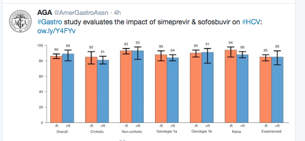
  • P Kwo et al. Hepatology 2016; 64: 370-80 (OPTIMIST-1) This study showed that 12 weeks of simeprevir+sofusbuvir for 12 weeks was highly effective (97% SVR) and that 8 weeks of this therapy was inferior (83% SVR).  N=310 with genotype 1 (w/o cirrhosis).  No patients stopped therapy due to adverse effects.
  • E Lawitz et al. Hepatology 2016; 64: 360-69 (OPTIMIST-2) This study showed that simeprevir+sofusbuvir for 12 weeks was effective in genotype 1 patients (n=103) with cirrhosis.  For treatment-naive, the SVR was 88% and for treatment-experienced patients, the SVR was 79%.

Also in Hepatology:

  • S Heibani et al Hepatology 2016; 64: 549-55. This study looked at 1-week versus 2-week intervals for endoscopic ligation.  While 1-week ligation eradicated varices more quickly, neither approach was associated with differences in number of endoscopies, complications (including rebleeding) or other clinical outcomes.
From earlier study of "real-world" treatment of Genotype 1. Gastroenterol 2016; 150: 419-29.

From earlier study of “real-world” treatment of Genotype 1. Gastroenterol 2016; 150: 419-29. (Full text link)


Dubin-Johnson Syndrome

From NEJM:

A 48-year-old woman scheduled to receive a laparoscopic cholecystectomy underwent a preoperative evaluation that disclosed conjugated hyperbilirubinemia…

A biopsy specimen revealed coarse, deep-brown, pigmented granules on periodic acid–Schiff staining (Panel B), primarily at the canalicular pole of the hepatocytes and especially in the pericentral zones, with otherwise well-preserved lobular architecture. Expression of the multidrug-resistance–associated protein 2 (MRP2) was absent on anti–MRP2 immunohistochemical analysis (Panel C; see also comparison with control specimen [inset]). A diagnosis of the Dubin–Johnson syndrome was confirmed. This syndrome is an autosomal recessive disorder that is caused by a mutation in MRP2 that results in deficient canalicular expression of MRP2 and impaired secretion of conjugated bilirubin into the bile. Such mutations cause an isolated increase in serum levels of conjugated bilirubin and the appearance of a black liver, without associated sequelae.

Dubin-Johnson

Do You Know When Hepatitis C Virus Transmission Peaked?

According to a recent study (AC Spaulding, LS Miller. Lancet Infect Dis. 2016 Mar 30. doi: 10.1016/S1473-3099[16 …), peak transmission of hepatitis C virus (HCV) peaked about 1950, likely due to reuse of metal and glass syringes.

This study counters the idea that HCV transmission was “primarily due to injection drug use, unsafe tattooing, high-risk sex, and travel to high endemic areas during youth,” according to the researchers. Here’s the link: Apportioning blame in the North American Hepatitis C virus epidemic

My take: Will this take away some of the stigma of HCV infection? Probably not.  But, hopefully as the costs for treatment reduce, more individuals can be infection-free and avoid complications related to infections.

HCV Infections

Vedolizumab for Primary Sclerosing Cholangitis (with IBD)?

“The monoclonal antibody vedolizumab may reduce biliary inflammation in patients with primary sclerosing cholangitis and comorbid inflammatory bowel disease, according to early, open-label study findings reported at the meeting sponsored by the European Association for the Study of the Liver”  –according to GIHepNews: Biliary inflammation reduced by IBD drug

“Vedolizumab given to 27 patients with primary sclerosing cholangitis (PSC) and inflammatory bowel disease (IBD) resulted in a 50% reduction or normalization of serum alkaline phosphatase levels in 17 cases (63%).”

This was an open-label, proof-of-concept study involving 27 patients aged 25-30 years with PSC and comorbid IBD.

My take: This is interesting but needs a lot more study.

Atlanta Zoo 2016

Atlanta Zoo 2016

Elafibranor Study & “My compliments to the photographer”

A while back, I remember seeing a cartoon with a dissatisfied patron leaving a restaurant and saying “my compliments to the photographer.”

Sometimes reading journal titles has the same feel.  The title does not always indicate what you are really going to get.  A recent study (V Ratziu et al. Gastroenterol 2016; 150: 1147-59) has the following title: “Elafibranor, an Agonist of the Peroxisome Proliferator –Activated Receptor –α and –β, Induces Resolution of Nonalcoholic Steatohepatitis Without Fibrosis Worsening.”  Sounds great –a new effective treatment for NASH, right?

Here’s are the results:

  • “In intention-to-treat analysis, there was no significant difference between elafibranor and placebo groups in the protocol-defined primary outcome.”
  • However, based on a post-hoc analysis with a modified definition, the treatment group had  a 19% NASH resolution compared with 12% of the placebo group.

This study examined 276 patients in a randomized double-blind placebo-controlled trial.

To me these results are not impressive.  The associated editorial (pg 1073) expresses more optimism and indicates that there have been evolving outcome measures in NASH studies to look for the combination of NASH resolution without worsening fibrosis.  Thus, prior studies that used only NASH resolution, such as pioglitazone (47%), vitamin E (36%) and obeticholic acid (22%) cannot be compared to his current study.

My take: Pretty picture or not, what this really means -is that we need more studies, including the outcome of phase III studies of this medication.

Georgia Terrace

The Georgian Terrace

Bring Out the Big Guns: Treating Infections with Cirrhosis

A recent study (M Merli et al. Hepatology 2016; 1632-39) indicates that health-care associated infections (HCA) in the setting of cirrhosis respond more favorably to broad-spectrum antibiotics.  In this prospective study of 96 randomized patients, in-hospital mortality was improved in the broad-spectrum group (6%) compared to the standard group (25%).  There was a similar multidrug-resistnace rate (50% broad spectrum compared with 60% in standard group).

Table 1 lists the antibiotic selection.  In the broad spectrum treatment, this almost always included imipenem/cilastin (I/C); with spontaneous bacterial peritonitis (SBP), I/C was combined with vancomycin, and with pneumonia it was combined with both vancomycin and azithromycin.  In contrast, the standard group’s main medication was augmentin (with added azithromycin for pneumonia) or cefotaxime for SBP.

My take: Does this study show that infections in the setting of cirrhosis are becoming more difficult to treat? Probably. How much these findings can be extended to the pediatric population remains uncertain.

Somewhat related topic: Primary prophylaxis of Variceal Bleeding in Children –Summary of the Baveno VI Pediatric Satellite Symposium.  BL Shneider et al. Hepatology 2016; 63: 1368-80. Key point: “there are few pediatric data…therefore, no recommendations for primary prophylaxis with endoscopic variceal ligation, sclerotherapy, or nonspecific beta-blockade in children was proposed.”

Silver Comet Trail

Silver Comet Trail

Interesting Fatty Liver Articles -Spring 2016

J Bousier et al. Hepatology 2016; 63: 764-75.  This study showed an association between the severity of nonalcoholic fatty liver disease and gut dysbiosis/shift in gut microbiome in 57 patients.  Specifically, Bacteroides was independently associated with NASH and Ruminococcus with significant fibrosis.

V WS Wong et al. Hepatology 2016; 63: 754-63. This study showed that NAFLD (identified by ultrasonography screening) was frequent (58.2%) among 612 consecutive patients who were undergoing coronary angiogram. During a followup (3679 patient-years), NAFLD patients had a lower adjusted HR of death (0.36).  Older age and diabetes were indepenently associated with cardiovascular events.  In addition, during f/u NAFLD patients in their cohort rarely developed liver cancer or cirrhotic complications.  Thus, NAFLD is common among patients with coronary artery disease but did not predict a worsened outcome.

F Piscaglia et al. Hepatology 2016; 63: 827-38. This report was a study of 756 patients with liver cancer (HCC) due to either NAFLD (145) or HCV (611). HCC in NAFLD patients had a larger volume, was more infiltrative, and was detected outside surveillance.  NAFLD-HCC was associated with a lower survival (25.5 months compared with 33.7 months for HCV-HCC). The authors note that after patient matching for tumor stage, the survival rate was similar. The difference in survival does not account for lead-time bias (What’s More Important: Improving Mortality Rate or Survival …).  Overall, the study indicates that without surveillance, HCC is detected later.  Due to the frequency of NAFLD, it is unclear which patients would benefit from surveillance and what type of surveillance should be recommended.

Related blog posts:

Farjado, Puerto Rico

Farjado, Puerto Rico


CDC: Increase in Acute Hep B in Appalachia

MMWR 2016; 65: 47-50. Increases in Acute Hepatitis B Virus Infections — Kentucky, Tennessee, and West Virginia, 2006–2013

An excerpt:

  • During 2006–2013, a total of 3,305 cases of acute HBV infection were reported to CDC from Kentucky, Tennessee, and West Virginia. During 2009–2013, incidence of acute HBV infection increased 114% in these three states, but remained stable in the United States overall
  • Among cases in which at least one risk factor was reported, the proportion of persons reporting injection drug use as a risk factor was significantly greater in 2010–2013, compared with 2006–2009 (75% versus 53%; p<0.001)…the increase was statistically significant only among cases occurring in non-urban counties
  • The findings in this report are subject to …limitations. First, NNDSS is a passive surveillance system, and therefore, unreported cases might have been missed. Second, the current case definition for acute HBV infection captures only symptomatic persons and excludes persons with asymptomatic HBV … Third, … certain populations at high risk (e.g., persons who are incarcerated, homeless, and uninsured) with limited access to care could potentially be underrepresented

My take: Increased drug use appears to be driving an increase in acute HBV in Appalachia. “Evidence-based prevention strategies, including increasing hepatitis B vaccination coverage, testing and linkage to care activities, and education campaigns targeting persons who inject drugs are urgently needed.”

Gibbs Gardens, Ball Ground

Gibbs Gardens, Ball Ground

Drug-Induced Liver and Skin Reactions

A recent study (H Devarbhavi et al. Hepatology 2016; 63: 993-99 & associated editorial 700–2) provide insight into outcomes and causative agents in patients who had both drug-induced liver injury (DILI) along with severe skin reactions.

With regard to the skin reactions, the authors were specifically focused on Stevens-Johnson syndrome (SJS) and toxic epidermal necrolysis (TEN).  SJS indicates an area of skin detachment of <10% and TEN involves >30%.  SJS/TEN overlap is 10-30%.

The study reviewed a single center DILI registry over 18 years with 748 patients.  There was prospective recruitment during the final 10 years of the study period (1997-2015). 36 (4.8%) had either SJS or TEN (mean age 32 years, 53% females).  9/36 (25%) were <18 years.

Causative agents:

  • Antiepileptics 47%
  • Sulfonamides 18%
  • Nevirapine 16%
  • Multiple agents 61%

Key points:

  • Median duration between drug initiation and onset of rash was 24 days
  • 13/36 (36%) died. 77% of those who died had jaundice.
  • 14/36 (39%) received steroids including 10 survivors and 4 who died.

While a mortality of 36% among those with both DILI and SJS/TEN is high, the discussion notes that the mortality is high even in those without DILI (~18% in ones study).  There were 8/36 in the study with HIV which is associated with a much higher risk of DILI.  There was a lower mortality in the pediatric age group (1 child 11%) and in those with HIV (1 patient 12.5%).

Related blog posts:

Walnut Street Bridge

Walnut Street Bridge

Liver Transplant Recipients Are Getting Older

Data(F Su et. al. Gastroenterol 2016; 150: 441-53) from 2002 thru 2014 indicate that liver transplant recipients are getting older.

The researchers reviewed data from the United Network for Organ Sharing (UNOS), including 60,820 adults who underwent liver transplantation and 122,606 listed for transplantation. Key findings:

  • Mean age of those listed increased from 51.2 to 55.7 years.  This trend was more prominent among those with hepatitis C (50.9 –>57.9).
  • The proportion of listed patients ≥60 years increased from 19% to 41%.
  • There were no differences in 5-year transplant-related survival “benefit”

The topic of survival “benefit” is reviewed in the discussion and the associated editorial (pg 306).  The survival benefit is calculated as the difference between life expectancy with and without liver transplantation. So, even though older transplant recipients have worse post-transplantation survival, this is counterbalance by the increased risk of waitlist mortality.  It is quite likely, however, that with more time (>5 year followup) that the survival benefit for younger patients would be more apparent.  In addition, the idea that the survival benefit could be equivalent could be influenced by selection bias.  Many transplant centers may be more selective when deciding to place older patients (>70 years) on the waitlist.

My take: The steady increase in age in adult liver transplant recipients is a concern due to worse outcomes in older patients.  This trend could be reversed if hepatitis C becomes a less frequent indication for liver transplantation.

Related blog posts: