Methods: The authors examined the predictive value of serum bile acids (sBA) patients (n=57) with biliary atresia who underwent a successful Kasai Procedure (KP), defined as a total serum bilirubin level of ≤25 μmol/L within 6 months after KP.
Key findings:
sBA thresholds of 56 and 30 μmol/L at a median time of 6 and 11 months after KP predicted HRV within 5 years after KP with a sensitivity of 100%.
No patient with sBA <18 μmol/L (between 4-9 months after KP) developed portal hypertension (PH) at 3 and 5 years after Kasai, whereas sBA >41 μmol/L predicted PH with PPV of 91% and 97% at 3 and 5 years after KP, respectively
From editorial on pages 1-2: “Persistent elevation f=of sBA likely reflects ongoing intrahepatic cholestasis and bile-acid mediated hepatocellular and fibrogenic injury, even in the setting of ostensibly adequate biliary drainage.”
My take: This study is in agreement with others regarding the predictive value of sBA. It suggests the current definition of a successful KP needs modification to incorporate sBA. Those with good bilirubin parameters but with persistent elevation of sBA could be considered partially-successful KP.
AM Upton et al. J Pediatr Gastroenterol Nutr. 2025;81:212–216. The “maximum echogenicity” at the right portal vein: Biliary atresia versus Alagille syndrome
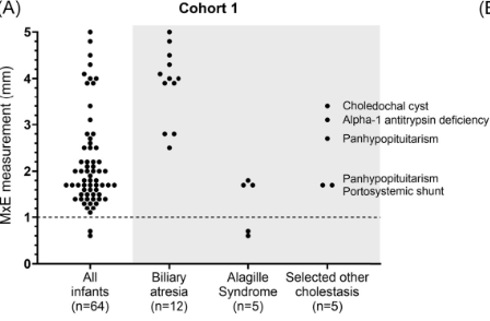
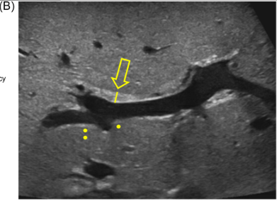
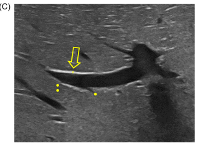
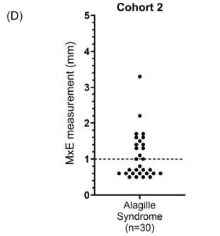
Background/Methods: One way clinicians can distinguish between biliary atresia and Alagille syndrome is with a positive “triangular cord sign.” This ultrasound finding refers to a thickened echogenicity at the anterior aspect of the right portal vein…the maximum echogenicity at the anterior aspect of the right portal vein (“maximum echogenicity” or “MxE”) was measured in a group of infants with cholestasis (Cohort 1, n=64) and in another group of infants with Alagille syndrome (Cohort 2, n=30).
Key findings:
“Thin echogenicity at the anterior aspect of the right portal vein may help distinguish between biliary atresia and Alagille syndrome…None of the 12 infants with biliary atresia in Cohort 1 had a MxE < 1.0 mm”
“A MxE < 1.0 mm could help identify Alagille syndrome. 2 of the 64 infants with cholestasis in Cohort 1 had a MxE < 1.0 mm. Both infants were eventually diagnosed with Alagille syndrome. In the Cohort 2 infants with Alagille syndrome, 16 of 30 infants had a MxE < 1.0 mm”
Infant with biliary atresia Infant with Alagille Syndrome
Discussion Point:
“Infants with Alagille syndrome can have smaller bile ducts which may be inapparent on invasive testing such as cholangiography. As a result, they may be presumptively diagnosed with biliary atresia and inappropriately treated with the Kasai portoenterostomy. Unfortunately, these infants have poorer outcomes compared to infants with Alagille syndrome who do not receive the Kasai portoenterostomy.” Thus, distinguishing Alagille from biliary atresia is very important.
My take: This study shows that MxE (a refinement of what has previously been called the triangular cord sign) on ultrasound may help distinguish biliary atresia from Alagille syndrome. As this is a single-center study, it will be important to determine if this ultrasound finding can be replicated in other centers and whether the finding is operator-dependent.
Disclaimer: This blog, gutsandgrowth, assumes no responsibility for any use or operation of any method, product, instruction, concept or idea contained in the material herein or for any injury or damage to persons or property (whether products liability, negligence or otherwise) resulting from such use or operation. These blog posts are for educational purposes only. Specific dosing of medications (along with potential adverse effects) should be confirmed by prescribing physician. Because of rapid advances in the medical sciences, the gutsandgrowth blog cautions that independent verification should be made of diagnosis and drug dosages. The reader is solely responsible for the conduct of any suggested test or procedure. This content is not a substitute for medical advice, diagnosis or treatment provided by a qualified healthcare provider. Always seek the advice of your physician or other qualified health provider with any questions you may have regarding a condition
Recently Dr. Balistreri gave our group an excellent lecture. I have taken some notes and shared some slides. There may be inadvertent omissions and mistakes in my notes.
Key Points:
Producing enough bile acids and recycling bile acids in enterohepatic circulation is crucial for bile acid flow. In addition, there are ‘good’ bile acids like cholic acid that have trophic properties and ‘bad’ bile acids like lithocholic acid that cause liver toxicity
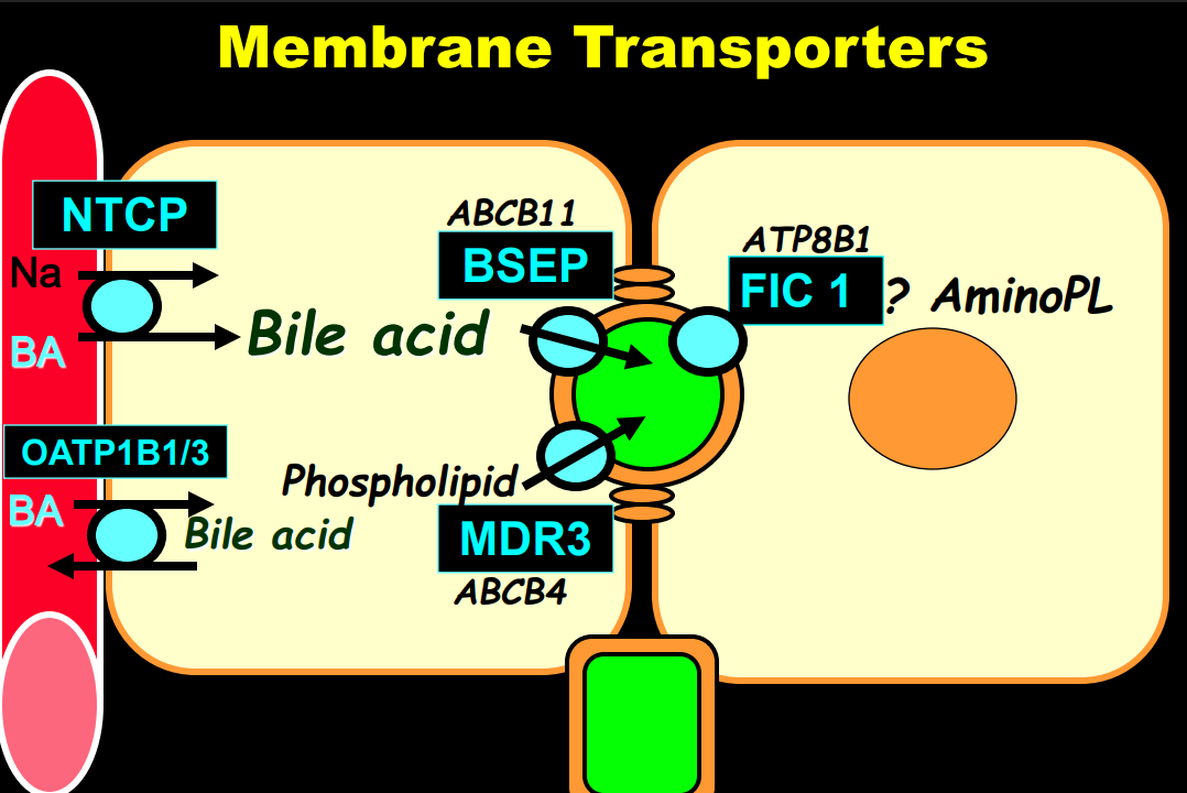
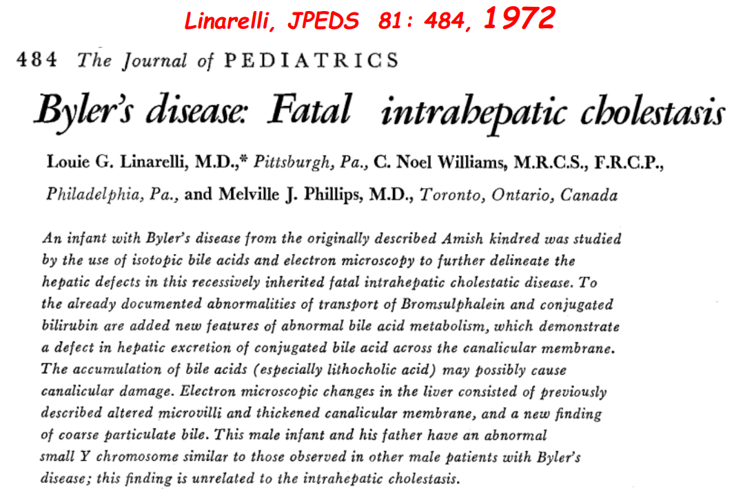
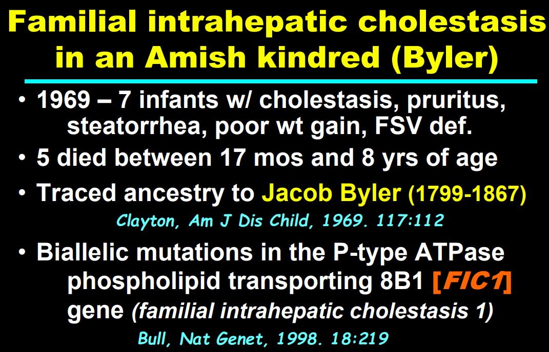
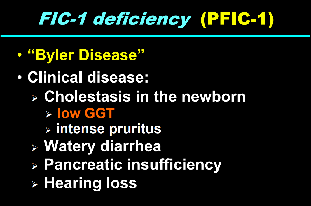
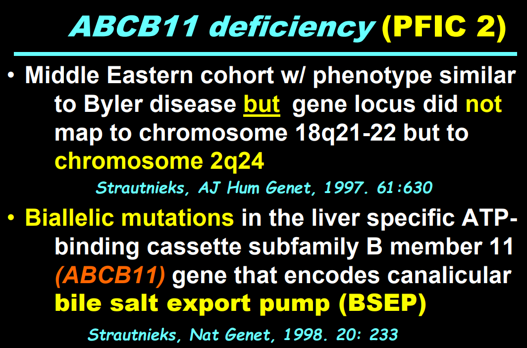
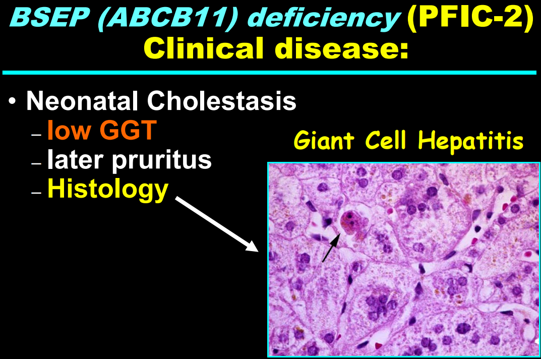
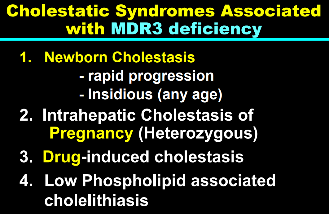
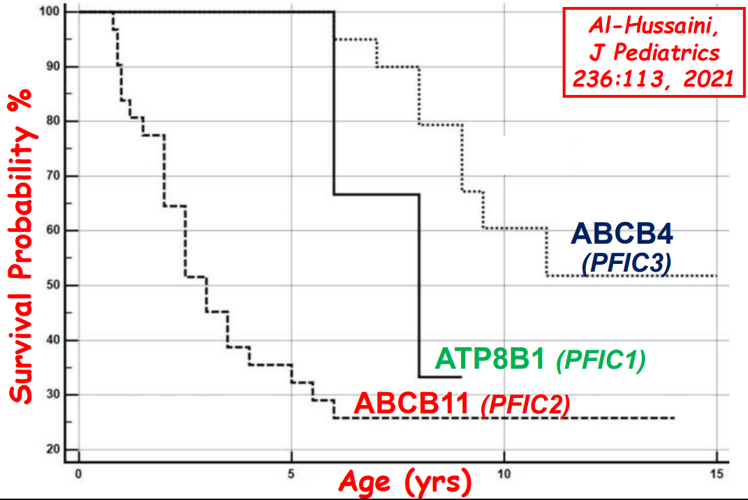
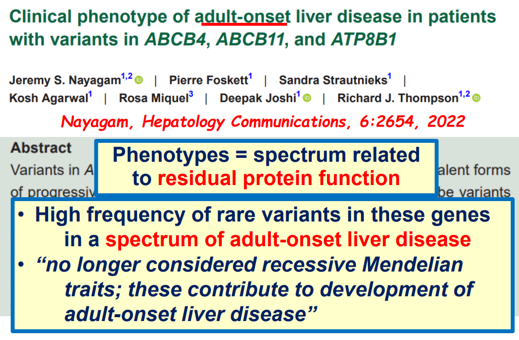
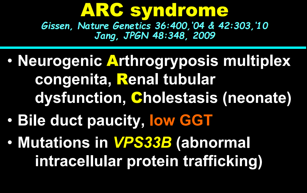
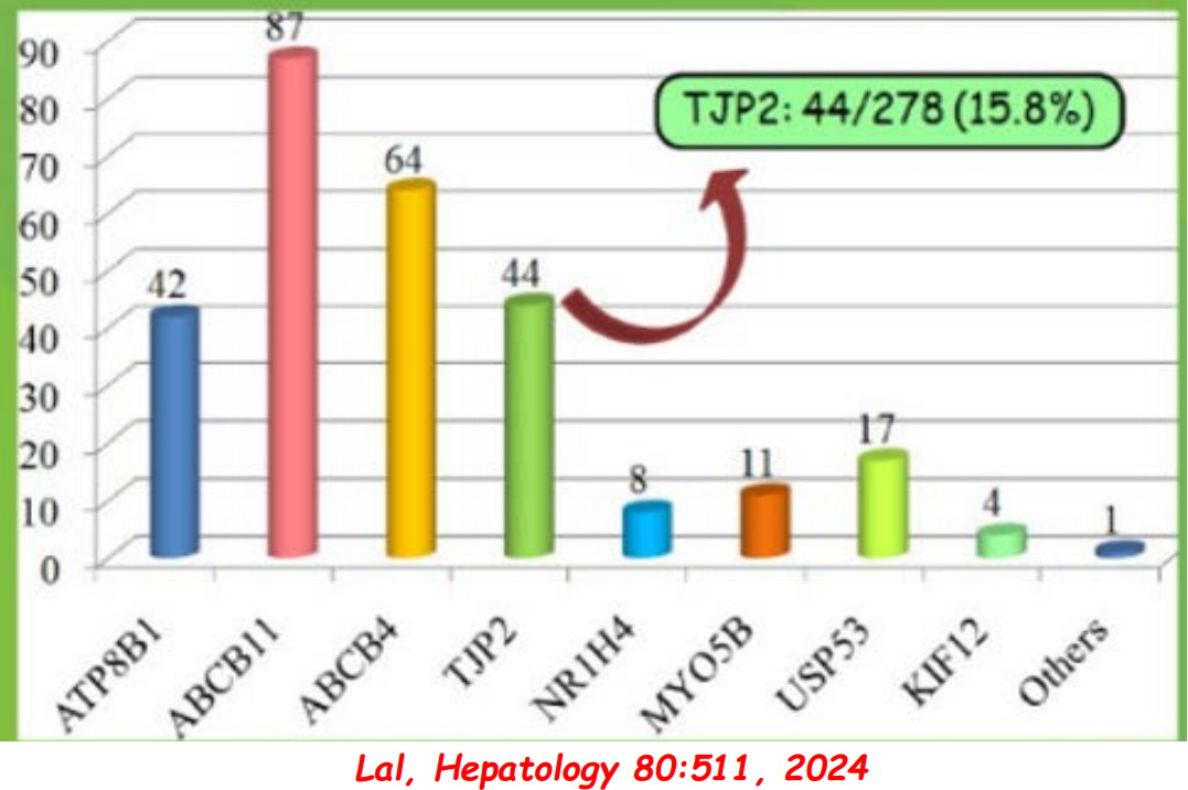
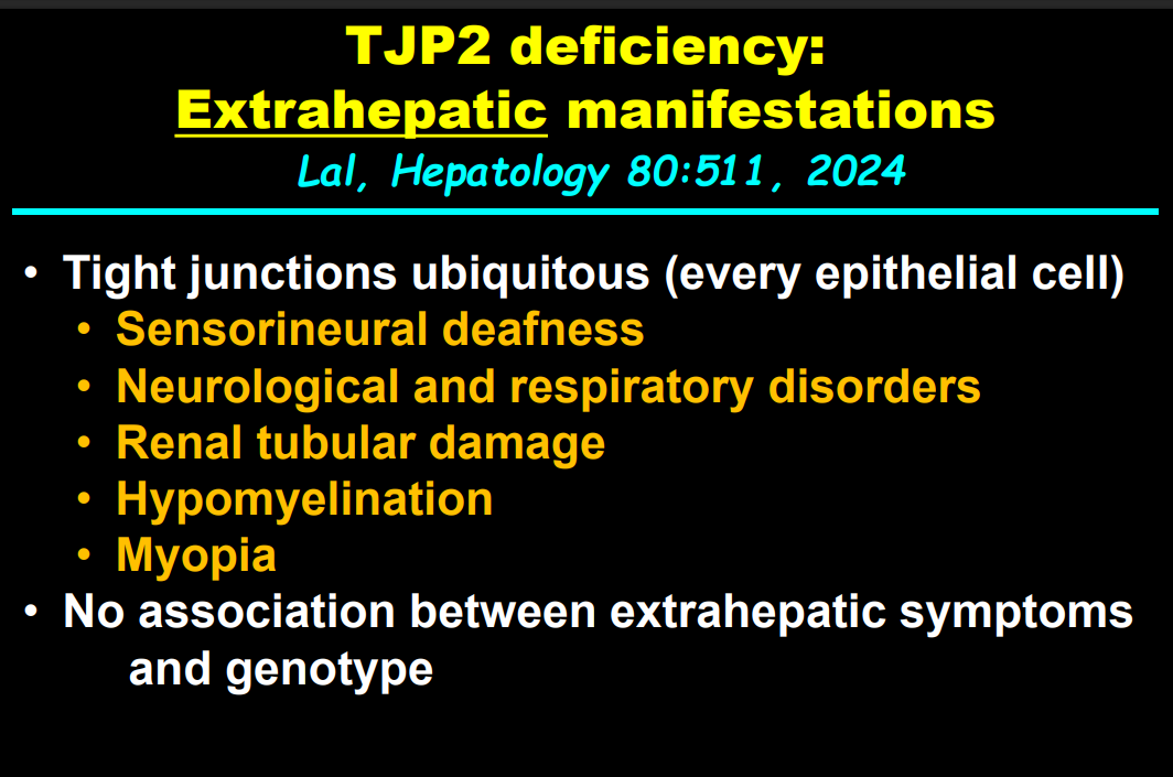
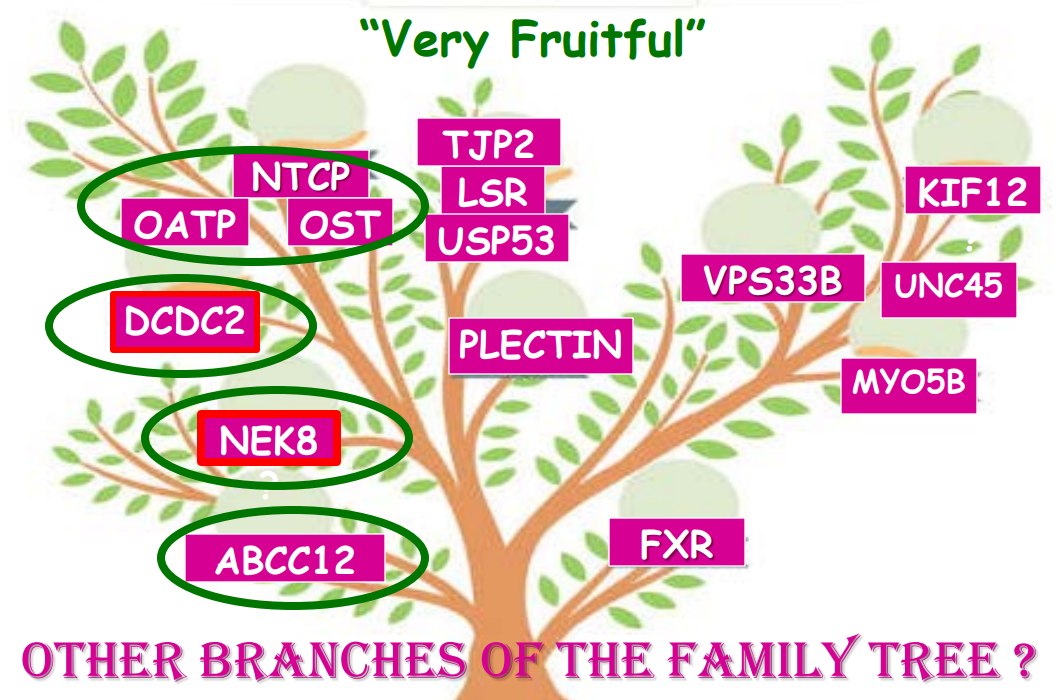
In addition to defects in the metabolic pathway of bile acids, discoveries identified defects in the membrane transporters (eg. FIC1, BSEP, MDR3), trafficking proteins (eg. MYO5B, VPS33B), nuclear control receptors (eg. FXR), and tight junction proteins (eg. TJP2). Tight junction protein defects are associated with bile leakage from bile canaliculus
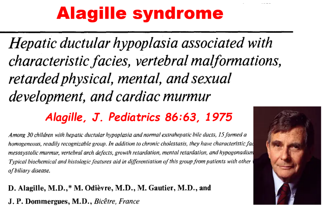
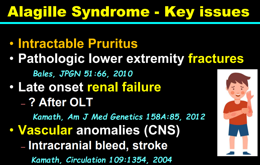
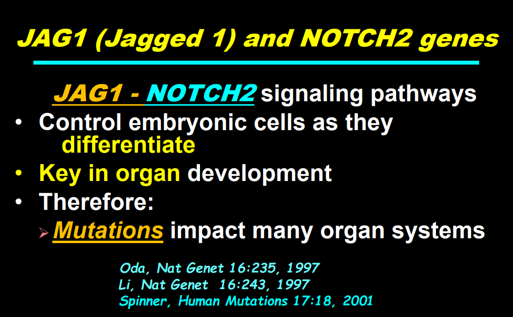
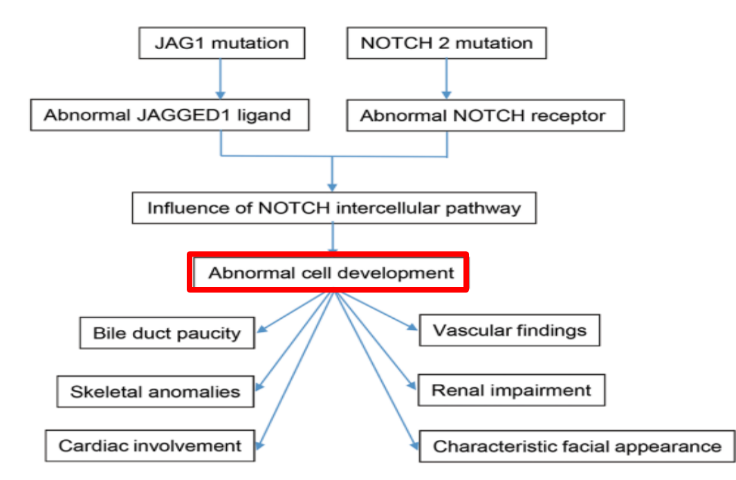
Alagille syndrome, a disorder of embryogenesis, related to JAG1-NOTCH2 signaling pathways affects organs throughout the body
Many of these genetic mutations are now being identified in adults with unexplained liver diseases (eg. intrahepatic cholestasis of pregnancy and cryptogenic cirrhosis)
Cholestasis panels and whole exome sequencing are important tools
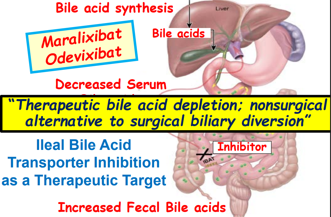
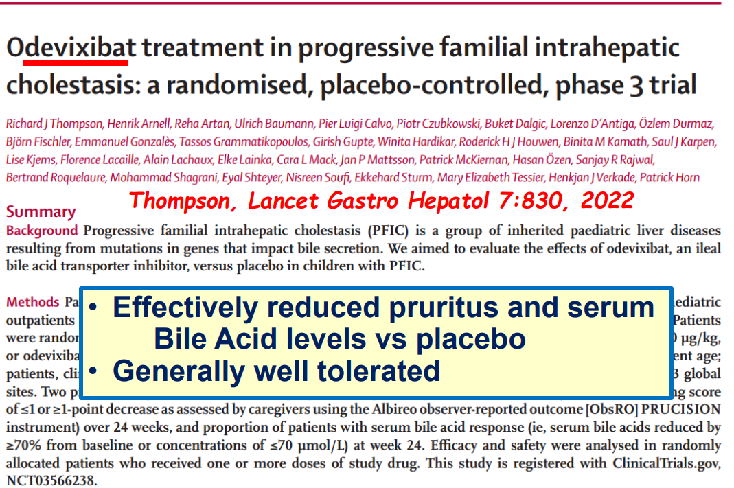
Ileal bile acid transporter (IBAT) inhibitors have emerged as important therapies for conditions like Alagille which were previously treated with biliary diversion
My take: This lecture really shows how the field of pediatric liver disease has been a puzzle. Now one can see how almost all of the pieces of the puzzle work together.
This retrospective study examined the Biliary atresia and Related Diseases (BARD) criteria for diagnosis of acute cholangitis in BA patients within the first year following Kasai hepatoportoenterostomy (HPE).
Key findings:
Of 185 consecutive BA patients, 59 (32%) had at least one episode of cholangitis within the first year after HPE
The correlation between the clinician’s impression and the standardized BARD definition was very strong (r = 0.8)
Only 41% of patients believed by their physicians to have cholangitis had fever
70% had increased WBC and/or CRP, and/or procalcitonin
90% had increased bilirubin/GGT, 68% had increased transaminases
Only one (1/59) patient in their cohort had a positive blood culture and only one (1/59) patient had bile lakes identified
56/59 children (94.9%), at least one laboratory or radiological item (group B) was pathologic at cholangitis diagnosis
My take: There really is not a precise way to diagnose cholangitis following HPE. Given how infrequently they are identified, it looks like both blood culture and bile lakes are not useful in establishing the diagnosis given. Overall, these criteria correlate well with how clinicians establish the diagnosis of cholangitis in at-risk children.
The Standardized Biliary Atresia and Related Diseases (BARD) cholangitis guidelines for the diagnosis of suspected and confirmed cholangitis within the first year following hepatoportoenterostomy for biliary atresia. *Vomiting, poor feeding, irritability. PCT, procalcitonin.
This single center retrospective study compared 20 consecutive infants underwent hepatoportoenterostomy (HPE) (beginning in 2017) for biliary atresia (BA) to a historical cohort. Analysis of successful biliary drainage 3 months after HPE (defined as serum total bilirubin (TB) <2 mg/dL) was the primary endpoint; survival with native liver at 2 years was the secondary endpoint.
Protocol:
Cefoxitin was administered to all infants following HPE for 3-4 days.
Standard protocol: If the stool color normalized (pigmented), the infant received “conventional” treatment with trimethoprim-sulfamethoxazole cholangitis prophylaxis, fat-soluble vitamin supplementation with DEKAsPlus or AquaADEKs (1 mL daily), and ursodeoxycholic acid (5 mg/kg twice daily).
Customized protocol: If the stools were acholic (or not consistently pigmented) and </=45 days, the infants received intravenous cefoxitin or piperacillin-tazobactam and methylprednisolone, initial dose 5 mg/kg/day and decreased by 1 mg/kg/day each day thru day 5; then orally treated with dose dropped 0.25 mg/kg weekly. When switched to oral steroids, IV antibiotics were stopped and infant was placed on amoxicillin-clavulanate which was continued until TB <2 mg/dL or discontinuation of corticosteroids (whichever came first).
If stools were acholic and infant was >45 days, then the same treatment was given if there was liver inflammation on histology.
Key findings:
8 had pigmented stools after HPE and received standard protocol.
12 had acholic/inconsistent stools. All of those >45 days had liver inflammation; thus, all 12 received the customized protocol. Two infants had two cycles of steroids/antibiotics who had initial response to treatment and then worsened.
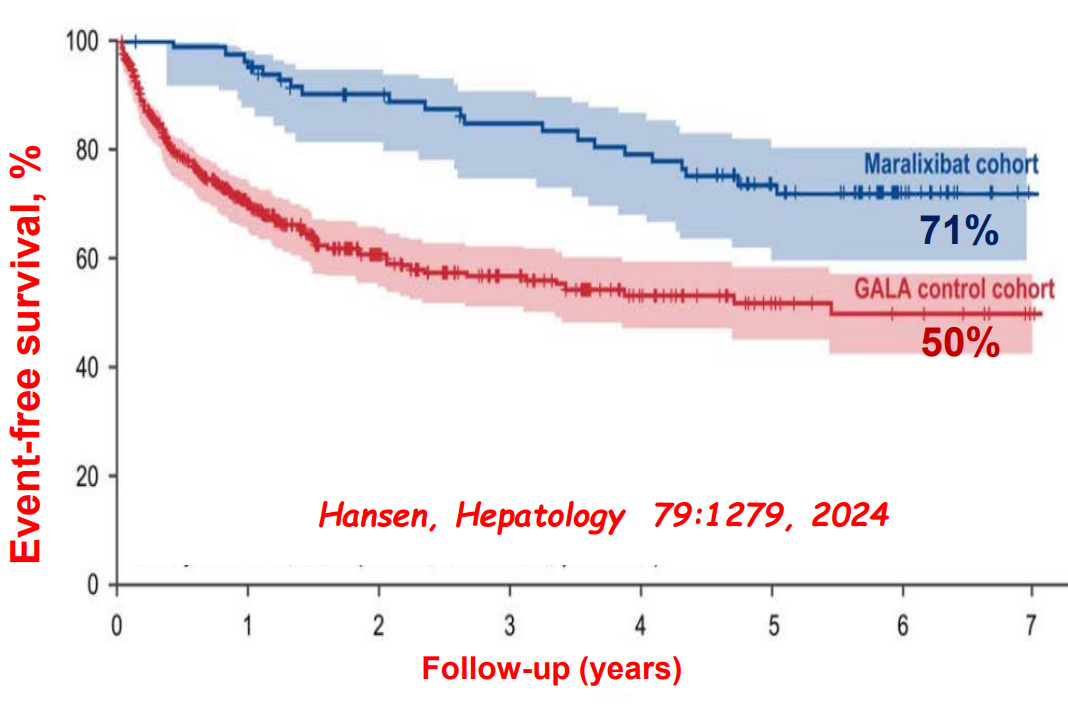
Sixteen of 20 (80%) infants had successful bile drainage, compared to 8 of 20 (40%) infants in the historical cohort (P = 0.0225)
Among the sixteen who have reached two years of age, 11 (68.8%) are alive with native livers versus 10 of 20 (50%) in the historical cohort (P = 0.0970). This did not achieve statistical significance.
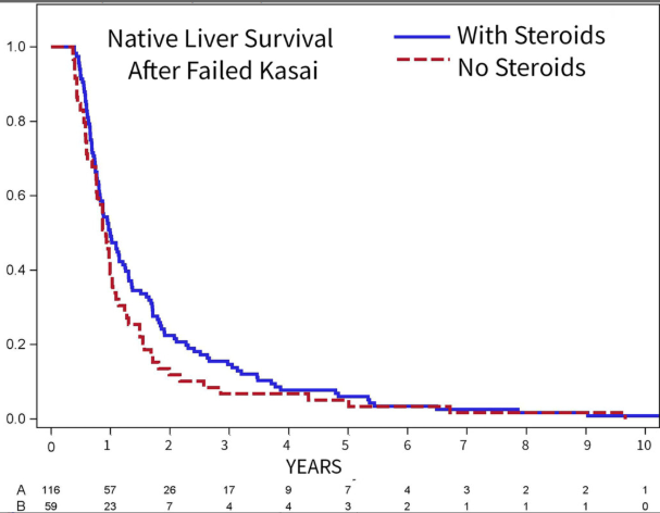
The authors established their protocol based on data from Kings College in 2016 suggesting that steroids appeared effective in younger patients who underwent HPE prior to 45 days (Peg Surg Int 2016; 32: 193-200). The START study showed no significant improvement in biliary drainage between patients receiving corticosteroids and placebo. However, in the group <70 days, 72% of infants receiving corticosteroids achieved biliary drainage compared with 57% of the placebo group (P=0.36).
My take: This is a small sample size. Perhaps, this protocol will help improve outcomes. If so, we still don’t know which factor is more important —the IV antibiotics or the high dose steroids. If these agents are helpful, are there other predictive factors –microbiome? MMP-&?
START Study: Steroids Not Effective For Biliary Atresia (After Kasai) In this study, the steroid intervention did not affect transplant-free survival which was 58.7% in the steroid group and 59.4% in the placebo group at 24 months of age. In addition, steroids were associated with an earlier onset of first serious adverse events.
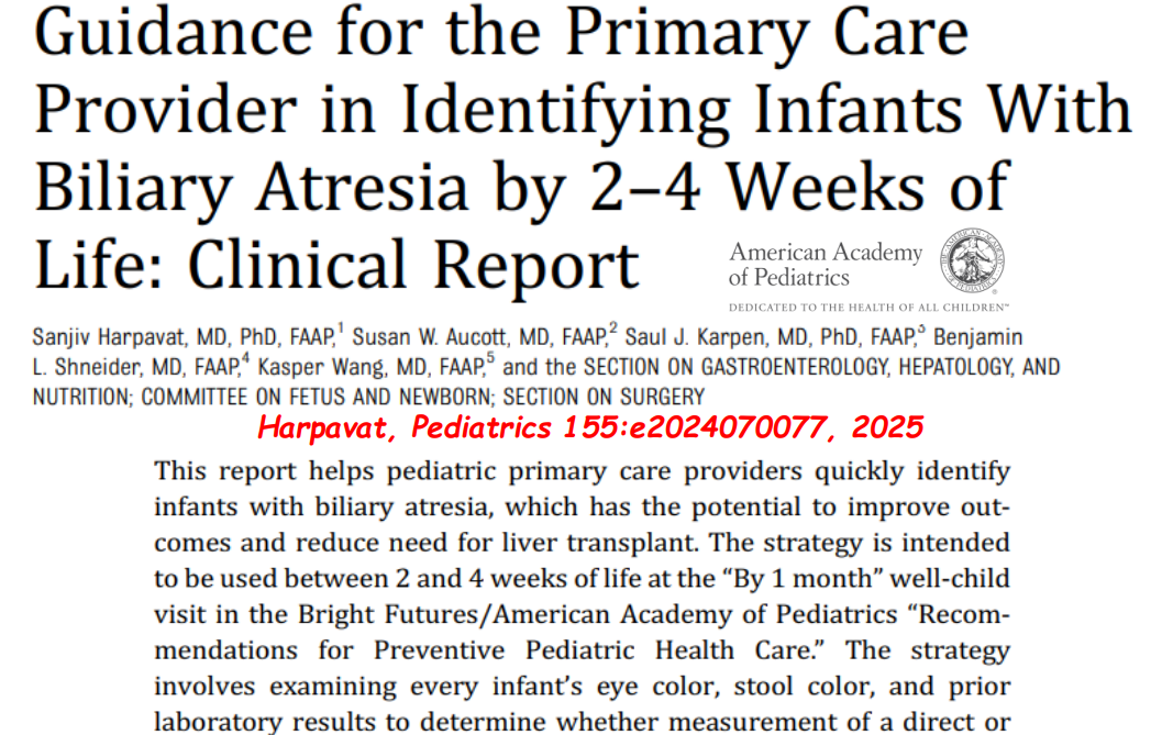
This retrospective study analyzed data from 11,965 infants who had fractionated bilirubin obtained in the nursery (2016-2019). Key findings:
DB of 0.6 mg/dL was chosen as the cut-off based on a high sensitivity (100%) and specificity (99%) for screening newborns for CLD
Out of 60 infants who met criteria for DB ≥0.6 mg/dL, only 15 (25%) had a repeat level drawn after nursery discharge; 3 (5%) were eventually diagnosed with CLD (2 with BA and 1 with Alagille syndrome)
It is fairly easy to get fractionated bilirubins on infants. Many need to get a bilirubin check and in many centers, a fractionated bilirubin is automatically generated at no additional costs. The hard part is making sure that those with abnormal values receive timely followup.
My take: It is a mistake to get fractionated bilirubins in newborns unless one has developed a plan/infrastructure to make sure those with abnormal values receive appropriate followup.
This retrospective study from Finland details the bone health of children with biliary atresia (BA). Key findings:
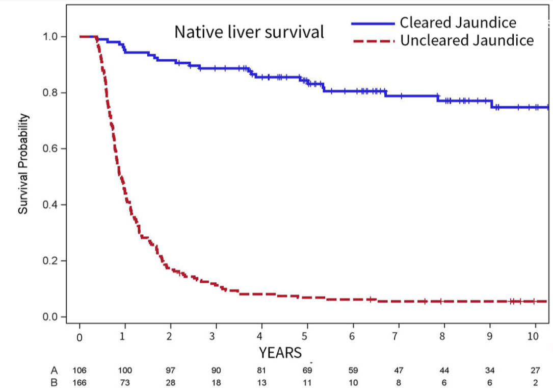
Out of 49 patients, 7 (14%) were diagnosed with rickets during infancy. Clearance of jaundice [odds ratio 0.055, 95% confidence interval [CI] 0.00266–0.393; P < 0.01] was a protective factor against rickets.
In DXA measurements, median lumbar spine aBMD anthropometrically adjusted z-scores were as follows:
in native liver survivors 0.8 (interquartile range [IQR] −1.9 to 1.4) at 5 and −0.3 (IQR −1.3 to 0.8) at 10 years
in liver transplanted patients 0.4 (IQR −0.2 to 1.1) at 5 and 0.6 (IQR −0.1 to 1.3) at 10 year.
Most BA patients have aBMD within normal range between 5 and 10 years of age irrespective of liver transplantation status.
My take: This study shows that early in life there is frequent bone impairment in children with BA. This generally improves in most children as cholestasis resolves (with or without liver transplantation).
This study, which relied on data from a pediatric database (PHIS) with 48 pediatric centers, identified 1112 subjects with biliary atresia (2004-2013).
Key findings:
Median age at time of Kasai (hepatoportoenterostomy) procedure: 63 days
Median number of admissions for cholangitis within 2 years was 2 episodes. The presence of portal hypertension (OR 2.24) and black race (OR 1.51) were associated with higher risk of cholangitis
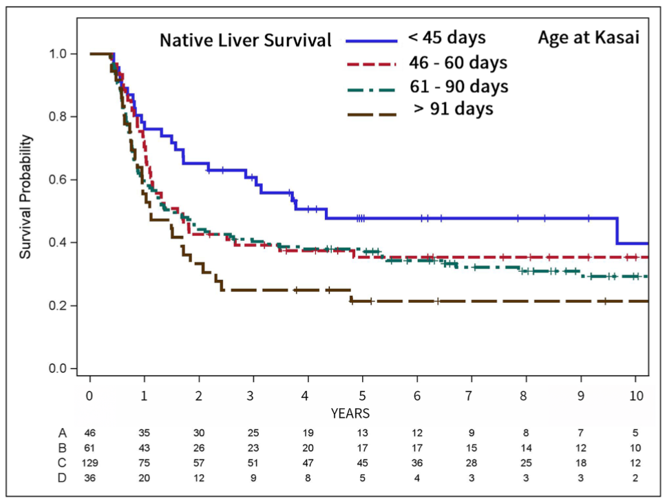
When Kasai was performed at >90 days, this lowered the likelihood of cholangitis (OR 0.46)
With regards to those with 5 or more bouts of cholangitis, risk factors included Asian ethnicity (OR 2.66), public insurance (OR 1.72), and portal hypertension (OR 2.88)
56% of patients had portal hypertension and 15.6% had esophageal varices
Neither steroids nor ursodeoxycholic acid were found to affect patient outcome
Limitations: lack of clear definition for cholangitis diagnosis and episodes of cholangitis may not have been captured if patients received care outside the participating centers
My take: Cholangitis is a common problem following hepatoportoenterostomy. Earlier diagnosis of biliary atresia provides the best opportunity for improving long-term outcomes.
Disclaimer: This blog, gutsandgrowth, assumes no responsibility for any use or operation of any method, product, instruction, concept or idea contained in the material herein or for any injury or damage to persons or property (whether products liability, negligence or otherwise) resulting from such use or operation. These blog posts are for educational purposes only. Specific dosing of medications (along with potential adverse effects) should be confirmed by prescribing physician. Because of rapid advances in the medical sciences, the gutsandgrowth blog cautions that independent verification should be made of diagnosis and drug dosages. The reader is solely responsible for the conduct of any suggested test or procedure. This content is not a substitute for medical advice, diagnosis or treatment provided by a qualified healthcare provider. Always seek the advice of your physician or other qualified health provider with any questions you may have regarding a condition.

As noted in previous post (Learning a lot from ChiLDREN (part 1) | gutsandgrowth), several recent studies highlight the benefits of multisite collaboration to study infrequent pediatric liver problems. In these studies, the Childhood Liver Disease Research and Education Network (ChiLDREN) has collected prospective longitudinal observational date from multiple centers; data from 10 centers provides useful information on the frequency of portal hypertension (PHT) in young adults with biliary atresia (BA) (JPGN 2012; 55: 57-73).
163 subjects were enrolled between May 2006 and December 2009. Seven patients were excluded due to the presence of polysplenia which interferes with the assessment of PHT.
Demographics: subjects ranged in age from 1 to 25 years with a mean of 9.2 years. 56% were female, 75% were caucasian.
PHT was considered definite if there was either a history of a PHT complication (variceal bleeding, ascites) or if there were clinical findings c/w PHT (both splenomegaly and thrombocytopenia). PHT was considered “possible” if either splenomegaly or thrombocytopenia was present and “absent” if no criteria were met.
PHT: definite in 80 (49%), possible in 27 (17%) and absent in 56 (34%)
43 subjects had a history of PHT complications: 32 with esophageal variceal (EV) bleeding, 14 with ascites, and 8 with hepatopulmonary syndrome.
Of the patients with EV, only 3 had normal platelet count and normal spleen size.
Teaching points:
One-third of subjects with EV bleeding survived with their native liver for at least 5 years.
EV age of onset was highly variable; 7 had bleeding in the first two years of life.
Growth parameters were fairly unremarkable in those with definite PHT.
Long-term followup will be needed to identify factors which predict progression of PHT and the development of adverse outcomes.
Biliary atresia (BA) is often suspected among infants with prolonged jaundice. In fact, efforts have been underway for a long time to encourage fractionation of the bilirubin values to look for conjugated hyperbilirubinemia, especially in infants that remain jaundiced at three weeks of life. While this is still good advice, given the lack of success in implementation, there is good evidence that obtaining a fractionated bilirubin at any time point can help identify cholestasis associated with BA.
A recent article by Karpen et al (Pediatrics 2011; 128:. e1428 -e1433) indicates that direct bilirubin values are elevated beginning within the first one to two days in patients with BA. In their cohort of 61 BA subjects, 56% had newborn fractionated bilirubin values. Every BA patient had elevated direct bilirubin, on average 1.4 ± 0.43 mg/dL (normal <0.5) (compared with control patients: 0.19 ± 0.075 mg/dL, P < .0001). Also, another important finding was that early on the ratio of direct bilirubin to total bilirubin was normal in 79%; normally this ratios is ≤0.2. As such, all patients with increased direct bilirubin need to be followed closely.