Lower Endoscopic Thresholds for Thrombocytopenia

According to an advances in endoscopy report (Ross, WA. Gastroenterology Hepatology 2015; 11: 115-17), lower platelet thresholds are indicated for many endoscopic procedures.  The author works at MD Anderson Cancer Center in Houston.

Key points:

  • “We feel that the traditional threshold of 50,000 platelets/microliter that many doctors adhere to or aim for should be put aside, and a lower platelet threshold of perhaps 25,000 or 30,000 platelets/microliter should be employed for endoscopic procedures, including biopsies.“
  • “We found that therapeutic maneuvers could be performed to control bleeding.”
  • “This change would require fewer platelet transfusions to prepare a patient for endoscopy.”
  • Based on their published experience (Krishna SG, et al. “Saftey of endoscopic interventions in patients with thrombocytopenia.” Gastrointest Endosc 2014; 80: 425-34), the author notes that “polypectomy could probably be performed with a platelet count under 50,000/microliter, likely in the 30,000-40,000/microliter range, particularly if the polyp was small (<10 mm).”  They caution that cold snare technique may be safer in this setting but is not suitable for larger polyps.
  • Other preventative measures include stopping aspirin use, limiting the number and size of biopsies, and using non thermal means to help stop bleeding, such as clips or injections.
  • “Performing an endoscopic procedure in a patient with an extremely low platelet count, such as 5000/microliter, is associated with a high risk of bleeding.”

“If the procedure is just a simple biopsy, a platelet count of 25,000/microliter to 30,000/microliter should suffice.”

Take-home message: While the data that the author references is derived from adults, it is likely that in pediatrics that endoscopy, if needed, can be performed in patients with platelet counts less than 50,000/microliter.

More on Adalimumab (Humira) in Pediatrics

A recent ‘observational cohort’ study (Cozijnsen M, et al. JPGN 2015; 60: 205-10) provides more information regarding the use of adalimumab in pediatrics.  The authors attempted to identify all Dutch patients <18 years of age with Crohn’s disease who received adalimumab after infliximab therapy.  Excluded patients were those with “bad treatment adherence,” conflicting comorbidity, and previous participation in prospective study by Hyams et al Gastroenterol 2012; 143: 365-74 (Related blog post: Adalimumab for children with Crohn’s disease | gutsandgrowth).

Key findings:

  • 53 patients met the inclusion criteria.  12 received monotherapy with adalimumab; cotherapy in 21 with thiopurines, 11 with methotrexate, 7 with steroids, and 2 with enteral nutrition.
  • Median followup was only 12 months.
  • Remission noted in 34 (64%) patients based on either wPCDAI or PGA (physician global assessment) after a median of 3.3 months.  Remission was durable in 50% of these patients for 2 years.
  • Adalimumab failure was noted in 18 (34%).  Only 3 patients in this study were primary infliximab nonresponders and only 1/3 responded to adalimumab.
  • One serious adverse event (infection) was reported.

One weakness of this study (& many others) is its reliance on clinical disease activity indices rather than more precise measures of mucosal healing.

Take-home point: This small study provides some information about the utility of adalimumab in clinical practice in pediatrics.  As noted in other studies, those with a loss of response to infliximab, rather than primary nonresponders, appear to have a more favorable response to adalimumab. In addition, the glaring weakness in this study (i.e. small number of participants) validates the rationale behind efforts like ImproveCareNow which can generate information quickly from a large patient population.

Related study: “Levels of Drug and Antidrug Antibodies are Associated with Outcome of Interventions After Loss of Response to Infliximab or Adalimumab.” Yanai H, et al. Clin Gastroenterol Hepatol 2015; 13: 522-30. Retrospective study of 247 adult and pediatric patients. Key findings:

  • Patients with adequate trough levels (>4.5 mcg/mL for adalimumab, >3.8 mcg/mL for infliximab) “identified patients who failed to respond to an increase in dosage or a switch to another anti-TNF agent with 90% specificity.”
  • “Levels of antibody against adalimumab >4 mcg/mL-eq or antibodies against infliximab >9 mcg/mL-eq identified patients who did not respond to an increased drug dosage with 90% specificity.” These patients were likely to benefit by switching to an alternative anti-TNF.

Related study: Clin Gastroenterol Hepatol 2015; 13: 539-47.  Trough levels of infliximab (>3 mcg/mL) at week 30 was associated with improved outcomes, including mucosal healing and corticosteroid-free remission.

Briefly noted from ImproveCareNow: Dotson JL et al. “Feasibility and Validity of the Pediatric Ulcerative Colitis Activity Index in Routine Clinical Practice.” JPGN 2015; 60: 200-04.  This study, with 2503 patients, found that the PUCAI was completed in 96% if visits.  The PUCAI correlated with PGA chain scores.

Related blog posts:

Don’t be Fooled About Withdrawing Immunomodulator Cotherapy -Look Past the Headline

The coverage on a recent study (Clinical Gastroenterology and Hepatology 2015: 13(3): 514-521.e4) suggests that it should be fine to stop immunomodulator co-therapy.  I recommend reading the entire study (or at least this blog post)–you will probably come to a different conclusion.

“The addition of immunomodulators increases the efficacy of maintenance therapy with infliximab for up to one year in patients with Crohn’s disease who have not been previously treated with immunomodulators. However, there are questions about the effect of withdrawing immunomodulator therapy from these patients. David Drobne and colleagues studied the effects of treatment with infliximab and immunomodulators (co-treatment) and then immunomodulator withdrawal on long-term outcomes of patients, as well as trough levels of infliximab and formation of anti-infliximab antibodies (ATI). Reporting in Clinical Gastroenterology and Hepatology, they find that, in a retrospective analysis, withdrawal of immunomodulators after at least six months (median, 13 months) of co-treatment with infliximab does not reduce the trough levels of infliximab in patients with Crohn’s disease. Detectable trough levels of infliximab at the time of immunomodulator withdrawal are associated with long-term response.”

Clinical Gastroenterology and Hepatology 2015: 13(3): 514-521.e4

Some additional details:

This was a retrospective open-label cohort study with 223 patients and median followup of 34 months. At baseline, 65 received infliximab (IFX) monotherapy and 158  received co-therapy with an immunomodulator (46 methotrexate, 112 thiopurine).  Immunomodulators were withdrawn “only in patients with durable response (ongoing clinical benefit with lasting disease control with low C-reactive protein [CRP] [below 10 mg/L]).”  Among the 158 on co-therapy, 117 reached a durable response and had withdrawal of immunomodulator after >6 months of combination therapy (median time 13 months).

Key findings:

  • At baseline, co-therapy patients, compared to monotherapy patients, had higher IFX trough levels (adjusted mean increase of 1.44-fold) and lower likelihood of antibodies to infliximab (ATI): 35/158 (22%) compared with 25/65 (38%), P=.01.
  • When immunomodulator was withdrawn, IFX levels remained stable: before 3.2 mcg/mL compared with after 3.7 mcg/mL. However, 45 of 117 (38%) required increasing doses of IFX and 21 of 117 (18%) discontinued IFX.
  • Trough levels of IFX and CRP  were most strongly associated with response to IFX dosing on monotherapy.
  • “Only 9 of 74 patients (12%) with detectable IFX trough levels at the time of immunomodulator withdrawal developed undetectable IFX trough levels during the subsequent follow-up.”
  • None of the 27 patients with IFX trough level >5 mcg/mL at time of immunomodulator withdrawal lost response to IFX during median follow-up of 29 months.

Though the headlines covering this article have suggested that IFX levels will stay stable when immunomodulators are withdrawn after >6 months, the authors proposed algorithm only recommends withdrawal for those with IFX trough level >5 mcg/mL.  In addition, the data showed that a large number of patients required dose escalation and/or lost detectable IFX levels. Despite their proposed algorithm to withdraw in this small group, the authors further backtrack in their conclusion:  “a prospective parallel group trial during a period of 5-10 years in a large group of patients is required to ascertain the real long-term benefit to risk ratio of continuing combined infliximab and immunomodulator treatment.”

Bottomline: If a patient is doing well, withdrawing immunomodulator co-therapy still has risks. I worry that the misleading reporting of this article will result in detrimental outcomes.

Related blog posts:

Disclaimer: These blog posts are for educational purposes only. Specific dosing of medications (along with potential adverse effects) should be confirmed by prescribing physician.  This content is not a substitute for medical advice, diagnosis or treatment provided by a qualified healthcare provider. Always seek the advice of your physician or other qualified health provider with any questions you may have regarding a condition.

 

Methotrexate Dosing in Dual Therapy

A recent retrospective study (DOI: http://dx.doi.org/10.1093/ecco-jcc/jjv027 first published online: 23 January 2015 -reference from KT Park’s twitter feed) has suggested that high-dose methotrexate (MTX) (15-25 mg/week) is more effective than low-dose MTX (≤12.5 mg/week) as part of dual therapy for inflammatory bowel disease.

Here’s the abstract: Optimal Doses of Methotrexate

Background and Aims: Methotrexate is sometimes used as part of combination therapy for the treatment of inflammatory bowel disease (IBD), however the optimal MTX dose for combination therapy has not been established. This study compared the efficacy of lower dose and higher dose methotrexate with anti-TNF therapy among IBD patients.

Methods: Retrospective chart review was performed of 88 IBD patients at our center between 2010-2013. Low-dose methotrexate was defined as ≤ 12.5mg/week and high-dose methotrexate as 15-25mg/week. Patients who met the criteria for clinical remission (Harvey-Bradshaw Index ≤ 4, Simple Clinical Colitis Activity Index ≤ 2) at baseline were followed for up to 42 months. Chart review occurred in six-month intervals. The primary outcome was consecutive months in remission prior to relapse. Secondary outcomes included other indicators of worsening disease (endoscopic inflammation, steroid use, therapy escalation/addition, or surgery) and adverse events. Regression analysis and Kaplan-Meier survival analysis were completed.

Results: We identified 73 (83%) dual-therapy patients, of which 32 low-dose and 14 high-dose individuals achieved remission. When compared with high-dose patients, low-dose patients were more likely to relapse (log-rank test, P<0.01). Secondary indicators of worsening disease occurred during 34.4% of low-dose review periods and 31.4% of High-dose review periods (P=0.67). 3/52 (6%) low-dose patients and 3/21 (14%) high-dose patients (P=0.34) discontinued methotrexate therapy due to adverse events.

Conclusions: When combined with anti-TNF therapy, methotrexate at doses of >12.5mg/week were more effective at maintaining clinical remission than lower doses. These findings will guide management of combination therapy in IBD patients.

Related blog posts:

Emigration -One Way to Acquire Inflammatory Bowel Disease

A recent study (Shitrit AB, et al. Inflamm Bowel Ds 2015; 21: 631-35) highlights the phenomenon of acquiring inflammatory bowel disease (IBD) by moving from a non-developed country to a developed country; the implication is that the changes in environment and diet predispose towards the development of IBD.

This study examined Ethiopian Jews who migrated to Israel.  Using a case-control study, the authors compared 32 Ethiopian immigrants to 33 Ashkenazi patients with IBD.

Key findings:

  • No Ethiopian immigrants had a positive family history compared with 42% of Ashkenazi group.
  • Crohn’s disease was more prevalent in the Ethiopian immigrants: 94% versus 73%.
  • The Ethiopian immigrants lived in Israel for at least 8 years before developing IBD an da median duration of 13 years.

The study discusses the difficulty of diagnosing IBD in rural Africa but speculates that rather than an underdiagnosis of IBD, it is likely to have a true low prevalence of IBD.

Take-home message: It takes many years for the environment exposures to allow for the development of IBD.  Additional work is needed to establish the clinical, genetic, and microbial factors that influence the acquisition of IBD in immigrants to developing countries.  Understanding the susceptibility of immigrants would have widespread application.

Related blog posts:

How Common are Clostridium difficile infections?

In 2011, Clostridium difficile was estimated, by a recent report from the CDC (NEJM 2015; 372: 825-34) to result in 453,000 infections in the U.S. based on surveillance data from 10 geographic regions.

Other key findings:

  • Approximately 29,000 deaths were attributed to Clostridium difficile infections
  • 65.8% of cases were health care associated.
  • 24.2% had onset in the hospital
  • Increased risk was particularly notable among the elderly, with an rate ratio of 8.65 in those 65 years of age or older
  • The aggressive NAP1 strain was evident in 30.7% of health-care associated infections compared with 18.8% of community-associated infections

Related posts:

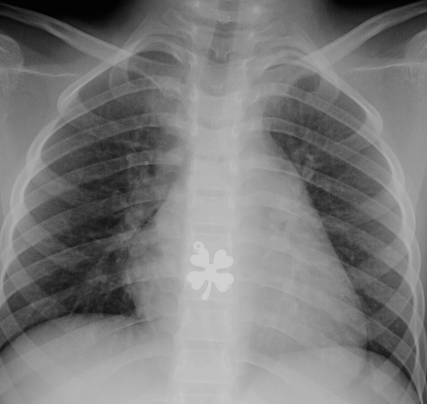
Note to blog followers: Yesterday’s blog was retitled to “Radiologic Image for St. Patrick’s Day” rather than “Endoscopic Image for St. Patrick’s Day”

Withdrawing Therapy Leads To Relapse, Even if in Deep Remission

A recent study, presented as an oral abstract (thanks to Jeff Lewis for forwarding this reference), indicates that even in patients in deep remission, withdrawal of anti-TNF therapy leads to relapse in about 50% even when thiopurines are continued; this is in agreement with previous posts (see below).

Full abstract: OP007 Relapse after Deep Remission in Crohn’s disease. Here are the results and conclusion from the abstract:

Results

Sixty one patients were included and followed-up for a median of 28 months (range 7-47). After withdrawal of anti- TNFa therapy (44 infliximab and 17 adalimumab) 47 (77%) patients continued thiopurines. 32 (52.5%) patients relapsed until the end of follow-up with a median time to relapse of 8 months (range 1-25). The cumulative probability of maintaining remission was 82% at 6 months, 59% at 1 year and 51% at 2 years. Analysis of 28 patients who were in deep remission (endoscopic healing; faecal calprotectin <150mg/kg; CRP <5mg/l) revealed no better survival (82%, 64% and 40% at 6 months, 1 and 2 years, respectively). Four (8%) of relapsing CD patients required surgery 5 to 19 months after anti-TNFa cessation (2 for new stricture development, 1 for medically refractory flare and 1 for high grade dysplasia). In multivariate model only disease localization was risk factor of disease relapse (colonic vs. ileal/ileocolonic: OR 0.16, 95%CI: 0.03-0.72; p=0.02). Type of anti- TNFa preparation, smoking, disease behaviour, corticosteroid or thiopurine therapy, biological markers and anti-TNFa trough levels did not impact disease relapse.

Conclusion

Approximately half of CD patients relapsed within 2 years after anti- TNFa discontinuation despite being in endoscopic remission when anti-TNFa was stopped. The highest relapse rate was observed during the 1st year. Ileal disease increased the risk of disease flare, while no other risk factor was identified.

Related blog posts:

Bryce Canyon

Bryce Canyon

Breath Testing for Bacterial Overgrowth

A recent review (Clin Gastroenterol Hepatol 2014; 12: 1964-72) provides useful advice on testing for bacterial overgrowth and explains the limitations/obstacles in determining whether bacterial overgrowth is present.

Their recommendations:

  • Preparation: avoid antibiotics for 4 weeks prior to testing, avoid bismuth for 2-4 weeks, avoid probiotics for 2-3 weeks before testing, avoid consumption of non absorbable carbohydrates (eg. pasta, bread, fiber cereal, beans) the night prior
  • Glucose substrate for hydrogen breath testing is likely most suitable.  50 g in 250 mL.  Check baseline and then every 15 minutes over 120 minute testing interval.  Positive study: increase of 12 ppm (or more) over baseline and/or baseline >20 ppm (if proper test conditions)
  • Glucose breath test (GBT): sensitivity 20-93%, specificity 30-86%.  False-positive results can occur with rapid small-bowel transit.  GBT may not detect distal small bowel bacterial overgrowth.  Lactulose breath test (LBT) generally has lower specificity.
  • If breath testing is not available, small-bowel aspiration for quantitative culture is a reasonable consideration (challenging methodology).  Alternatively, a trial of empiric antibiotics may be considered if pretest probability is high.

Related blog post:

In NASH, is ALT Wrongly Used as a Marker of Liver Injury?

According to a recent report (Hepatology 2015; 61: 153-60), elevation of alanine aminotransferase (ALT) which is frequently used as an indicator to select patients for further investigations (eg. liver biopsy) is NOT a good indicator of liver parenchymal injury in patients with nonalcoholic fatty liver disease (NAFLD).

The researchers enrolled 440 patients and divided them into three groups: no NAFLD (n=60), NALFLD with normal ALT (n=165), and NAFLD with elevated ALT (n=215). The patients were overweight/obese patients prospectively recruited from newspaper ads, general medicine clinics and hepatology clinics at several VA hospitals. Those with history of alcohol abuse were excluded.

Numerous investigations were performed including liver fat by proton magnetic resonance spectroscopy (H-MRS), liver biopsy (n=293), and insulin resistance measurements.

Key findings:

  • NAFLD & NASH patients with elevated ALT had higher liver triglyceride content (P<0.0001), worse adipose tissue insulin resistance (ATIR) (P<0.0001), and lower plasma adiponectin (P<0.05).
  • Steatosis was worse on liver biopsy in those with NASH and elevated ALT (P<0.0001).
  • There were no differences in liver inflammation (P=0.62), ballooning (P=0.13), or fibrosis (P=0.12). Thus, patients with normal versus elevated ALT had similar severity of NASH liver histology.

Take-home message:  In adults, ALT values are “poor surrogate markers of disease activity” in NAFLD.  ALT values, in these patients, are indicative of metabolic disease.  Given the staggering numbers of individuals, adults and children, with fatty liver disease, the lack of simple screen tool is quite problematic.  Equally problematic is a lack of a simple treatment approach.

Related blog posts: