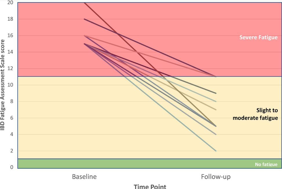
Lean Patients with Inflammatory Bowel Disease Have Higher Risk of Steatotic Liver Disease Than Lean Patients Without IBD

SJ Martinez-Dominguez et al. Inflammatory Bowel Diseases, Volume 30, Issue 8, August 2024, Pages 1274–1283https://doi.org/10.1093/ibd/izad175 Open Acess! Inflammatory Bowel Disease Is an Independent Risk Factor for Metabolic Dysfunction–Associated Steatotic Liver Disease in Lean Individuals 

Methods: This was a cross-sectional, case-control study including 300 lean cases with IBD and 80 lean controls without IBD, matched by sex and age (median age ~45 yrs). All participants underwent a liver ultrasound, transient elastography, and laboratory tests. All patients with current or previous use of systemic steroid in the last 2 years were excluded from the analysis

Key Findings:

  • The lean IBD group showed a significantly higher prevalence of MASLD compared with lean non-IBD group (21.3% vs 10%; P = .022), but no differences were observed in the prevalence of significant liver fibrosis (4.7% vs 0.0%; P = 1.000)
  • No differences were found between the prevalence of MASLD in IBD and non-IBD participants who were overweight/obese (66.8% vs 70.8%; P = .442)
  • IBD was an independent risk factor for MASLD in lean participants (odds ratio [OR], 2.71) after adjusting for classic metabolic risk factors and prior history of systemic steroid use
Prevalence of metabolic dysfunction–associated steatotic liver disease (MASLD) in cases and controls according to body mass index (BMI) status. Blue bars: cases (inflammatory bowel disease). Red bars: controls (non–inflammatory bowel disease). P values in bold indicate statistical significance (P < .05).

My take: This study suggests that “chronic inflammation could play a role in MASLD development.” Also, this indicates that MASLD could be a reason for elevated LFTs in patients with IBD, even in lean patients.

Related blog posts:

Fibrosis and Steatotic Liver Disease -Who Needs to be Followed by Hepatology?

N Ma et al. JPGN 2024; 79:229–237. Fibrosis and steatotic liver disease in US adolescents according to the new nomenclature

Methods: Among 1410 adolescents (12–19 years) in NHANES (2017-March, 2020), the controlled attenuation parameter (CAP) of transient elastography (TE) was used to define steatosis and fibrosis (TE ≥ 7.4 kPa). Obesity and alanine aminotransferase (ALT) ≥ 80 U/L were used to identify adolescents qualifying for hepatology referral according to practice guidelines.

Key findings:

  • At the supplier (EchoSens)-recommended CAP threshold of 240 dB/m, 30.5% of adolescents had steatotic liver disease (SLD) and about 85% of adolescents with NAFLD met criteria for MASLD. At a CAP threshold of 270 dB/m, SLD prevalence was about 16% in adolescents. The other 15% of NAFLD patients do not meet diagnostic criteria MASLD and would receive a diagnosis of cryptogenic SLD or possible MASLD
  • At higher CAP thresholds, MASLD/NAFLD concordance increased and approached 100%.
  • Among adolescents with MASLD-fibrosis, only 8.8% had overweight/obese and ALT ≥ 80 U/L. Thus, more than 90% of adolescents in this group would not merit hepatology evaluation based on current guidelines.

My take: This study identifies potential problems with current thresholds for which patients need to be seen by pediatric hepatologists. This will be even more important as effective pharmaceuticals become available.

Related blog posts:

Channel Islands off the California coast

Selecting Patients with Biliary Atresia for Variceal Endoscopy Screening

Y-C Ling et al. JPGN 2024;79:222–228. Performance of Baveno VII criteria for the screening of varices needing treatment in patients with biliary atresia

Methods: This retrospective study enrolled 48 BA patients (23 females and 25 males) who underwent an esophagogastroduodenoscopy (EGD) and transient elastography at a mean age of 11.18 ± 1.48 years. Transient elastography (Fibroscan® 502 Touch; Echosens) was applied for the LSM assessment in all BA patients recruited in this study.

Clinically-significant portal hypertension (CSPH) of Baveno VI criteria recommend avoiding upper endoscopies for cirrhotic patients with liver stiffness <20 kPa and platelets>150 × 10-9 cells/L (favorable Baveno VI status), and the CSPH of the expanded Baveno VI criteria as the exclusion of subjects with LSM < 25 kPa and platelet count >110 × 10-9 cells/L. (Ref: D Thabut et al. Gastroenterol 2019. Validation of Baveno VI Criteria for Screening and Surveillance of Esophageal Varices in Patients With Compensated Cirrhosis and a Sustained Response to Antiviral Therapy)

CSPH of Baveno VII criteria was defined as LSM ≥ 25 kPa and excluded patients with LSM < 15 kPa and platelet count ≥150 × 10-9 /L. Subjects with LSM between 20 and 25 kPa and platelets <150 × 10-9 /L or LSM between 15 and 20 kPa and platelets <110 × 10-9/L are also defined as CSPH. (Ref: Baveno VII criteria Ref: M Mendizabal et al. Annals of Hepatology; 2024: 29: 101180. Evolving portal hypertension through Baveno VII recommendations)

Key findings:

  • The sensitivity and negative predictive value of Baveno VI and Baveno VII criteria for the prediction of varices needing treatment (VNT) in BA patients were both 100% and100%, respectively

In the discussion, the authors note that the utility of the Baveno VII criteria for adults. “The real‐world data showed the CSPH defined by Baveno VII criteria predicts a five‐times increase in the risk of liver decompensation in chronic active liver disease patients.”

My take: This study shows that the combination of LSM and platelet counts using the Baveno VI or VII criteria help select patients with BA who need upper endoscopy to screen for varices needing treatment. These criteria also identify patients needing liver transplantation.

Related blog posts:

Channel Islands off California coast
View from Griffith Park, Los Angeles

Disclaimer: This blog, gutsandgrowth, assumes no responsibility for any use or operation of any method, product, instruction, concept or idea contained in the material herein or for any injury or damage to persons or property (whether products liability, negligence or otherwise) resulting from such use or operation. These blog posts are for educational purposes only. Specific dosing of medications (along with potential adverse effects) should be confirmed by prescribing physician.  Because of rapid advances in the medical sciences, the gutsandgrowth blog cautions that independent verification should be made of diagnosis and drug dosages. The reader is solely responsible for the conduct of any suggested test or procedure.  This content is not a substitute for medical advice, diagnosis or treatment provided by a qualified healthcare provider. Always seek the advice of your physician or other qualified health provider with any questions you may have regarding a condition.

ENTERPRET: Vedolizumab Optimization Study in Ulcerative Colitis

V Jairath et al. Clin Gastroenterol Hepatol 2024; 22: 1077-1086. Open Access! ENTERPRET: A Randomized Controlled Trial of Vedolizumab Dose Optimization in Patients With Ulcerative Colitis Who Have Early Nonresponse

Methods: ENTERPRET was a phase 4, open-label, randomized, controlled trial (n=278) that included patients with moderate to severe UC who had high drug clearance at week 5 (serum concentration, <50 μg/mL) and nonresponse to standard vedolizumab treatment at week 6. At week 6, eligible patients were randomized 1:1 to receive standard dosing (300 mg every 8 weeks) or dose-optimized vedolizumab (600 mg at week 6, then 300 mg every 4 weeks; or 600 mg at week 6, then 600 mg every 4 weeks [based on week 5 serum concentration]). 

After an initial clinical response of 47.5% (132/275) at week 6, 108 patients were subsequently randomized into either standard (n = 53) or dose-optimized vedolizumab (n = 55). The majority (86%) with nonresponse at week 6 had high drug clearance

Key findings at week 30:

  • Endoscopic improvement: 10 patients (18.9%) with standard vedolizumab vs 8 patients (14.5%) with dose-optimized vedolizumab
  • Clinical remission: Five patients (9.4%) with standard vedolizumab vs 5 patients (9.1%) with dose-optimized vedolizumab
  • Clinical response: 17 (32.1%) with standard vedolizumab vs 17 (30.9%) patients (30.9%) with dose-optimized vedolizumab

In their discussion, the authors note the gain of clinical response of ~16% in both groups is consistent with other studies showing “8% to 16% increase in the proportion of patients who had a clinical response between 6 and 14 weeks.” Thus, duration of treatment may be more important that serum drug level.

My take (borrowed in part from authors): “In patients with early nonresponse and high drug clearance, vedolizumab dose optimization is probably not required.” At week 30, both standard dosing and dose-optimized groups had a significant response despite lack of response at 6 weeks. So, it may be best to give a longer treatment course before reaching a conclusion on vedolizumab’s efficacy.

c Mayo endoscopic subscore </= 1.
d Reduction in total Mayo score of >/=3 points and >/= 30% from baseline with an accompanying decrease in rectal bleeding subscore of >/=1 or absolute rectal subscore of </= 1

Related blog posts:

Overuse of Tongue-Tie Surgeries

J Thomas et al. Pediatrics 2024; https://doi.org/10.1542/peds.2024-067605. Open Access! Identification and Management of Ankyloglossia and Its Effect on Breastfeeding in Infants: Clinical Report

To examine the tongue, the authors recommend the following:

  • Assessment of tongue movement and coordination with a clean, gloved finger in the mouth to test the suck reflex and to palpate the hard and soft palate
  • Appearance of lingual frenulum, including the inability of the infant to extend tongue over the lower alveolar ridge or lift tongue midway to the palate, or a heart-shaped tongue on extension

Key Recommendations:

  • Ankyloglossia is a variation of a normal oral structure. Symptomatic ankyloglossia is defined as a restrictive lingual frenulum that causes problems with breastfeeding that are not improved with lactation support. Infants with ankyloglossia and normal feeding patterns need no intervention. Frenotomy for other problems or to prevent issues such as speech articulation or obstructive sleep apnea in the future is not evidence based.18 
  • Posterior ankyloglossia is a poorly defined term, lacking agreement from experts, and should not be used as a reason to perform surgical intervention on an infant.
  • Labial and buccal frenae are normal oral structures unrelated to breastfeeding mechanics and do not require surgical intervention to improve breastfeeding. Sucking blisters are a normal finding in newborn infants, and as such, are not suggestive of pathology.
  • Suboptimal breastfeeding is a complex issue and every nursing dyad with painful or ineffective feeding should have a complete breastfeeding assessment before any treatment is offered.65,66  Here, multidisciplinary communication and management between lactation specialists, feeding therapists, surgeons, and pediatricians are paramount for the best outcome for the family.
  • Newborn infants with possible symptomatic ankyloglossia need close monitoring, support of breastfeeding while in the hospital, early postdischarge follow-up, and monitoring of weight gain in their medical home.
  • Surgical intervention for symptomatic ankyloglossia, versus laser, can reasonably be offered after other causes of breastfeeding problems have been evaluated and treated. Frenotomy may decrease maternal nipple pain.6,17,67  Although the evidence is not strong, addressing pain is important for successful continued breastfeeding.
  • Frenotomy should be performed by a trained professional, either the medical home provider or another to whom the medical home refers the patient. The performing professional should be experienced in the medical care of newborns and older infants and should maintain needed privileges for the procedure. As with any surgical procedure, before performing a frenotomy, the performing provider should take a “time out” to:
  • Obtain a signed consent
  • Discuss alternatives, risks, and benefits of the procedure
  • Discuss and provide pain control options
  • Document previous receipt of intramuscular vitamin K
  • Provide information on postsurgical care and follow-up
  • Attention to prevention of surgical complications, hemorrhage risk, pain mitigation, and evidence-based postsurgical care is recommended. Postoperative stretching exercises are not evidence-based and are not recommended.
Number of inpatient newborn ankyloglossia diagnoses in United States by year from Wei EX, Tunkel D, Boss E, Walsh J. Ankyloglossia: update on trends in diagnosis and management in the United States. 2012–2016. Otolaryngol Head Neck Surg. 2020;163(5):1029–1031.
“(A) An example of anterior ankyloglossia with the lingual frenulum attaching at the tongue tip, limiting tongue mobility. (B) Posterior attachment of lingual frenulum. In the AAO-HNS consensus statement, consensus was not reached regarding the definition of posterior ankyloglossia. Some in the consensus group would describe this figure as an example of posterior ankyloglossia if there are objective findings of restricted tongue mobility caused by the lingual frenulum”

My take: Everyday I see infants with feeding problems that were attributed to being tongue-tied who do not improve after frenotomy. Most often, ankyloglossia does not need any intervention.

Related article: NY Times 7/2/24: Pediatricians Warn Against Overuse of Tongue-Tie SurgeriesThe tongue procedures, which often cost several hundred dollars, should be done only to the small fraction of infants with severely tethered tongues, the report said.”

The Link Between Ultra-Processed Foods and Irritable Bowel

S Wu et al. Clin Gastroenterol Hepatol 2024; 22: 1497-1507. Ultra-Processed Food Consumption and Long-Term Risk of Irritable Bowel Syndrome: A Large-Scale Prospective Cohort Study

Methods: Participants (N = 178,711) who completed 24-hour dietary recalls during 2009 to 2012 from the UK Biobank, and free of IBS, celiac disease, inflammatory bowel disease, and any cancer at baseline, were included. During a median of 11.3 years of follow-up, 2690 incident IBS cases were identified.

Key findings:

  • The mean UPF consumption was 21.0% (SD, 11.0%) of the total diet
  • An 8% higher risk of IBS (hazard ratio, 1.08; 95% CI, 1.04–1.12) was associated with every 10% increment of UPF consumption
  • Compared with the lowest quartile of UPF consumption, the highest quartile was associated with a significantly increased risk of incident IBS (hazard ratio, 1.19)

Discussion:

  • “The worsening global epidemic of IBS has coincided with increased dietary consumption of UPF over the past few decades…A similar positive association with IBS (odds ratio of quartile 4 vs quartile 1 = 1.25; 95% CI, 1.12–1.39) was detected in…the French NutriNet-Santé study”
  • High UPF consumption has been associated with “a 42% greater risk of nonalcoholic fatty liver disease and a 22% greater risk of depression.”
  • The authors note that with an observational study, this limits the determination of causality between UPF and IBS

My take: Yet, another study showing that UPF are associated with negative health outcomes. This study reported that UPF consumption was 21% of participants’ diet. In U.S., the current estimate is 60-70% (Source: NPR May 2023: What we know about the health risks of ultra-processed foods). It would be helpful if these foods that are bad for our health did not taste so good!

Related blog posts:

Bryce Canyon

Bloating, Belching –Bowel Sounds Podcast with Dr. Jose Garza

A recent episode of Bowel Sounds Podcast with my colleague Dr. Jose Garza was terrific. I scribbled a few notes afterwards but I usually listen while on a ride to an outreach clinic. So, there may be errors in omission, and in transcription.

Here’s the link: buzzsprout.com/581062/15556601

Key points:

  • Toddlers frequently have aerophagia and asymptomatic bloating
  • Nitrogen gas needs to be expelled from GI tract
  • Bloating symptoms can occur with and without distention
  • Bloating without other symptoms usually does not require an extensive workup.  Bloating with diarrhea could indicate malabsorption
  • Bloating symptoms can worsen due to constipation
  • Chronic symptoms are usually reassuring; acute changes are more concerning
  • Intermittent distention is less concerning than persistent distention
  • Abdomino-phrenic dyssynergia occurs when the diaphragm pushes down on the abdomen and results in distention (see reference below)
  • Bloating symptoms frequently occur in those with only a small amount of excess air (less than 1 oz).  There is often a degree of hypersensitivity. 
  • Treatments for bloating could include diaphragmatic breathing, peppermint products, neuromodulators and sometimes diet.  Diets can result in adverse effects including eating disorders, nutrient deficiencies, and cost
  • SIBO is over diagnosed in those without risk factors
  • pH-Impedance study, read manually, is a good test for belching: differentiates aerophagia, supragastric belching, and gastric belching
  • The podcast reviewed the inability to burp due to rRetrograde cricopharyngeus dysfunction (see prior post: Jose Garza: What’s New in Motility (Part 2))

Dr. Garza also previously did a Bowel Sounds Podcast on Why Kids Poop Their Pants

Related blog posts:

Celiac Disease: Pro Tips (Part 4)

In June 2024 (special issue), Gastroenterology published an entire issue (193 pages) focused on celiac disease. There was a lot of useful information on almost every aspect of this disease. Below I have summarized some of the points –this is the last of 4 posts on this issue.

In addition to these articles, another good update on aspects of CeD was the Bowel Sounds podcast with Arun Singh. Link: Clinical Conundrums in Celiac Disease.

DW Adams et al. Gastroenterol 2024; 167: 51-63. Clinical Presentation and Spectrum of Gluten Symptomatology in Celiac Disease

This article highlights the myriad presentations of CeD. Due to screening of at-risk groups. fewer patients are presenting with malabsorption and many patients at diagnosis are overweight or obese. In adults, 15-28% are overweight and 7-11% are obese.

  • Hematology: Among adults with iron-deficiency anemia, 1 in 31 have histologic evidence of CeD
  • Skin: Dermatitis herpetiformis
  • Neurologic: Headache, brain fog, peripheral neuropathy, ataxia
  • Oral: dental enamel defects, aphthous stomatitis
  • Musculoskeletal/rheumatologic: osteopenia/osteoporosis/fractures, arthralgias/arthritis. Low bone mass with “z-scores less than -2 SD for age is present in 6-16% of children” with CeD
  • Growth in children: pubertal delay, short stature, growth failure
  • Fertility: female infertility, spontaneous abortions, premature births, hypogonadism (both sexes)
  • GI: stomach pain, diarrhea, bloating/distention, constipation, weight loss
  • Liver: elevated liver enzymes in up to 40% of patients with CeD at diagnosis. 1 in 20 patients with cryptogenic cirrhosis have CeD. In children, ~25% have “modest elevations in liver enzymes” at diagnosis. “Normalization …is generally seen within 6-12 months of a GFD.”
  • Endocrine: higher rates of type 1 diabetes and autoimmune thyroid disease
  • Asymptomatic: “10-27% of CeD patients are asymptomatic at diagnosis…Asymptomatic patients with CeD were taller, had lower TTG-IgA levels, and had less severe intestinal damage.” However, “patients reporting to be asymptomatic often record improvement” with GFD.

The authors detail how gluten exposure triggers symptoms. Gluten ingestion activates gluten-specific T cells and acute increases in interleukin-2 (IL-2). IL-2 is postulated to cause neuro-enteric symptoms and leads to release of serotonin. Serum IL-2 is a biomarker for acute gluten exposure.

MI Pinto-Sanchez et al. Gastroenterol 2024; 167: 116-131. Nutrition Assessment and Management in Celiac Disease

  • This article provides specific nutritional recommendations for patients with CeD including involvement of a dietitian. The authors note that some studies in children have found low iron, vitamin D, calcium, vitamin B12, vitamin B6, folate, zinc and magnesium in their GFD. They recommend checking for iron, ferritin, folate, vitamin B12, vitamin D, vitamin A, zinc, selenium, copper, and chromium at diagnosis.
  • The adoption of a GFD “has been associated with an increased risk of MASLD likely related to the nutritional composition of packaged gluten-free foods.” (see: Gluten-Free Diet Can Be Unhealthy)
  • 30% of CeD adult patients experience persistent symptoms. The most common cause is continued gluten exposure (often inadvertent). Gluten contamination elimination diet can be implemented in this setting “though not feasible as a long-term solution.” (See: What To Do For Pediatric Patients with Non-Responsive Celiac Disease)

My take: More recent studies have indicated that more limited nutrient testing is appropriate in the pediatric population (see: How to Provide More Cost-Effective Celiac Care and Nutrient Deficiencies with Celiac Disease). Many nutrient deficiencies (in pediatrics) improve with institution of GFD and other nutrient deficiencies are similar in frequency as the general population.

U Volta et al. Gastroenterol 2024; 167: 104-115. Diagnosis of Seronegative and Ultrashort Celiac Disease

  • “Seronegative villous flattening is a very rare condition” in children…and is “often attributable to other diagnosis (eg. inflammatory bowel disease…). The authors provide a diagnostic algorithm. In this setting, first step is to check HLA-DQ2/DQ8. A negative test “virtually rules out CeD.” A positive test indicates CeD is possible if other conditions are excluded. This includes autoimmune enteropathy, immune deficiencies, medication effects, infections, and other conditions. (See:Seronegative Villous Atrophy and@AmyOxentenkoMD: Celiac Disease and Mimics and How Many Cases of Celiac Disease Are We Missing?)
  • CeD confined to 1st portion of duodenum is considered ultra-short CeD. Algorithm for ultra-short CeD (Figure 2). The disease is supported by positive serology and positive HLA DQ2/DQ8. If serology and/or HLA DQ2/DQ8 are negative, it is very unlikely to be CeD and other diagnosis need to be identified (eg. peptic duodenitis).
Flowers at Getty
Angeles Crest (near Los Angeles)

Celiac Disease: Pro Tips (Part 3)

In June 2024 (special issue), Gastroenterology published an entire issue (193 pages) focused on celiac disease. There was a lot of useful information on almost every aspect of this disease. Below I have summarized some of the points.

ME Robert et al. Open Access! Opportunities for Improving Biopsy and Non–Biopsy-Based
Diagnosis of Celiac Disease

This article has the most pragmatic advice in the entire issue.

Key points regarding the Non-Biopsy Approach should be applicable in adults:

  • “Retrospective studies in adults found that, in different clinical settings, TTG IgA raised to 10 or more times the upper normal value predicts the presence of mucosal atrophy from 95.2% in low-risk populations to 100% in high-risk populations. There is evidence that the magnitude of the TTG increase correlates with the risk of more severe damage at histology.”
  • “A subsequent prospective multicenter, international study added further evidence for a non-biopsy approach. This study confirmed that a 5-fold and a 10-fold increase of TTG in a high-risk population predicted mucosal atrophy in 97.4% and 97.5%, respectively, when the biopsy was interpreted locally. However, after a central expert pathologist re-evaluated local histology, the PPV of a 10-fold TTG elevation was 99.4%…the only patient [without initial diagnosis of CeD] was eventually diagnosed as having CeD.”
  • The authors discount the rationale for endoscopy in those with TTG IgA >10-fold ULN. 1. Missed CeD-related diagnosis (eg. EoE, lymphocytic gastritis): “The concern about missed endoscopic diagnoses in a biopsy-free approach appears to be a theoretical concern without empiric data.” 2. Concern for CeD complications: “Ulcerative jejunitis and enteropathy-associated T cell lymphoma are rare and, generally, not detected by the initial endoscopy” 3. Overdiagnosis (identification of Potential CeD):”PCeD is rarely found in cases of very high levels of TTG…Moreover, concern about the long-term implications of untreated PCeD may lead to a recommendation for a GFD, rendering moot the need for a biopsy.”
  • Certain populations like those with type 1 diabetes should undergo a biopsy as the serology has a “lower specificity” in this group.
  • There is concern that widespread adoption of a biopsy-free approach may lead to an over-reliance on serologies that fall short of the criteria that would lead to an accurate diagnosis.

Key points about Biopsy-Based Approach:

  • At present, there is broad consensus that among individuals with an elevated TTG IgA that falls short of a 10-fold elevation, a biopsy-based approach is necessary.
  • Obtain appropriate biopsies: guidelines recommend 2 biopsies from the first portion of duodenum and 4 biopsies from the distal duodenum (1 biopsy per pass recommended)

Related blog posts:

SR Bozorg et al. Open Access! The Economic Iceberg of Celiac Disease: More Than the Cost of Gluten-Free Food

Key points:

  • Long-term data from Sweden have revealed a persisting excess use of health care, with health care costs estimated to be 1.79 times higher than in reference individuals up to 5 years after diagnosis. Similar observations were made by Violato et al, who found UK primary health care costs in patients with incident CeD to be approximately 1.9 times higher than in reference individuals from 5 to 10 years after diagnosis.
  • Data collected from the Swedish national social insurance register showed that working-age patients with prevalent CeD had 1.49 times more work loss than matched reference individuals (42.5 days vs 28.6 days), equivalent to $2800 in lost productivity in 2015

Related blog post: Work Disability with Celiac Disease

Celiac Disease: Pro Tips (Part 2)

In June 2024 (special issue), Gastroenterology published an entire issue (193 pages) focused on celiac disease. There was a lot of useful information on almost every aspect of this disease. Below I have summarized some of the points.

V Disepolo et al. Open Access! How Future Pharmacologic Therapies for Celiac Disease Will Complement the Gluten-Free Diet

  • This article focuses on the emerging pharmacologic treatments
One of the treatment strategies has been to try to sequester gluten. This has included using enzymes to degrade dietary gluten. Other approaches include genetically modifying diet, tight junction modulation, immune modulation and tolerance induction.
Some therapies being developed in adults may have unacceptable risk profiles for children. This could include blocking cytokine signaling (anti–IL-15, anti–IL-15/IL-21) and blocking intestinal T-cell recruitment

Related blog posts:

G Malamut et al. Advances in Nonresponsive and Refractory Celiac Disease