Celiac Advocacy: Food Labeling Modernization Act

Gluten-Free Watchdog: Let’s Make Some Noise! Everything you need to contact your members of Congress asking them to support the Food Labeling Modernization Act of 2021

There are multiple ways to reach out to representatives and senators. Choose whatever method works best for you–mail, email, phone call, or in-person. Below is some information to help make the process easier.

  • Find representatives HERE.
  • Find senators HERE.
  • Sample phone scripts and letters noted on webpage link

More information from Celiac Disease Foundation: Food Labeling Modernization Act of 2021 – Marilyn’s Message August 2021

“On August 3, 2021, the Food Labeling Modernization Act (FLMA) of 2021 (H.R.4917 and S.2594) was introduced by House Energy and Commerce Committee Chairman Frank Pallone, Jr. (D-NJ), House Appropriations Committee Chairwoman Rosa DeLauro (D-CT), and Senators Richard Blumenthal (D-CT), Sheldon Whitehouse (D-RI) and Ed Markey (D-MA). This legislation would update front-of-package food labeling requirements, require updates to the ingredients list on packaged foods, and apply consumer friendly labeling requirements, including the disclosure of gluten-containing grains...

As we know from the FDA’s Gluten-Free Labeling rule, food labels play an important role in managing celiac disease, yet federal labeling rules still do not require that food ingedients disclose if they containing barley or rye. This labeling change will allow concerned consumers to know, for example, if the malt syrup or natural flavorings in their food contains barley.

Food labels need to provide the simple, straightforward information that celiac patients need to evaluate products and make healthy choices.

Please take one minute to email your Members of Congress to support the Food Labeling Modernization Act of 2021 to make it easier and safer for individuals with celiac disease or gluten sensitivity to purchase food items by disclosing if foods contain gluten.”

Engineering New Treatments for Celiac Disease

This month’s Gastroenterology featured two new approaches for the treatment of celiac disease.

CP Kelly et al. Gastroenterol 2021; 161: 66-80. Full text: TAK-101 Nanoparticles Induce Gluten-Specific Tolerance in Celiac Disease: A Randomized, Double-Blind, Placebo-Controlled Study

IS Pultz et al. Gastroenterol 2021; 161: 81-93. Full text: Gluten Degradation, Pharmacokinetics, Safety, and Tolerability of TAK-062, an Engineered Enzyme to Treat Celiac Disease

In the first study, Kelly et al used TAK-101 nanoparticles in Phase 1 and Phase 2a trials. In the Phase 2a trial with 33 patients, TAK-101 induced an 88% reduction in change from baseline in interferon-γ spot-forming units vs placebo (2.01 vs 17.58, P = .006). Vh:Cd deteriorated in the placebo group (−0.63, P = .002), but not in the TAK-101 group (−0.18, P = .110) Overall, TAK-101 was well tolerated and prevented gluten-induced immune activation.

Graphical abstract from CP Kelly et al. Gastroenterol 2021; 161: 66-80.

In the second study, Pultz et al developed TAK-062 which is a novel, computationally designed endopeptidase to break down gluten under simulated gastric conditions in vitro and in healthy participants in the phase I study.  Residual gluten (collected through gastric aspiration in the phase I study) was quantified using R5 and G12 monoclonal antibody enzyme-linked immunosorbent assays. Key finding: In vitro, TAK-062 degraded more than 99% of gluten (3 g and 9 g) within 10 minutes. In the phase I study, administration of TAK-062 was well tolerated and resulted in a median gluten degradation ranging from 97% to more than 99% in complex meals containing 1–6 g gluten at 20–65 minutes postdose.

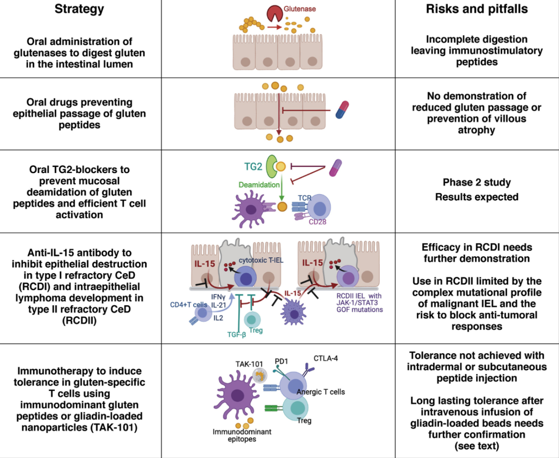
The associated editorial highlights these studies and reviews their limitations; in addition, the authors review the current non-dietary strategies (see below), pg 21-24: Full text: The Promise of Novel Therapies to Abolish Gluten Immunogenicity in Celiac Disease

From editorial, Gastroenterol 2021; 161: 21-24.

My take: These studies indicate that non-dietary treatments may be effective at some point, but not in the near future.

Related blog posts:

Oral Treatment of Celiac Disease & Research Optimist

A long time ago I heard a joke from a mentor about how can you tell if a person is an optimist.  An optimist is a person who finds a pile of manure under the tree on Christmas morning and declares: ‘Oh boy, I’m getting a pony.’

Researchers who are trying to identify oral treatments for celiac disease are probably true optimists. Yet, despite my skepticism, a recent study (D Schuppan et al. NEJM 2021; 385: 35-45. A Randomized Trial of a Transglutaminase 2 Inhibitor for Celiac Disease) provides the best proof yet that an oral treatment may be helpful.

In this 6-week randomized, double-blind, placebo-controlled study with 159 participants, treatment with ZED1227, a selective oral transglutaminiase 2 inhibitor reduced histologic injury compared to placebo; all patients were receiving a diet with 3 grams of daily gluten. Key findings:

  • Treatment with ZED1227 at all three dose levels attenuated gluten-induced duodenal mucosal injury. The estimated difference from placebo in the change in the mean ratio of villus height to crypt depth from baseline to week 6 was 0.44 (95% confidence interval [CI], 0.15 to 0.73) in the 10-mg group (P=0.001), 0.49 (95% CI, 0.20 to 0.77) in the 50-mg group (P<0.001), and 0.48 (95% CI, 0.20 to 0.77) in the 100-mg group (P<0.001)
  • The estimated differences from placebo in the change in intraepithelial lymphocyte density were −2.7 cells per 100 epithelial cells (95% CI, −7.6 to 2.2) in the 10-mg group, −4.2 cells per 100 epithelial cells (95% CI, −8.9 to 0.6) in the 50-mg group, and −9.6 cells per 100 epithelial cells (95% CI, −14.4 to −4.8) in the 100-mg group
  • Adverse events were similar to placebo; 3 (8%) patients in the 100 mg group developed a rash

The need for a treatment besides a gluten-free diet is significant; among adults, 40-50% do not achieve mucosal healing/recovery despite GFD institution; in addition, the diet is difficult and costly.

My take: I think it is still a long journey to find an effective & safe oral treatment for celiac disease.

Related blog posts:

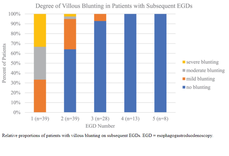
Persistent Villous Atrophy in Celiac Disease Despite a Gluten-Free Diet

A recent study (F Fernandez-Banares et al. Am J Gastroenterol 2021; 116: 1036-1043. Persistent Villous Atrophy in De Novo Adult Patients With Celiac Disease and Strict Control of Gluten-Free Diet Adherence: A Multicenter Prospective Study (CADER Study) shows that there is a high likelihood of persistent villous atrophy among adults with celiac disease (CD) despite adherence with a gluten-free diet (GFD). Thanks to Ben Gold for showing me this paper.

Key findings:

  • Among 76 patients (median age 36.5 years) who were prospectively followed for 2 years, persistent villous atrophy was observed in 40 (53%). In this group, 72.5% were asymptomatic (based on Likert scales) and 75% had negative serology
  • Detectable fecal gluten immunogenic peptides (f-GIPs) were present in at least one sample in 69% of patients. (Two samples obtained at f/u visits which were ~every 6 months during study)
  • Excellent or good adherence to GFD was demonstrated in 68.4% of patients based on dietetic evaluations. Only 6 (8%) were clearly nonadherent
  • “There were no significant differences in the rate of clinical and serological remission between patients with villous atrophy and those with mucosal recovery”
  • The authors did not find potentially modifiable predictive factors

Discussion:

  • The authors note that serology is “not useful for monitoring patients on a GFD.” Anti-TTG2 and EMA, in a recent meta-analysis, had a pooled sensitivity of around 50%.
  • “Adults are significantly less likely than children to normalize their duodenal histology.”

Editorial:

  • The associated editorial by Rej et al (pg 946-948) outline a personalized approach for dealing with persistent villous atrophy:
    • In those with persistent symptoms/positive GIPs/elevated serology/micronutrient deficiency, the first step is careful dietetic assessment. After this, endoscopy could be considered to confirm presence or absence of mucosal healing.
    • In those with no symptoms and no abnormalities, use of monitoring endoscopy needs to be weighed against the costs as well as potential complications.
    • Other points in the editorial: 1. GIPs have poor concordance with mucosal healing and 2. causes of poor mucosal healing include the following: natural slow healing process, super sensitive to gluten, ongoing gluten exposure, and refractory celiac disease.

My take: This study shows that there is ongoing gluten exposure in the majority of patients even in those with excellent or good adherence to a GFD; in addition, it shows that clinical/serological markers are NOT effective in predicting mucosal healing in adults. Nevertheless, it is not clear that followup endoscopy is beneficial.

Related blog posts:

Forbes (7/1/21): 99.5% Of People Killed By Covid In Last 6 Months Were Unvaccinated, Data Suggests

Disclaimer: This blog, gutsandgrowth, assumes no responsibility for any use or operation of any method, product, instruction, concept or idea contained in the material herein or for any injury or damage to persons or property (whether products liability, negligence or otherwise) resulting from such use or operation. These blog posts are for educational purposes only. Specific dosing of medications (along with potential adverse effects) should be confirmed by prescribing physician.  Because of rapid advances in the medical sciences, the gutsandgrowth blog cautions that independent verification should be made of diagnosis and drug dosages. The reader is solely responsible for the conduct of any suggested test or procedure.  This content is not a substitute for medical advice, diagnosis or treatment provided by a qualified healthcare provider. Always seek the advice of your physician or other qualified health provider with any questions you may have regarding a condition

Persistently Abnormal Celiac Labs =High Likelihood of Celiac Disease

CM Trovato et al JPGN 2021; 72: 712-717. Diagnostic Value of Persistently Low Positive TGA-IgA Titers in Symptomatic Children With Suspected Celiac Disease

This retrospective study provides insight into the predictive value of persistently abnormal celiac labs in symptomatic children.

Inclusion criteria:  not eligible for a non-biopsy diagnosis AND children with at least 2 TGA-IgA measurements, endomysial antibody (EMA) assessment and esophagogastroduodenoscopy with biopsies

Methods: Patients were classified in groups according to median TGA-IgA values: Group A (TGA-IgA>1 ≤ 5 × ULN; defined as “low-positive”), Group B (TGA-IgA > 5 < 10 × ULN; “moderate-positive”), and C (controls).

Key findings:

  • In group A, CD was diagnosed in 142/162 (87.7%)
  •  In group B, all 62 children (100%) received a CD diagnosis

My take: In individuals with mild elevation of celiac serology, it is reasonable to recheck prior to confirming with endoscopy. However, those with persistently abnormal values are very likely to have celiac disease.

Related blog posts:

  • If TTG IgA at 1-fold ULN, then PPV 61%, NPV 98%, Sens 90%, Spec 90%
  • If TTG IgA at 2-fold ULN, then PPV 79%, NPV 97%, Sens 82%, Spec 96%
  • If TTG IgA at 5-fold ULN, then PPV 93%, NPV 94%, Sens 62%, Spec 99%
  • If TTG IgA at 7-fold ULN, then PPV 96%, NPV 91%, Sens 41%, Spec 100%
Near Hahn Woods, Atlanta

Presentation and Knowledge of Celiac Disease

Two recent JPGN articles from the same researchers highlight changes in presentation and deficits of knowledge with celiac disease (CD). Interestingly, the authors chose to spell celiac disease differently in the two articles.

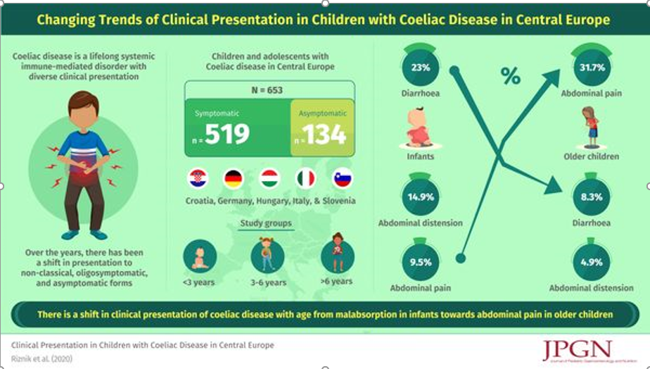
P Riznik et al. JPGN 2021; 72: 546-551. Clinical Presentation in Children With Coeliac Disease in Central Europe.

This retrospective study included data from 653 children and adolescents (median age 7 years 2 months; 63.9% girls) from Croatia, Germany, Hungary, Italy, and Slovenia were available for the analysis. Key findings:

  • One fifth (N = 134) of all children were asymptomatic.
  • In symptomatic children, the most common leading symptom was abdominal pain (33.3%), followed by growth retardation (13.7%) and diarrhoea (13.3%). Many children (47.6%; N = 247) were polysymptomatic.
  • Symptoms and signs of malabsorption (eg. diarrhea and distention) were significantly more common in younger (P < 0.001)

P Riznik et al. JPGN 2021; 72: 552-557 The Knowledge About Celiac Disease Among Healthcare Professionals and Patients in Central Europe

This study surveyed 1381 HCPs and 2262 patients with CD. Key findings.

  • Overall knowledge of CD was considered poor. Scores on web-based questionnaire were 51% for HCPs, 56% for patients, and 69% for patients
  • The authors recommend an EU e-learning program, for patients and HCPs: Celiac Facts Focus IN CD. This site has information/video course specific for patients. Celiac Facts for Patients

Related blog posts:

Real-World = Partially-Treated Celiac Disease

A recent prospective observational study reinforces the idea that most people with celiac disease are unable to accomplish a strict gluten-free diet (GFD): JP Stefanolo et al. Clin Gastroenterol Hepatol 2021; 19: 484-491. Real-World Gluten Exposure in Patients With Celiac Disease on Gluten-Free Diets, Determined From Gliadin Immunogenic Peptides in Urine and Fecal Samples

The investigators enrolled 53 adults with celiac disease (CD) for at least two years and followed symptoms as well as stool/urine testing for gluten immunogenic peptide (GIP). “GIP in stool can detect gluten consumption of more than 40 mg/d and the urine tests are positive from 40 and 500 mg/d of gluten.”

Key findings:

  • Over the 4-week study period, weekend samples (urine) identified 70% of patients excreted GIP at least once, compared with 62% during weekdays (stool).
  • Patients had a median of 3 exposures during the 4 weeks.
  • Also, the authors noted increases in GIP excretion towards the end of the study. “This suggests a potential Hawthorne effect that could be explained by a decrease in hypervigilance that often is seen in a context of research studies.”

The authors note that GIP “excretions of greater than 2 mcg/g in stool or greater than 12 ng/mL in urine can induce mucosal damage in almost 100% of patients.”

My take: This study adds to the body of literature emphasizing the high rate of inadvertent gluten exposure.

Related blog posts:

Before and After at Lake Michigan shoreline (1 month apart in Evanston, IL)

Early January -Evanston, IL
Early February -Evanston, IL

How a Gluten Challenge Could Change

A recent randomized double-blind study (MM Leonard et al. Gastroenterol 2021; 160: 720-733. Full Text: Evaluating Responses to Gluten Challenge: A Randomized, Double-Blind, 2-Dose Gluten Challenge Trial) provide evidence that a gluten challenge could detect evidence of celiac disease in hours, not weeks.

This study consisted of 14 adults with biopsy-proven celiac disease (CD) who were randomized to 3 g or 10 g gluten/day intake for 14 days. Each participant underwent extensive studies to detect histological, visible, and biochemical changes associated with gluten introduction. Data required multiple endosopic duodenal biopsies, VCEs and blood collection.

Key findings:

  • Symptoms and plasma interleukin-2 levels “increased significantly or near significantly at both doses.”
  • Interleukin-2 appeared to be the earliest, most sensitive marker of acute gluten
    exposure. IL-2 increases were observed 4 hours after exposure in patients with CD but not in healthy controls.
  • Intestinal damage is more complex and requires a longer duration and higher dose of gluten exposure. In this study, the higher dose (10 g) of gluten exposure was required for enteropathy within the study time frame.

My take: These study findings need to be confirmed in a broader patient cohort. However, in patients needing a gluten challenge, IL-2 response after a single-dose (measured at 4 hours) could be helpful. Those without IL-2 response are unlikely to have CD. Those with an IL-2 response at 4 hours, could confirm CD by completing a gluten challenge.

Related blog posts:

Seronegative Villous Atrophy

A recent large retrospective study (R Mandile et al. JPGN 2021; 72: 282-287. Seronegative Villous Atrophy in Children: Clinical and Immunohistochemical Features) provides information about conditions, besides celiac disease (CD) which present with villous atrophy. 64 of 1282 pediatric patients were seronegative with villous atrophy; seronegative was defined as testing negative twice for serology (TTG IgA/EMA or if IgA-deficient, IgG antibody serology).

Key findings:

  • Diagnoses were: inflammatory bowel diseases (IBD) (21/64), food allergy (8/64), infections (7/64, of which 3 HIV infections), immune deficiency (3/64), short bowel syndrome (3/64), congenital diarrhea (2/64), other/inconclusive diagnosis (8/64). In addition, there were 12 with Gastro-Esophageal Reflux Disease (GERD) & the authors speculate that perhaps hyperacidity could play a role in some of these cases.
  • Only one quarter of the seronegative patients had an increased number of intraepithelial lymphocytosis (IELs)
  • Among those with villous atrophy attributed to IBD, this was nearly equally-split between Crohn’s disease and ulcerative colitis, 10 and 11 patients respectively (according to Table 1)
  • The authors note that the ~5% of patients with seronegative villous atrophy with alternative diagnosis than Celiac disease may be an overestimation as more individuals are being diagnosed without biopsy based on serology
  • Despite the large cohort, there are still other rare conditions that were not identified in this study (eg. autoimmune enteropathy, CTLA4B deficiency,drug-induced enteropathy, and tropical sprue)

My take: This article provides a good starting point for patients with villous atrophy and negative serology.

Related article: J Devara et al. JPGN 2021; 72: 288-293. The Significance and Clinical Outcome of Lymphocytic Duodenosis in Children: Mayo Clinic Experience and Systematic Review Background: Lymphocytic duodenosis (LD) defined as increased intraepithelial lymphocytes >25 intraepithelial lymphocytes (IELs) per 100 epithelial cells with normal villous architecture is associated with many gastrointestinal (GI) disorders.

Key findings:

  • During the study period 12,744 children underwent an EGD with biopsies. Of those, we identified 426 children with LD (3%).
  • Among the LD (compared to control group), 5% had celiac disease (vs 0%, P < 0.001), 9% had Crohn disease (3%, P = 0.003) and 3% had Helicobacter pylori gastritis (1%, P = 0.021).

Related blog post: @AmyOxentenkoMD: Celiac Disease and Mimics

Does It Make Sense to Look for Celiac Disease in Children with Functional Constipation?

A recent study (AC Fifi et al. J Pediatr 2020; 227: 77-80.Full text PDF: Celiac Disease in Children with Functional Constipation: A School-Based Multicity Study) shows that celiac disease was not more prevalent in Colombian children with functional constipation(n=203) than in matched healthy controls (n=419). Patients were recruited from public schools.

Key finding:

  • The overall prevalence of celiac disease in the entire cohort was 0.6%. Of those with functional constipation, 1 (0.5%) was diagnosed with celiac disease, and 3 (0.7%) of the control patients

The authors note that some prior publications (references 11 and 12) have found a slight increase risk of celiac disease in children with constipation.

My take: In children with functional constipation, the yield from testing for celiac disease is very low and probably not significantly greater than the general population. In children with irritable bowel syndrome (which is often confused with constipation), the yield is probably a bit higher.

Related blog posts:

Chicago