Unknown's avatar

About gutsandgrowth

I am a pediatric gastroenterologist at GI Care for Kids (previously called CCDHC) in Atlanta, Georgia. The goal of my blog is to share some of my reading in my field more broadly. In addition, I wanted to provide my voice to a wide range of topics that often have inaccurate or incomplete information. Before starting this blog in 2011, I would tear out articles from journals and/or keep notes in a palm pilot. This blog helps provide an updated source of information that is easy to access and search, along with links to useful multimedia sources. I was born and raised in Chattanooga. After graduating from the University of Virginia, I attended Baylor College of Medicine. I completed residency and fellowship training at the University of Cincinnati at the Children’s Hospital Medical Center. I received funding from the National Institutes of Health for molecular biology research of the gastrointestinal tract. During my fellowship, I had the opportunity to work with some of the most amazing pediatric gastroenterologists and mentors. Some of these individuals included Mitchell Cohen, William Balistreri, James Heubi, Jorge Bezerra, Colin Rudolph, John Bucuvalas, and Michael Farrell. I am grateful for their teaching and their friendship. During my training with their help, I received a nationwide award for the best research by a GI fellow. I have authored numerous publications/presentations including original research, case reports, review articles, and textbook chapters on various pediatric gastrointestinal problems. In addition, I have been recognized by Atlanta Magazine as a "Top Doctor" in my field multiple times. Currently, I am the vice chair of the section of nutrition for the Georgia Chapter of the American Academy of Pediatrics. In addition, I am an adjunct Associate Clinical Professor of Pediatrics at Emory University School of Medicine. Other society memberships have included the North American Society for Pediatric Gastroenterology Hepatology and Nutrition (NASPGHAN), American Academy of Pediatrics, the Food Allergy Network, the American Gastroenterology Association, the American Association for the Study of Liver Diseases, and the Crohn’s and Colitis Foundation. As part of a national pediatric GI organization called NASPGHAN (and its affiliated website GIKids), I have helped develop educational materials on a wide-range of gastrointestinal and liver diseases which are used across the country. Also, I have been an invited speaker for national campaigns to improve the evaluation and treatment of gastroesophageal reflux disease, celiac disease, eosinophilic esophagitis, hepatitis C, and inflammatory bowel disease (IBD). Some information on these topics has been posted at my work website, www.gicareforkids.com, which has links to multiple other useful resources. I am fortunate to work at GI Care For Kids. Our group has 17 terrific physicians with a wide range of subspecialization, including liver diseases, feeding disorders, eosinophilic diseases, inflammatory bowel disease, cystic fibrosis, DiGeorge/22q, celiac disease, and motility disorders. Many of our physicians are recognized nationally for their achievements. Our group of physicians have worked closely together for many years. None of the physicians in our group have ever left to join other groups. I have also worked with the same nurse (Bernadette) since I moved to Atlanta in 1997. For many families, more practical matters about our office include the following: – 14 office/satellite locations – physicians who speak Spanish – cutting edge research – on-site nutritionists – on-site psychology support for abdominal pain and feeding disorders – participation in ImproveCareNow to better the outcomes for children with inflammatory bowel disease – office endoscopy suite (lower costs and easier scheduling) – office infusion center (lower costs and easier for families) – easy access to nursing advice (each physician has at least one nurse) I am married and have two sons (both adults). I like to read, walk/hike, bike, swim, and play tennis with my free time. I do not have any financial relationships with pharmaceutical companies or other financial relationships to disclose. I have helped enroll patients in industry-sponsored research studies.

What Went Wrong with EMRs: Death by a Thousand Clicks

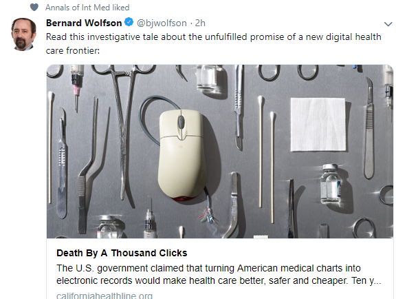
Link: Death by a Thousand Clicks Where Electronic Health Records Went Wrong

This lengthy article highlights a lot of issues with EMRs/EHRs including data sharing between systems, pulldown menus, disruption of physician-patient interactions, upcoding, safety risks and provides numerous personal examples.

An excerpt:

The U.S. government claimed that turning American medical charts into electronic records would make health care better, safer, and cheaper. Ten years and $36 billion later, the system is an unholy mess…

Instead of reducing costs, many say, EHRs, which were originally optimized for billing rather than for patient care, have instead made it easier to engage in “upcoding” or bill inflation…

More gravely still, a months-long joint investigation by KHN and Fortune has found that instead of streamlining medicine, the government’s EHR initiative has created a host of largely unacknowledged patient safety risks…

Compounding the problem are entrenched secrecy policies that continue to keep software failures out of public view. EHR vendors often impose contractual “gag clauses” that discourage buyers from speaking out about safety issues and disastrous software installations…

EHRs promised to put all of a patient’s records in one place, but often that’s the problem. Critical or time-sensitive information routinely gets buried in an endless scroll of data, where in the rush of medical decision-making — and amid the maze of pulldown menus — it can be missed…

[Problem with scrolldown options]: [doctors] had to read the list carefully, so as not to click the wrong dosage or form — though many do that too..

The numbing repetition, the box-ticking and the endless searching on pulldown menus are all part of what Ratwani called the “cognitive burden” that’s wearing out today’s physicians and driving increasing numbers into early retirement…

Beyond complicating the physician-patient relationship, EHRs have in some ways made practicing medicine harder,.. “Physicians have to cognitively switch between focusing on the record and focusing on the patient,” … “Texting while you’re driving is not a good idea.a.. But in medicine … we’ve asked the physician to move from writing in pen to [entering a computer] record, and it’s a pretty complicated interface.

My take: This article makes many good points.  Though, if you polled physicians in our group, hardly any would choose to go back to what we had before EMRs.

Related blog posts:

Best Predictor for Mortality from Biliary Atresia Liver Transplantation Candidates –Cardiomyopathy?

Briefly noted: A recent study (NM Gorgis et al. Hepatology 2019; 69: 1206-18, editorial 940-2 by Elizabeth Rand) indicates that cirrhotic cardiomyopathy (CCM) is very important factor for survival for biliary atresia (BA) patients requiring liver transplantation.

CCM was defined based on two-dimensional echocardiographic criteria: LV mass index ≥95 g/meter-squared or relative wall LV thickness of LV ≥0.42.

Key points:

  • Overall, 11 of 69 patients died, 4 while awaiting liver transplantation and 7 following transplantation.
  • 34 of 69 BA patients in this cohort had BA-CCM
  • All 11 who died had BA-CCM compared with no deaths in the 35 patients without CCM.

My take: Severe BA-CCM needs to be examined further; if severe, it may merit changing allocation policy.

Related blog posts:

Joshua Tree National Park

Getting the Most Out of Vedolizumab

A recent cross-sectional study (B Al-Bawardy et al. Inflamm Bowel Dis 2019; 25: 580-6) correlated vedolizumab (VDZ) trough drug levels (VDT) and clinical outcomes in 171 patients (62% Crohn’s disease (CD), 31% ulcerative colitis (UC), and 7% indeterminate colitis (IC)).

Key findings:

  • Median VDT was 15.3 microgr/mL.
  • Median VDT was 17.3 microgr/mL for patients with normal CRP compared with 10.7 for patients with high CRP.  This differnece was noted significantly for CD (20.3 vs 10.4) but not for UC.
  • No relationship  between VDT and mucosal healing was noted.
  • Shorter dose intervals and lower BMO resulted in higher VTLs
  • Only 1 patient had detectable antibodies to VDZ

A second systematic review (L Peyrin-Biroulet et al. Clin Gastroenterol Hepatol 2019; 17: 838-46) analyzed data from 10 cohorts who had received vedolizumab.  Most had prior anti-TNF exposure. Key finding: the pooled incidence rates of loss of response were 47.9 per 100 person-years of follow up among patients with CD and 39.8 per 100 person-years of follow up among patinets with UC.  Dose intensification restored response to the drug in 53.8% of secondary non-responders.

My take: While VDZ dose intensification can restore response, the utility of therapeutic drug monitoring is unclear with VDZ therapy.

Related blog posts:

Biliary Atresia and Bile Lakes

A retrospective study (DA Ginström et al. JPGN 2019; 488-94) review cholangitis associated with biliary atresia and the role of bile leaks.  This study encompasses a ~30 year period (1987-2016) and 61 patients.

Key findings: 

  • 54% had survived with their native liver at a median of 7.3 years; 28 (46%) had ≥ 1 cholangitis episodes/year
  • Cholangitis bouts were >5 times higher among patients with bile lakes
  • Management of bile lakes after 1995 (n=6) was with operative intestinal drainage (bile-lake jejunostomy) which resulted in disappearance or regression of bile lakes in all patients.  Associated with this, yearly cholangitis bouts decreased from 8.8 (4.2-14) to 1.1 (0.2-3.2). Prior to 1995, two patients were managed with PTCD; both died within one year.
  • Among patients who had surgical drainage of their bile lakes, 4 have survived jaundice-free and 2 received liver transplants at 1.3 and 4.9 years after intestinal drainage.

My take: This report provides insight into the management of bile lakes (also referred to as intrahepatic biliary cysts) indicating that bile-lake jejunostomy is effective in symptomatic patients.

Related blog posts:

Joshua Tree National Park

ESPGHAN Juvenile Polyposis Syndrome in Children –Position Paper

Management of Juvenile Polyposis Syndromes in Children and Adolescents: A Position Paper from the ESPGHAN Polyposis Working Group: Link: JPGN 2019; 68 (3): 453-462

Diagnosis: JPS is diagnosed by use of the following criteria in the absence of  extraintestinal features consistent with PHTS (Cowden syndrome [CS] or Bannayan-Riley-Ruvalcaba syndrome) (6):
1. Five or more JPs of the colon or rectum, or
2. JPs in other parts of the GI tract, or
3. Any number of JPs and a positive family history.

SUMMARY OF SOME OF THE RECOMMENDATIONS
1. Routine predictive genetic testing for paediatric patients at risk of developing JPS should start at 12 to 15 years of age. Children that develop rectal bleeding earlier than this age should undergo colonoscopy and then proceed to genetic testing if polyps are identified. Weak recommendation, very low quality of evidence.
2. Colonoscopic surveillance should commence from age 12 to 15 years, or earlier if symptomatic. Once polyps (>10 mm) are detected they should be removed and olonoscopy repeated annually until polyps >10 mm have been resected, then
repeated every 1 to 5 years. Weak recommendation, very low quality of evidence.
3. Surveillance of the upper GI tract in affected or at-risk JPS patients is not required in childhood or teenage years, unless there is unexplained anaemia or upper GI symptoms. Weak recommendation, very low quality of evidence.
4. Paediatric patients with SMAD4 mutation should be evaluated for HHT including screening and preventative treatment for cerebral and pulmonary AVMs. Weak recommendation, very low quality of evidence.
5. In JPS patients with an isolated BMPR1A gene mutation, there are no additional investigations required beyond the endoscopic procedures described above. Children with BMPR1A mutation and early onset polyposis and/or a severe phenotype and/or
extraintestinal manifestations should be evaluated for PTEN mutation. Weak recommendation, very low quality of evidence.
6. In a child with a single JP, a repeat colonoscopy is not routinely required. Weak recommendation, very low quality of evidence.
7. If a specific gene mutation has been detected in a child, then genetic testing should be offered to all first-degree family members. If no specific gene mutation was detected, then first degree relatives should be referred for screening colonoscopy at the age of 12 to 15 years. Weak recommendation, very low quality of evidence.
8. There is no role for chemoprevention in JPS. Weak recommendation, very low quality of evidence.

Related blog postUpdated Guidelines on Genetic Testing/management for Hereditary GI Cancer Syndromes

Atlanta Zoo

Disclaimer: These blog posts are for educational purposes only. Specific dosing of medications (along with potential adverse effects) should be confirmed by prescribing physician.  This content is not a substitute for medical advice, diagnosis or treatment provided by a qualified healthcare provider. Always seek the advice of your physician or other qualified health provider with any questions you may have regarding a condition.

Eosinophilic Esophagitis and Inflammatory Bowel Disease

A recent study (Limketkai BN, et al. Gut. 2019;doi:10.1136/gutjnl-2018-318074.) shows that the likelihood of eosinophilic esophagitis (EoE) is higher in patients with inflammatory bowel disease (IBD) and that the likelihood of IBD is higher in EoE patients.

Summary from Healio Gastroenterology –Risk for EoE higher in patients with IBD, and vice versa:

  • Researchers conducted a prospective cohort analysis using the Truven MarketScan database from 2009 to 2016 to define the epidemiology and clinical implications of concurrent EoE and IBD diagnoses.
  • Among their cohort comprising 134,013,536 individuals, the incidence of EoE was 23.1 per 100,000 person-years, CD was 51.2 and ulcerative colitis was 55.2.
  • Compared with patients without either diagnosis, the risk for EoE was higher in patients with CD (IRR = 5.4, P < .01; prevalence ratio [PR] = 7.8, P < .01) and UC (IRR = 3.5, P < .01; PR = 5, P < .01). Meanwhile, the risk for IBD was higher among patients with EoE (CD: IRR = 5.7, P < .01; PR = 7.6, P < .01; UC: IRR = 3.4, P < .01; PR = 4.9, P < .01).

CHOP QI: Anemia in IBD Pathway

A recent article in Gastroenterology & Enoscopy News, “QI Pathway Improves Anemia Management in Pediatric IBD” (also presented at NASPGHAN 2018 -abstract 7, J Breton et al), discusses anemia and provides a link to CHOP QI Pathway for Anemia

This link contains useful information regarding treatment options and links to recommendations on management.  This algorithm suggests using intravenous iron for anemia in all IBD patients with active disease as well as using intravenous iron for those with moderate to severe anemia.  The rationale for parenteral iron in those with active disease is due to two factors:

  1. to overcome the block to intestinal iron absorption induced by hepcidin in the setting of inflammation (making oral iron less effective in active IBD regardless of disease location)
  2. due to data showing  that oral iron may aggravate intestinal inflammation by altering the gut microbiome and increasing intestinal permeability

My take: The CHOP initiative provides some clear cut recommendations.for anemia in IBD.  Parenteral iron is more efficacious in improving anemia; however, the effects of parenteral iron on the microbiome and other potential risks (eg. increased sepsis) are not clear. In my view, more information about outcomes and costs are needed to determine the optimal approach.

Related blog posts:

Disclaimer: These blog posts are for educational purposes only. Specific dosing of medications/diets (along with potential adverse effects) should be confirmed by prescribing physician/nutritionist.  This content is not a substitute for medical advice, diagnosis or treatment provided by a qualified healthcare provider. Always seek the advice of your physician or other qualified health provider with any questions you may have regarding a condition.

How Do Home Infusions Stack Up?

One of the advantages of infusions in the office (or hospital) compared to home infusions and home injections is close communication by those giving the infusion with the physician.  In addition, with each infusion, in these settings offers an opportunity to review the patient’s progress and adjust the patients orders.  A recent study (Fenster M, et al. Clin Gastroenterol Hepatol. 2019;10.1016/cgh.2019.03.030.) indicates that these advantages may make infusions more successful than infusions given at home.

A summary is offered by Healio Gastroenterology: Home biologic infusions in IBD suffer from lack of monitoring

Researchers conducted a matched retrospective cohort study of patients treated with infliximab or vedolizumab with home infusion (n = 69) compared with hospital infusion at a large, tertiary care IBD center.  The primary endpoint was a composite of adverse outcomes, including stopping biologic therapy, IBD-related emergency department visit or IBD-related hospitalization.

  • “Patients on home infusion were more likely to experience adverse outcomes compared with control patients (43.5% vs. 21.7%; P = .006), and they also had a shorter time to adverse outcomes than patients who got hospital infusions.”
  • “Patients with home infusions trended toward stopping therapy within 1 year (20.3% vs. 8.7%; P = .053) and stopping therapy within the complete follow-up window (27.5% vs. 15.9%; P = .099) compared with controls.”
  • Patients with home infusions had “more emergency department visits (30.4% vs. 7.2%, P < .001), they did not have significantly more hospitalizations (17.4% vs. 11.6%).”

The authors noted that the “increase in adverse events might have been related to a reduced level of monitoring observed in home infusion patients. In the year following home infusion initiation or matching, patients who persisted on home infusions had significantly fewer IBD clinic visits (1.23 vs. 1.75; P = .018) compared with controls.”

My take (borrowed from a previous post): In my experience, office-based infusions can be provided safely and in a cost-effective manner.  While the convenience and potential cost-savings of home-based infusion are desirable, before implementing broadly, issues regarding communication, safety protocols, and documentation to allow modifications in therapy need to be proactively addressed. These issues could affect a patient’s long-term response to biologic therapy.

Related blog posts:


Which is Safer and More Effective –Miralax (PEG 3350) or Lactulose?

A recent randomized multicenter study (D Jarzebicka et al. JPGN 2019; 68: 318-24) compared polyethylene glycol 3350 (PEG, aka Miralax) and lactulose for functional constipation in a cohort of 102 patients (12 weeks of treatment and 4 weeks of follow-up).

Dosing:

  • PEG 3350 dosing divided into 2 doses:
    • <8 kg:  5 g/day
    • 8-12 kg: 10 g/day
    • 12-20 kg: 15 g/day
    • >20 kg: 20 g/day
  • Lactulose was given as 1 mL/kg twice a day

A “good clinical outcome” was considered if there were three or more stools per week and an improvement in stool consistency of at least 2 types in the Bristol scale.

Key findings:

  • At week 12, a good clinical outcome was noted in 98% of PEG group compared with 90% in the lactulose group
  • PEG group had increased defecations relative to lactulose: 7.9 vs. 5.7, less stool retention: 7% vs 10%, and fewer hard stools 7% vs 13%
  • Side effects, mainly abdominal pain and bloating, were reported more frequently with lactulose than with PEG: 23 vs 15, P=0.02)

My take: This study showed that PEG 3350 was more effective and had fewer side effects than lactulose for constipation in children between 6 months of age and 6 years.

Related blog posts:

From Civil and Human Rights Museum, Atlanta

Which Symptom is Best for Indicating Reflux in Infants?

A recent study (CR Collins et al. J Pediatr 2019; 206; 240-7) showed that symptoms alone are not able to predict the degree/presence of reflux in infants.

Methods: The authors recruited ‘symptomatic’ infants with median 41 weeks postmenstrual age (median 29 weeks at birth) and employed a combination of 24-hour pH-impedance to determine acid reflux index (ARI) and subsequently used provocative esophageal infusions (with air, water then apple juice) to assess manometry and symptoms. In total, they analyzed 2635 separate esophageal stimula in 74 infants.

The authors considered ARI <3% as normal, 3-7% as indeterminate ARI, and >7% as abnormal ARI.

Symptoms that they recorded included arching, irritability, cough, gag, sneeze, gasp, bradycardia, desaturation, throat clearing, startle, grimace, grunting, mouthing, and yawning.

Key findings:

  • “The presence of physical symptoms (ARI <3: 80%; ARI 3-7: 77%; ARI >7: 81%; P=0.4), cardiorespiratory symptoms (ARI <3: 27%; ARI 3-7: 40%; ARI >7: 26%; P=0.4),or sensory symptoms (ARI <3: 26%; ARI 3-7: 22%; ARI >7: 35%; P=0.3) were also not significantly different by ARI groups”
  • All infants had a normal symptom index (SI) for acid, nonacid, or total reflex.
  • Accepted metrics, SI, SSI, and SAP, had higher association with nonacid events compared with acid events.
  • Cardiorespiratory symptoms are more likely to be elicited by esophageal infusions with both water and apple juice than air.  “Symptoms are indicators of esophageal dysmotility and maladaptive aerodigestive protective mechanisms.”
  • The authors in their discussion state that “GERD severity plays no role in the generation of symptoms.”

My take: All infants with and without reflux have a lot of “symptoms.”  Nonacid reflux is much more likely to provoke symptoms in this population than acid reflux reinforcing the idea that acid suppression is likely ineffective and potentially harmful.  “The findings of this study challenge the rationale for instituting anti-GERD therapies in neonates based on either symptoms alone or the existing acid-reflux indices or pH-impedance metrics.”

Related blog posts:

Link to article cited below from The Onion: Dog Feels Like He Always Has To Be ‘On’ Around Family Thanks to Jennifer for this reference.

Disclaimer: These blog posts are for educational purposes only. Specific dosing of medications/diets (along with potential adverse effects) should be confirmed by prescribing physician/nutritionist.  This content is not a substitute for medical advice, diagnosis or treatment provided by a qualified healthcare provider. Always seek the advice of your physician or other qualified health provider with any questions you may have regarding a condition.