
Unknown's avatar

About gutsandgrowth

I am a pediatric gastroenterologist at GI Care for Kids (previously called CCDHC) in Atlanta, Georgia. The goal of my blog is to share some of my reading in my field more broadly. In addition, I wanted to provide my voice to a wide range of topics that often have inaccurate or incomplete information. Before starting this blog in 2011, I would tear out articles from journals and/or keep notes in a palm pilot. This blog helps provide an updated source of information that is easy to access and search, along with links to useful multimedia sources. I was born and raised in Chattanooga. After graduating from the University of Virginia, I attended Baylor College of Medicine. I completed residency and fellowship training at the University of Cincinnati at the Children’s Hospital Medical Center. I received funding from the National Institutes of Health for molecular biology research of the gastrointestinal tract. During my fellowship, I had the opportunity to work with some of the most amazing pediatric gastroenterologists and mentors. Some of these individuals included Mitchell Cohen, William Balistreri, James Heubi, Jorge Bezerra, Colin Rudolph, John Bucuvalas, and Michael Farrell. I am grateful for their teaching and their friendship. During my training with their help, I received a nationwide award for the best research by a GI fellow. I have authored numerous publications/presentations including original research, case reports, review articles, and textbook chapters on various pediatric gastrointestinal problems. In addition, I have been recognized by Atlanta Magazine as a "Top Doctor" in my field multiple times. Currently, I am the vice chair of the section of nutrition for the Georgia Chapter of the American Academy of Pediatrics. In addition, I am an adjunct Associate Clinical Professor of Pediatrics at Emory University School of Medicine. Other society memberships have included the North American Society for Pediatric Gastroenterology Hepatology and Nutrition (NASPGHAN), American Academy of Pediatrics, the Food Allergy Network, the American Gastroenterology Association, the American Association for the Study of Liver Diseases, and the Crohn’s and Colitis Foundation. As part of a national pediatric GI organization called NASPGHAN (and its affiliated website GIKids), I have helped develop educational materials on a wide-range of gastrointestinal and liver diseases which are used across the country. Also, I have been an invited speaker for national campaigns to improve the evaluation and treatment of gastroesophageal reflux disease, celiac disease, eosinophilic esophagitis, hepatitis C, and inflammatory bowel disease (IBD). Some information on these topics has been posted at my work website, www.gicareforkids.com, which has links to multiple other useful resources. I am fortunate to work at GI Care For Kids. Our group has 17 terrific physicians with a wide range of subspecialization, including liver diseases, feeding disorders, eosinophilic diseases, inflammatory bowel disease, cystic fibrosis, DiGeorge/22q, celiac disease, and motility disorders. Many of our physicians are recognized nationally for their achievements. Our group of physicians have worked closely together for many years. None of the physicians in our group have ever left to join other groups. I have also worked with the same nurse (Bernadette) since I moved to Atlanta in 1997. For many families, more practical matters about our office include the following: – 14 office/satellite locations – physicians who speak Spanish – cutting edge research – on-site nutritionists – on-site psychology support for abdominal pain and feeding disorders – participation in ImproveCareNow to better the outcomes for children with inflammatory bowel disease – office endoscopy suite (lower costs and easier scheduling) – office infusion center (lower costs and easier for families) – easy access to nursing advice (each physician has at least one nurse) I am married and have two sons (both adults). I like to read, walk/hike, bike, swim, and play tennis with my free time. I do not have any financial relationships with pharmaceutical companies or other financial relationships to disclose. I have helped enroll patients in industry-sponsored research studies.

NASPGHAN Postgraduate Course 2017 (Part 2): Celiac, Fad Diets, and H pylori

Over the next 2 weeks or so, I am posting my notes/pictures from this year’s annual meeting.  This post reviews the 2nd module from the postgraduate course.

This blog entry has abbreviated/summarized these presentations. Though not intentional, some important material is likely to have been omitted; in addition, transcription errors are possible as well.

Here is a link to postgraduate course syllabus: NASPGHAN PG Syllabus – 2017

Celiac Disease Diagnosis: ESPGHAN vs. NASPGHAN Guidelines

Michelle Pietzak   Children’s Hospital of Los Angeles

This lecture started with a succinct history of celiac disease, including discussion of the banana diet and the observation that wheat deprivation during World War II was associated with improvement in children with celiac disease. The sequential diagnostic recommendations of ESPGAN and NASPGHAN were reviewed.

Key points:

  • ESPGHAN 2012 guidelines separated children in two groups: symptomatic and asymptomatic.  The guidelines indicated that symptomatic children with high titer serology (TTG IgA >10-fold ULN and positive EMA) could be diagnosed without an intestinal biopsy.
  • ACG 2013 Guidelines: TTG IgA preferred serologic test if over age 2 years,  TTG IgA along with deamidated gliadin (IgG and IgA) recommend if younger than 2 years. Even if negative serology, a biopsy was recommended if significant suspicion of diagnosis.
  • NASPGHAN, AGA, ACG all recommend small bowel biopsy to confirm this lifelong condition
  • The speaker did not critically discuss the Leonard et al study suggesting that 19% of individuals had persisting enteropathy on a gluten fee diet (see previous posts below)
  • Jimmy Fallon: “10% of the population is allergic to gluten and 90% are tired of hearing about it”

Related posts:

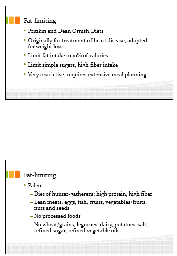
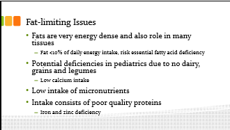
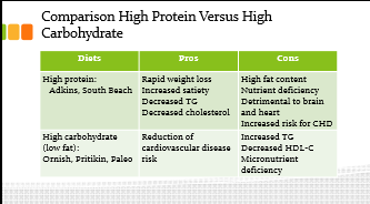
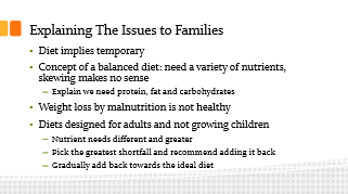
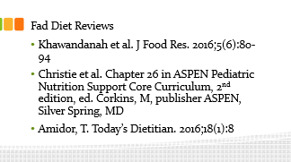
Fad Diets: The Good, the Bad, and the Just Plain Ugly

Mark Corkins      University of Tennessee Health Science Center

Key points:

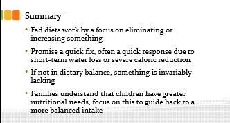
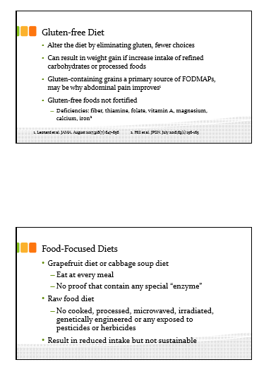
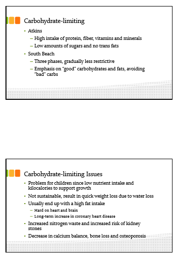
  • Fad diets driven in part by obesity
  • Gluten-free diet is the most trendy, ~30% of U.S. adults are ‘limiting’ gluten, though most do not understand what gluten is.  “Wheat Belly” book by Dr. William Davis has been influential despite lack of data.
  • According to marketing report, 35% who consumer gluten limited diet have no reason for this, 26% for ‘healthier option,’, 19% for ‘digestive health,’, 13% for weight loss, 8% for gluten sensitivity
  • Recommends: choosemyplate.gov. These feeding guidelines have been developed by nutrition experts.
Update on H pylori

Nicola Jones    Hospital for Sick Children (Toronto)

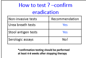
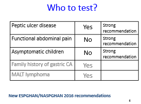
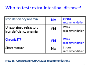
  • Reviewed current recommendations based on ESPGHAN/NASPGHAN 2016 recommendations.
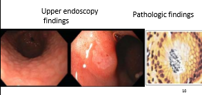
  • The speaker indicates that upper endoscopy is recommended for initial diagnosis of H pylori but testing is not recommended for functional abdominal pain.  The lecture did not address the issue that there can be an overlap in the presentations of these disorders.
  • Adherence is crucial for adequate eradication
  • Newer studies show improved eradication rates for quadruple therapy (bismuth-based) & in U.S. this is recommended as first-line treatment unless resistance patterns are known (which could allow for triple therapy)
 

NASPGHAN Postgraduate Course 2017 (Part 1): Strictures, GI Bleeding, Pancreatic Fluid Collections

Over the next 2 weeks or so, I am posting my notes/pictures from this year’s annual meeting.  The first few days will review the postgraduate course.  For the most part, I find the postgraduate course reassuring that I have kept up with current approaches; there is usually not a lot of new information but a solid review of the topics.

Here is a link to postgraduate course syllabus: NASPGHAN PG Syllabus – 2017

This blog entry has abbreviated/summarized these presentations. Though not intentional, some important material is likely to have been omitted; in addition, transcription errors are possible as well.

Strictures beyond the esophagus

Petar Mamula, Children’s Hospital of Philadelphia

Some useful points:

  • Fluoroscopy very useful with most strictures –may improve safety and effectiveness. Helps define anatomy
  • Reviewed strictures in stomach –rare. May be due to caustic ingestion, Crohn’s disease or chronic granulomatous disease
  • Intestinal/colonic strictures (or narrowing): duodenal webs -can be treated with needle knife, Crohn’s disease strictures -can be balloon dilated, Short gut syndrome, Graft versus host disease

GI Bleeding Update

Diana Lerner  Medical College of Wisconsin

Useful points

Upper GI Bleeding:

  • IV PPIs reduce risk of transfusion and reduce risk of re-bleeding
  • IV PPI BID treatment has been shown to be noninferior to continuous drip
  • Conservative transfusion therapy
  • Erythromycin can be helpful
  • Lecture had good videos with review of techniques: clipping, heater probe, epinephrine injection (not recommended as monotherapy), argon plasma coagulation, and bipolar electrocautery

Cleveland et al. World J Pediatr 2012

Lower GI Bleeding:

  • Etiologies include the followiing: Post-polypectomy, Solitary Rectal Ulcer syndrome, Blue Rubber Bleb syndrome, anastomotic ulcer bleeding, Meckel’s diverticulum
  • Lower GI evaluation is best after prep –much higher yield

Management of Pancreatic Fluid Collections

Matt Giefer Seattle Children’s Hospital

Key points:

  • Imaging in first 7 days of diagnosis may miss the development of fluid collections
  • With necrotizing pancreatitis, fluid collections are either ANC: acute necrotic collection (<4 weeks) or WON: walled off necrosis (>4 weeks); Bryan et al. Radiographics 2016; 36: 675
  • With interstitial edematous pancreatitis, fluid collections are either acute peripancreatic fluid collection (<4 weeks) or Pseudocyst: >4 weeks,
  • Fluid collections do not preclude feeding patients
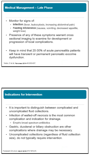
  • Drainage often needed if fluid collection becomes infected or if fluid collection causes obstruction
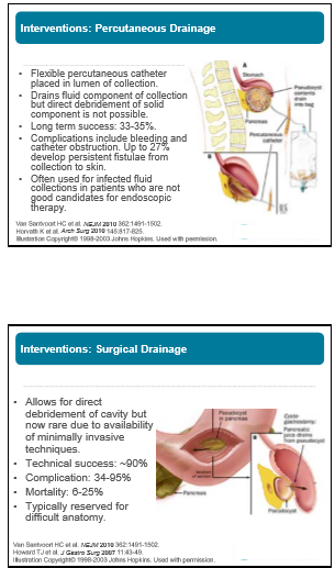
  • Endoscopic drainage is first-line approach: equally effective as surgery, fewer complications, equal efficacy, and lower cost


Three Studies Show the Benefit of Concomitant Therapy for Inflammatory Bowel Disease (Part 2)

Continued from yesterday…

The third study (HM Kansen et al. JPGN 2017; 65: 425-29) retrospectively (2009-2014) examined 162 children (with available data) with Crohn’s disease (CD) for the development of anti-infliximab antibody (ATI) while receiving either monotherapy or concomitant therapy. This was a collaborative study from the Kids with Crohn’s Colitis (KiCC) working group (Netherlands).  In the majority of their patients (222 of 229), IFX was initiated as step-up therapy. Key findings:

  • 15% developed ATIs
  • 6 of 62 (10%) developed ATIs while receiving ongoing concomitant immunosuppression
  • 11 of 81 (14%) developed ATIs after receiving early concomitant immunosuppression (median of 6.2 months of concomitant immunosuppression followed by IFX monotherapy) 10 of 11 who developed ATIs  within the first 12 months, compared to 1 of 26 (4%) after 12 months.
  • 8 of 19 (42%) developed ATIs on IFX monotherapy

In their discussion, the authors note concerns regarding the safety of thiopurines. However, they point out that “the benefit of combination therapy (reduction of ATI formation) relative to IFX monotherapy should outweigh the risk of serious infections and malignancies to achieve an optimal treatment strategy for paediatric CD.” The authors: “combination therapy for approximately 12 months from initiation of IFX, followed by IFX monotherapy, may be equally effective alternative to continuous combination therapy.”

Overall, the totality of the evidence favors combination therapy for most patients with CD.  Yet, there is wide variation in clinical practice. As I was thinking about this, I came across a recent commentary: “The Power of Regret” (J Groopman, P Hartzband. NEJM 2017; 377: 1507-9).  The authors note that “disappointment is an unavoidable aspect of making difficult choices…but disappointment is not associated with self-recrimination and thus differs notably from regret.”  They indicate that “process regret” occurs when patients do not consider information about all available choices before making a decision.  I wonder if many patients/families fear using concomitant therapy because they worry they will regret the decision if a complication occurs.  Perhaps, working with all available information, some reluctant patients/families will feel better about their decision if the process for their decision was thorough, considering the risks/benefits of the treatment but also the risks/benefits of not choosing the treatment. .

My take: Overall, for most pediatric patients with CD, to date, concomitant therapy has been the most effective treatment.  More prospective studies are needed to determine more conclusively the benefit and optimal duration/timing of combined therapy, particularly with the more frequent use of therapeutic drug monitoring. Also, as will be noted in future posts from annual meeting, thiopurine use is declining.

Related blog posts:

Three Studies Show Benefit of Concomitant Therapy for Inflammatory Bowel Disease (Part 1)

In the first study (J Cheng et al. Inflamm Bowel Dis 2017; 23: 1762-73), the authors retrospectively reviewed 148 children (113 with Crohn’s disease, 35 with ulcerative colitis). 90 patients received concomitant therapy (infliximab with either a thiopurine [n=67], methotrexate [n=23]) and 58 received infliximab monotherapy. Key findings:

  • Concomitant therapy >6 months  significantly lowered the risk of secondary loss of response in Crohn’s disease (CD) (HR =0.39) compared to monotherapy.   A similar trend was noted with ulcerative colitis (UC) but did not reach statistical significance.
  • Steroid-free remission rates at 1 year were 78% for CD patients with concomitant therapy compared with 54% on monotherapy
  • Among primary nonresponders, 67% of CD patients and 75% of UC patients were receiving IFX monotherapy.
  • No differences in adverse events were evident between patients receiving monotherapy compared with concomitant therapy. One patient (receiving azathioprine) developed a follicular lymphoma; this patient was well 10 years later.

The second study (Y Qui et al. Clin Gastroenterol Hepatol 2017; 15: 1359-72) was a systemic review of 35 studies that met the authors’ inclusion criteria. In total, 6790 patients with inflammatory bowel disease were enrolled in these studies. This study looked at multiple anit-TNF agents including infliximab, adalimumab, certolizumab, and golimumab. Key finding:

  • Antidrug antibodies were reduced by 51% in patients receiving concomitant therapy
  • Conclusion from authors: “concomitant use of immunomodulators should be considered in patients treated with anti-TNF treatment.”

My take: Overall, for most pediatric patients with CD, to date, concomitant therapy has been the most effective treatment.  More prospective studies are needed to determine more conclusively the benefit and optimal duration/timing of combined therapy, particularly with the more frequent use of therapeutic drug monitoring.  Also, as will be noted in future posts from annual meeting, thiopurine use is declining.

More on this topic tomorrow.

Related blog posts:

Disclaimer: These blog posts are for educational purposes only. Specific dosing of medications (along with potential adverse effects) should be confirmed by prescribing physician.  This content is not a substitute for medical advice, diagnosis or treatment provided by a qualified healthcare provider. Always seek the advice of your physician or other qualified health provider with any questions you may have regarding a condition.

Infliximab Infusions Without Premedication

Briefly noted:

A recent study (SQ Hutsell, M Wu, KT Park. JPGN 2017; 65: 430-31) examined two practice changes with regard to infliximab (IFX) infusions:

  1. 1-hour infusions
  2. Omission of premedications

The authors reviewed ~900 IFX infusions; though, only 111 infusions were administered without premedications.  These two changes resulted in a 51% decrease in infusion hours, despite a 9% increase in total number infusions. No increase in adverse reactions was identified.

The authors state that these changes improve patient experience, shorten monitoring time, and reduce costs.


More on It’s Past Time to Split

In followup to this morning’s post, Pediatric Liver Transplantation: It’s Past Time to Split, one reader pointed out an abstract by Emily R. Perito et al.presented at this year’s AASLD which showed that pediatric deaths would decrease if more livers were split.

ABSTRACT #137

Increasing split liver transplantation in the U.S. could decrease pediatric deaths on the liver transplant waiting list

Emily R. Perito1,3, Garrett Roll2, Jennifer L. Dodge2,

Background: In the United Kingdom, defaulting to split liver transplantation (LT) with suitable deceased donor grafts has virtually eliminated pediatric waitlist (WL) mortality. In the US, only <2% of LTs are split, but 1 in 10 infants die on the WL.

Methods: Using UNOS STAR data, livers for potential split LT were identified from all transplanted, deceased-donor livers 2010-15 who fit strict criteria: age 18-40y, BMI<30, recovered in US after donor brain death, 0-1 vasopressors, a <155meq/L, AST/ALT<100IU/L, bilirubin<3mg/dL, <7d hospitalized, cardiac arrest≤30min, HBV/HCV neg, not CDC high-risk, steatosis≤10% if biopsied, not multi-organ transplant, and no bloodstream infection. Livers allocated to patients high-risk for split LT were also removed: status 1A or MELD/PELD≥40 at WL removal, re-transplant, in the ICU, BMI>34, or >300mi from donor hospital. Pediatric WL deaths included deaths and removals for too sick to transplant, never relisted.

Results: Of 35,461 livers transplanted 2010-15, 6.7% were potentially utilizable for split LT based on donor characteristics. Of these, 95% were transplanted whole (n=2,253). 50% went to recipients deemed possibly high-risk for split LT. This left 1,116 potential livers for split LT (FIGURE); 78% of their primary recipients were listed as willing to accept a segmental liver, and 97% to accept cold ischemia time≥6h (CIT, median 12h). Median donor risk index for this subset was 1.06 (max 1.67). During the same 5y, 261 children died after ≥3d on the WL (median 57d, IQR 15-161)—87% of all pediatric WL deaths. Of these, 56% were <2y of age, 26% 2-12y, 18% 13-18y. Median weight was 9.2kg (IQR 5.9-29.4kg). 36% died at centers that reported doing no pediatric split LTs (15%) or ≤1/year (22%).

Conclusions: Increased utilization of split LT could decrease US pediatric WL mortality—without decreasing LT access for adults. Barriers are significant, but changes to  allocation policy, increasing centers with splitting experience, and splitting on normothermic perfusion could increase access and reduce WL mortality.

Jose Garza, Chelly Dykes, Elvis, and Jay Hochman at Cincinnati Children’s Reception

Pediatric Liver Transplantation: Past Time to Split

A recent study (EK Hsu et al. Gastroentorol 2017; 153: 988-95, editorial 888-89) exposes some deep flaws in organ transplantation in U.S.

The retrospective study examined children on the U.S liver transplant wait-list from 2007-14.  This included 3852 pediatric candidates.  Key findings:

  • Of 27,831 adults who underwent transplantation, 1667 (6%) received livers from pediatric donors (<18 years)
  • Of children who died or were delisted, the centers caring for 173 (55%) had received an offer of 1 or more livers that was subsequently transplanted into another pediatric recipient.  The remaining 45% died or delisted with no offers. High-volume (>15 transplants per year) centers were more likely to accept an organ than a low-volume center (<5 transplants per year).
  • Only 29% of children received a split graft.  When a splittable adult liver graft was allocated to an adult the chance of it being used as a split was 0.6%.

Background:

  • Children have much lower survival rate than adults on waiting list. Of adults who died or delisted, 85% receive at least one transplant offer; whereas, nearly half of all children never even receive an offer.  Children who died/delisted had wait-time of 33 days compared with 92 days for adults who died/delisted.
  • Less than 10% of all liver transplant recipients are pediatric transplants.  Per editorial, “a measure that improves pediatric access by 20% would only reduce adult access by 2%.”
  • There are more than 100 pediatric liver transplant centers in U.S. Certainly, this improves convenience; however, per editorial:  “three-fourths are very low volume centers, performing <5 liver transplantations per year…Death on waiting list” occur 5 times more at low-volume transplantation centers.
  • In this study, only 29% of children received split livers; in comparison, in the UK, >80% receive either a split graft or living donor graft.

The editorial points out that splittable livers that are allocated to adults are virtually never split; this is either due to inconvenience or lack of expertise.  A small increase in liver splitting would dramatically lower the pediatric mortality wait list.  There is no incentive in the current system to split a liver/save a child’s life.

My take: The data from this study points out glaring problems in pediatric liver transplantation.

  1. Children are dying due to lack of prioritization.  Pediatric livers are going to adults.
  2. There is practically no splitting when liver organs are allocated to an adult.  Incentives to increase organ splitting would save many children from dying waiting for an organ.
  3. Large volume pediatric centers are much more likely to accept a liver offer for patients waiting at their centers.  There is an increased wait-list mortality at very low volume centers, perhaps due to lack of expertise and passing up viable organs.  Do hepatologists/surgeons at these centers explain this risk to families at their centers?

Related blog posts:


>99% Accuracy in Non-Biopsy Diagnosis of Celiac Disease

Another large study (KJ Werkstetter et al. Gastroenterol 2017; 153: 924-35) shows a high accuracy of diagnosing celiac disease (CD) without a biopsy when very high celiac titers are identified in symptomatic patients. A previous study (n=898) this year also showed similar findings: The Non-Biopsy Diagnosis of Pediatric Celiac Disease

The current study (2001-2014) identified 743 consecutive pediatric patients with positive celiac serology (TTG-IgA). Key findings:

  • If TTG-IgA was higher than 10-fold the upper limit of normal and a separate sample tested positive for endomysial antibodies, then non-biopsy approach had a positive predictive value >99.6%.  The authors utilized a variety of TTG-IgA assays.
  • The authors noted that HLA-DQ2/DQ8 typing did not improve the accuracy of CD diagnosis.  “Negative results for HLA-DQ2/DQ8 in patients with TGA or EMA positivity are most likely false negative …or due to very rare risk-allele combinations not recognized by the test systems.”
  • “At least 50% of affected children in clinical practice will benefit from this nonbiopsy approach, which reduces burden and risks of endoscopy and anesthesia” and is more cost-effective.

The authors’ conclusion: “allowing omission of biopsies enables a correct diagnosis of CD in symptomatic children if TTG-IgA exceed 10xULN and positive EMA-IgA confirms celiac disease autoimmunity in a second blood sample. If one of these criteria is not fulfilled, biopsy should be performed to confirm the diagnosis.”

My take: This study provides convincing data that CD diagnosis does NOT require an intestinal biopsy under specific conditions.

Related blog posts:

Disclaimer: These blog posts are for educational purposes only. Specific dosing of medications (along with potential adverse effects) should be confirmed by prescribing physician.  This content is not a substitute for medical advice, diagnosis or treatment provided by a qualified healthcare provider. Always seek the advice of your physician or other qualified health provider with any questions you may have regarding a condition.