Aspen Webinar 2021 Part 3-IBAT Inhibitors

This blog entry has abbreviated/summarized this presentation. Though not intentional, some important material is likely to have been omitted; in addition, transcription errors are possible as well. Another great lecture from Dr. Suchy.

IBAT Inhibitors Frederick Suchy

Key  points:

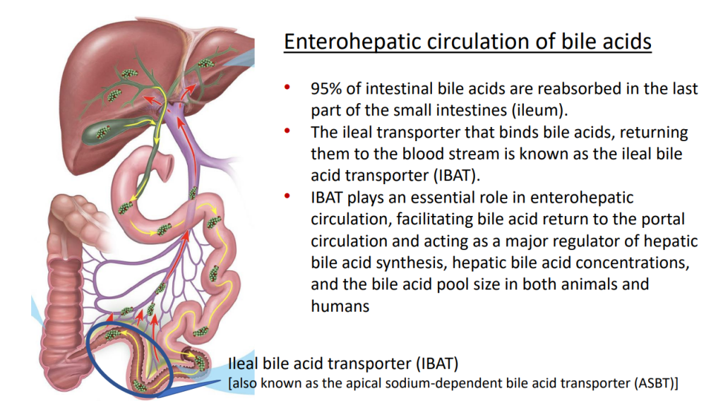
  • IBAT inhibitors block intestinal absorption of bile acids/disrupt enterohepatic circulation; this leads to augmented bile acid excretion in stools
  • IBAT inhibitors may reduce liver damage in the setting of cholestasis/accumulation of toxic bile acids
  • Potential diseases for IBAT inhibitors include Alagille syndrome and PFIC
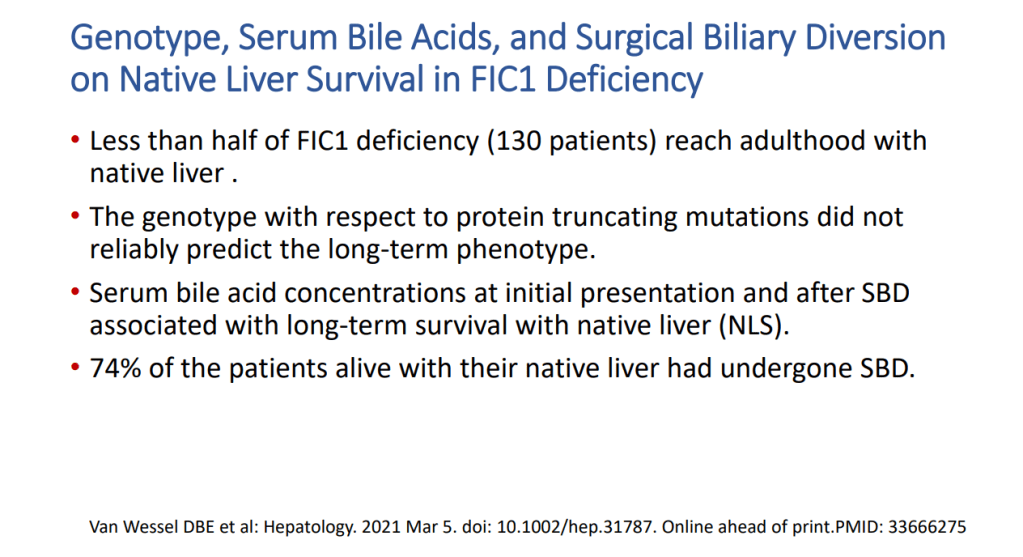
  • Van Wessel et al (J Hepatol 2020; 73: 84-93) correlated survival with PFIC1/PFIC2 with bile acid levels and showed improvement in survival in those with surgical biliary diversion
  • Goals for IBAT inhibitor trials: improvement in pruritus, bile acids, reduced ALT, hepatic fibrosis, HCC and need for liver transplantation
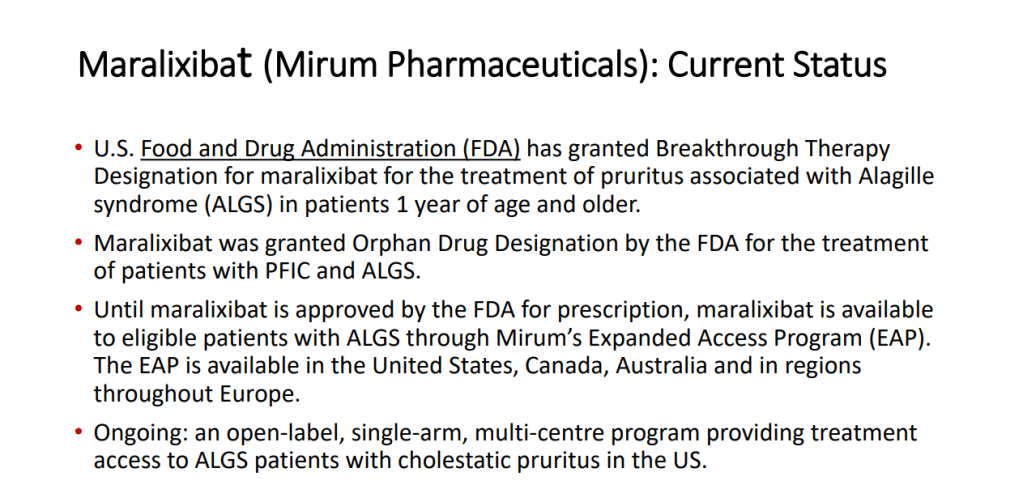
  • Marixibat is available for use as an FDA approved breakthrough medication for Alagille and PFIC2 in pediatric patients older than 1 year
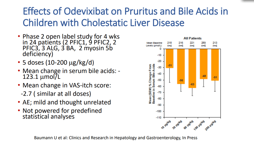
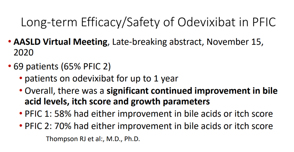
  • Odexibat is designated as an orphan drug for Alagille, PFIC, PBC, and biliary atresia
  • Safety appears good with IBAT inhibitors. Fat soluble vitamin monitoring is needed
This image has an empty alt attribute; its file name is image-132.png
This image has an empty alt attribute; its file name is image-134.png
This image has an empty alt attribute; its file name is image-136.png
This image has an empty alt attribute; its file name is image-138.png
This image has an empty alt attribute; its file name is image-140.png
This image has an empty alt attribute; its file name is image-142.png
This image has an empty alt attribute; its file name is image-144.png
This image has an empty alt attribute; its file name is image-146.png
This image has an empty alt attribute; its file name is image-148.png

Related blog posts:

Case report: Alejandro Velez Lopez

3 yo presented with fatigue and jaundice, 3 weeks after COVID-19 infection. She was not taking any medications.  Labs:  ALT 939, AST 1321, T bili 5.5, D bili 0.9, INR 2, Plts 174, Hgb 12.8, LDH 1297. remained positive for SARS-CoV2 by PCR. Acetaminophen -no exposure.  Evaluation: LKM 1:1280. Neg ANA, NL Ferritin, NL sIL2r, Other viral studies negative, NL IgG. Developed encephalopathy with NH4 317, INR peaked at 2.8.  Treated with steroids, rifaximin and lactulose.  Liver biopsy showed sub-massive necrosis and fibrosis (indicative of  autoimmune hepatitis, likely triggered or exacerbated by COVID-19).  Patient responded to medical therapy and did not require liver transplantation.
Aspen Webinar 2021 Part 2 -Nonalcoholic Steatohepatitis

From the 2nd lecture of the Aspen Webinar. This blog entry has abbreviated/summarized this presentation. Though not intentional, some important material is likely to have been omitted; in addition, transcription errors are possible as well. This was a terrific lecture!

NASH Update Stavra Xanthakos

Key points:

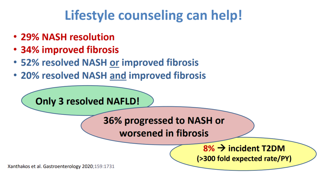
  • Lifestyle intervention is 1st line Rx (especially avoiding sugary beverages, and processed foods). This may lead to resolution of NASH in ~29%, fibrosis resolution in 34%; though, only 3% resolved fatty liver
  • Many have progressive disease despite lifestyle treatment recommendations
  • Consider adjunctive treatments if not improving with lifestyle intervention
  • Vitamin E for biopsy-confirmed disease (often for 1-2 years of therapy)
  • Pioglitazone -off-label for adults (18+). Can increase weight; a lot of trials in adults
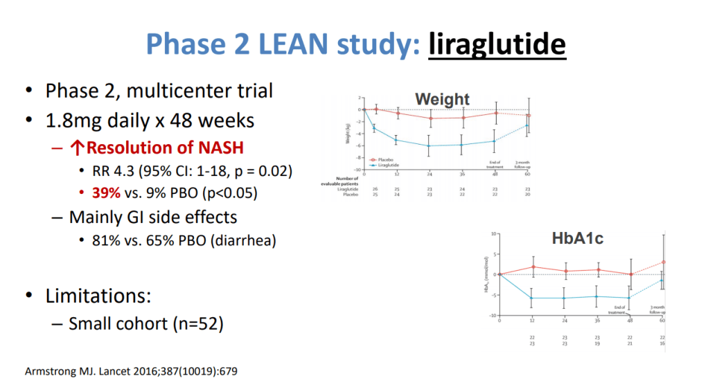
  • Several phase 3 trials in NASH -GLP1 agonists promising in phase 2 trials
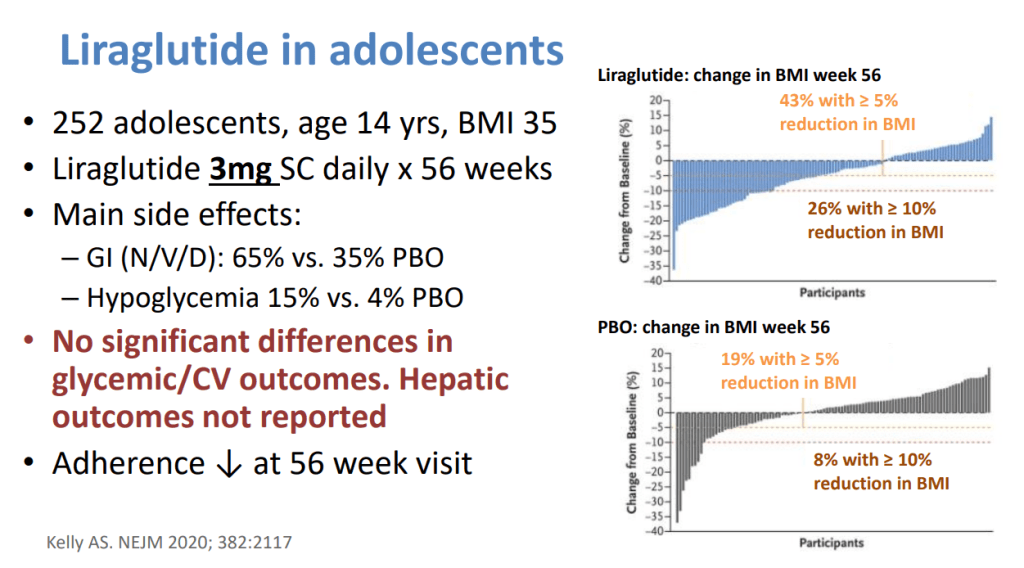
  • GLP1 agents being used in adolescents with T2DM who may also have NASH – monitor liver outcomes
  • Anti-obesity medications may help with weight and perhaps the liver. Approved agents for adolescents include 1) Orlistat -safe, but frequent side effects (eg. Diarrhea, greasy accidents) and 2) Liraglutide -daily SC.  Insurance coverage is limited.
  • Biopsy is important before implementing medications and may influence decision to pursue bariatric surgery.  
  • Fibroscan, if shows no significant fibrosis, can help limit biopsy.
This image has an empty alt attribute; its file name is image-230.png
This image has an empty alt attribute; its file name is image-232.png
This image has an empty alt attribute; its file name is image-234.png
This image has an empty alt attribute; its file name is image-236.png
This image has an empty alt attribute; its file name is image-240.png
This image has an empty alt attribute; its file name is image-238.png
This image has an empty alt attribute; its file name is image-242.png
This image has an empty alt attribute; its file name is image-244.png

Related blog posts:

Aspen Webinar 2021 Part 1-COVID-19 and the Liver

Notes from this year’s Aspen Webinar 2021. This blog entry has abbreviated/summarized some of these presentations. Though not intentional, some important material is likely to have been omitted; in addition, transcription errors are possible as well. The first talk was excellent.

What’s Hot -COVID-19 and the Liver  William Balistreri

This lecture covered a ton of information and publications with regard to COVID-19 and the liver.

Key points:

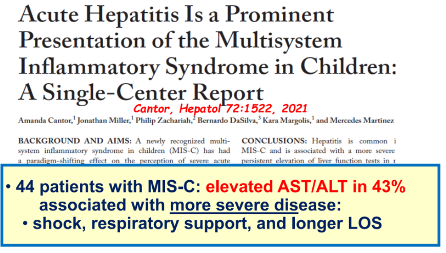
  • Numerous studies generally show that liver problems related to COVID-19 are mild, particularly in children
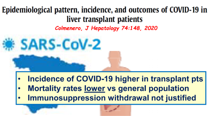
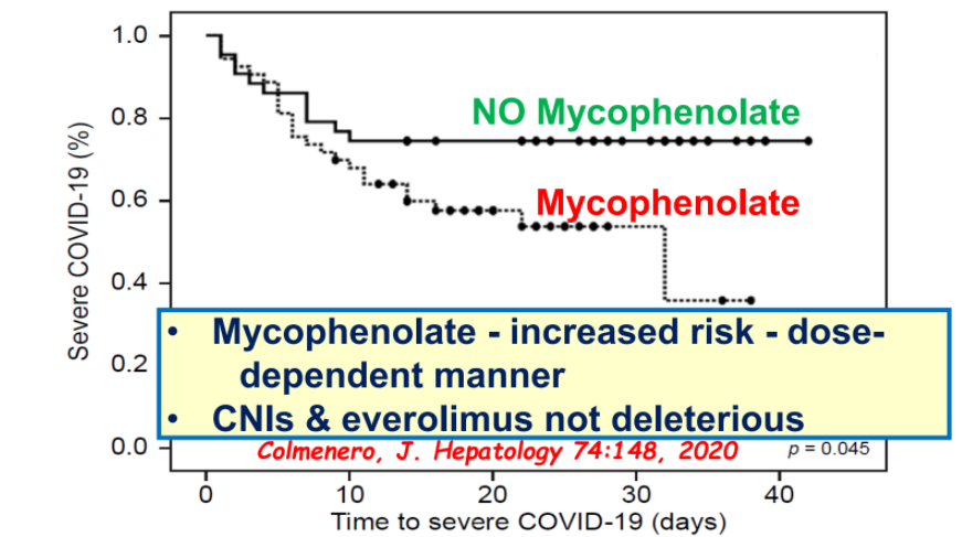
  • Most immunosuppression agents do not worsen outcomes with COVID-19 and should not be reduced in autoimmune hepatitis or liver transplantation; the exception, mycophenolate has been associated with worsened outcomes
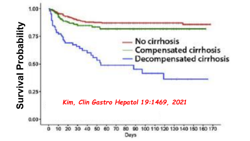
  • Cirrhosis is associated with worse outcomes in patients with COVID-19
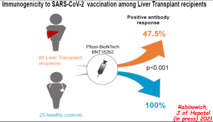
  • Vaccine response is blunted in immunocompromised patients with 40-50% developing antibody response after two doses of mRNA vaccines and generally lower titers.  3rd dose of vaccine may improve response.

Some of the slides:

Pediatric Data:

Hepatic Steatosis as an Incidental Finding

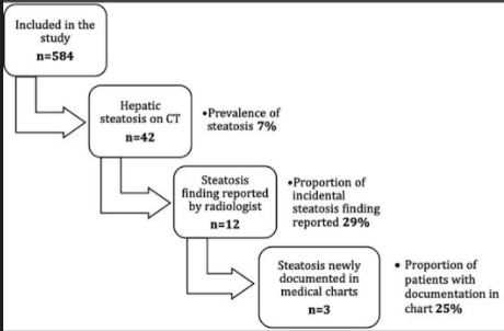
A recent report (H Okura et al. J Pediatr 2021; 234: 92-98. Under-reporting of Hepatic Steatosis in Children: A Missed Opportunity for Early Detection) showed that hepatic steatosis is under-reported as an incidental finding on CT scans which were ordered for nephrolithiasis.

Key findings:

  • Using absolute liver attenuation <48 Hounsfield units (HU), the prevalence was 7% (n = 42/584). Steatosis was reported for only 12 of 42 (28%) of these patients and was documented in clinical notes in only 3 of those cases
  • 232 (40%) had liver enzymes available within 24 hrs of CT scan. 79 had elevated ALT values; steatosis accounted for only 22% of those with elevated ALT values
  • In those with liver attenuation < 48 HU, nearly all had abnormal ALT values and the median was 52 U/L
  • Patients with steatosis had an 8-fold likelihood of overweight/obesity

Discussion:

  • These findings are similar to an adult study of 1290 patients in which 26% had steatosis but only 5% had those findings identified and documented beyond the radiology report. (N Kutaiba et al. J Med Imaging Radiat Oncol 2019; 3: 431-8)
  • CT findings are considered much more accurate than ultrasonography.
  • The authors argue that identification of NAFLD is “crucial” to allow for further specialty evaluation and to exclude secondary causes of steatosis.

My take: This study shows that there is an opportunity to improve identification of incidental steatosis. If identified, this can/should be addressed by their primary care team to emphasize improved diet choices and physical activity.

Related blog posts:

From Journal of Pediatrics Twitter Feed

Algorithm for Neonatal Acute Liver Failure

K Borovsky et al. JPGN 2021; 73: 80-85. Applying an Age-specific Definition to Better Characterize Etiologies and Outcomes in Neonatal Acute Liver Failure

This single-center retrospective study with 43 patients (over 11 year timeframe) identified etiology and outcomes for neonatal acute liver failure (NALF).

Key findings:

  • Etiologies included viral infection (23%), gestational alloimmune liver disease with neonatal hemochromatosis (GALD-NH) (21%), cardiac-associated ischemia (16%), other ischemia (14%), genetic etiologies (9%), Trisomy 21-associated myelodysplasia (TAM) (7%), hemophagocytic lymphohistiocytosis (HLH) (2%), and not identified (7%)
  • Infants with viral etiologies had the highest alanine aminotransferase (ALT) at presentation (1179 IU/L, interquartile range [IQR] 683–1585 IU/L) in contrast to low levels in GALD-NH (23 IU/L, IQR 18–64 IU/L)
  • Across all etiologies, only 33% were alive at 1 year
  • Among laboratory values at presentation, alpha-fetoprotein (AFP) was significantly higher in patients that survived with their native liver (P = 0.04)

Figure 4 provides a helpful management algorithm for NALF. Figure 4 is similar to the slide below (shared by lead author).

  • -Consider empiric acyclovir in those with INR >/= 2.0 in the first 30 days of life
  • -In those with normal to low elevation of aminotransferases, consider empiric IVIG while undergoing workup. Part of workup should include either MRI or lip biopsy for GALD
  • -In those with moderate to severe elevation of aminotransferases, workup should include assessment for viral, HLH and genetic etiologies. Fulminant viral hepatitis or HLH likely with Ferritin levels >10,000. Hypoglycemia and hyperammonemia is suggestive of metabolic/mitochondrial disorder
  • -Liver biopsy may be needed if etiologies not identified

Related blog posts:

Disclaimer: This blog, gutsandgrowth, assumes no responsibility for any use or operation of any method, product, instruction, concept or idea contained in the material herein or for any injury or damage to persons or property (whether products liability, negligence or otherwise) resulting from such use or operation. These blog posts are for educational purposes only. Specific dosing of medications (along with potential adverse effects) should be confirmed by prescribing physician.  Because of rapid advances in the medical sciences, the gutsandgrowth blog cautions that independent verification should be made of diagnosis and drug dosages. The reader is solely responsible for the conduct of any suggested test or procedure.  This content is not a substitute for medical advice, diagnosis or treatment provided by a qualified healthcare provider. Always seek the advice of your physician or other qualified health provider with any questions you may have regarding a condition.

Why It Is Hard to Stop Immunosuppression with Autoimmune Hepatitis and Lower Bone Density with Fatty Livers

C Schulthei et al. Hepatology 2021; 73: 1436-1448. Full text: Next-Generation Immunosequencing Reveals Pathological T-Cell Architecture in Autoimmune Hepatitis

This is highly technical study of 60 patients with AIH. “Our key finding was a clearly biased signature of TRBV-J gene usage in peripheral and liver-infiltrating T-cells of patients with AIH, independent of AIH predisposing HLA-DRB1 alleles. This signature was unaffected by immunosuppressive treatment and not related to complete biochemical disease remission. This suggests that treatment acted on T-cell functionality rather than on the underlying pathological T-cell architecture in this disease that has a high relapse rate.”

My take (borrowed from authors): “Patients with AIH show profound and persisting T-cell architectural changes that may explain high relapse rates after tapering immunosuppression.”

Related blog posts:

LF Chun et al. J Pediatr 2021; 233: 105-111. Hepatic Steatosis is Negatively Associated with Bone Mineral Density in Children

Key findings:

  • Using a community-based sample of 235 children, the authors found that there was a significant negative relationship between liver MRI-PDFF and BMD z score R = −0.421, P < .001).
  • There was no significant association between vitamin D status and BMD z score (P = .94).
  • Children with clinically low BMD z scores were found to have higher alanine aminotransferase (P < .05) and gamma-glutamyl transferase (P < .05) levels compared with children with normal BMD z scores.

My take: This study shows another organ that is affected in children with fatty liver disease; other associated problems include increased risk for cardiovascular disorders, pancreatic dysfunction/diabetes, cancer, polycystic ovary syndrome, and neurologic disorders

Related blog posts:

Image below near the Seven Mile Road on the Florida Keys:

Liver Shorts: Stopping immunosuppression after transplant, toxicity of acetaminophen at therapeutic dosing, best imaging of PSC

S Feng et al. Hepatology 2021; 73: 1985-2004. Efficacy and Safety of Immunosuppression Withdrawal in Pediatric Liver Transplant Recipients: Moving Toward Personalized Management. iWITH study found that in highly-selected patients (n=88), immunosuppression could be withdrawn in 38%. Criteria included consistently normal liver biochemistries, >4 years after transplantation, and transplants unrelated to autoimmune etiologies or HBV/HCV.

A Louvet et al. Hepatology 2021; 73: 1945-1955. Acute Liver Injury With Therapeutic Doses of Acetaminophen: A Prospective Study This prospective study (2002-2019) showed that 89 of 400 adult patients with acetaminophen-induced acute liver injury (ALI) had received therapeutic doses of acetaminophen (<6 g). Especially in individuals with underlying alcoholic liver disease or fasting, acetaminophen (more than 2 gm/day) can trigger liver injury.

JE Eaton et al. Hepatology 2021; 73: 1868-1881. Early Cholangiocarcinoma Detection With Magnetic Resonance Imaging Versus Ultrasound in Primary Sclerosing Cholangitis This multicenter retrospective study showed that MRI is superior to ultrasound for the detection of early-stage CCA in patients with PSC. Identification of CCA before the onset of symptoms with MRI is associated with improved outcomes. The authors note that individuals diagnosed with CCA while asymptomatic had a 36% reduction in 5-year mortality and that MRI allowed for a 77% reduction in 5-year mortality among asymptomatic persons. One important limitation of this study would be lead-time bias; that is, those with disease detected at an earlier stage will live longer than those detected at a later stage and thus earlier diagnosis may be ascribed as conferring a greater longevity even with no intervention.

Long-Term Outcomes in Children with Choledochal Malformations

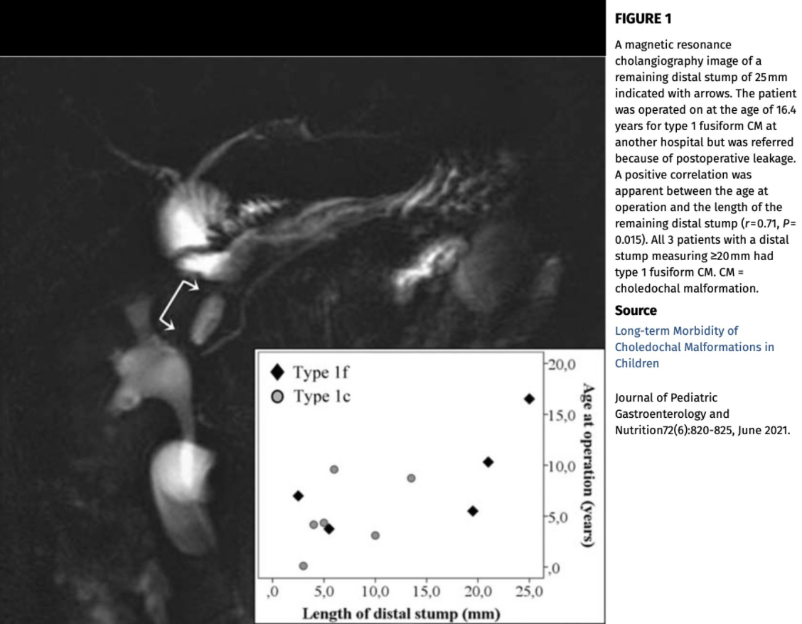
I Hyvarinen et al JPGN 2021; 72: 820-825. Long-term Morbidity of Choledochal Malformations in Children

This single-center retrospective study (n=55 median f/u 6 years) provides data on long-term morbidity of choledochal malformations. Key findings:

  • 21% had long-term complications including cholangitis in 9 (>2 episodes in 5) patients, anastomotic stricture in 2, adhesive volvulus in 1 and hepatocellular carcinoma in 1. 
  • Magnetic resonance cholangiography (MRCP) performed 6.4 (3.6–16) years after hepaticojejunostomy, diameters of both main intrahepatic ducts had decreased significantly to 3.0 (2.5–3.5) mm (P = 0.0001) but a distal cyst stump was remaining in 30% with a length of 6.0 (4.0–20) mm

My take: Despite surgical management (hepaticojejunostomy), biliary tract malignancy is still possible in patients with choledochal cysts. Regular CA 19-9 testing is probably worthwhile, especially in teens and older. The authors note that in patients with type 1 choledochal malformations, some have recommended annual liver biochemistries and ultrasonography following successful surgery (J Gastroenterol Hepatol 2019; 34: 966-974).

Figure from JPGNonline twitter feed:

What is the likelihood of an alternative liver etiology in children with obesity/overweight?

T Yodoshi et al. Pediatrics: 2021, 147 (4) e2020009829; DOI: https://doi.org/10.1542/peds.2020-009829. Alternative Etiologies of Liver Disease in Children With Suspected NAFLD

METHODS: Multicenter, retrospective cohort study of patients aged ≤18 years with overweight and obesity and evidence of elevated serum aminotransferases and/or hepatic steatosis on imaging, referred for suspected NAFLD to Cincinnati Children’s Hospital Medical Center (2009–2017) or Yale New Haven Children’s Hospital (2012–2017). Testing was performed to exclude the following: autoimmune hepatitis (AIH), Wilson disease, viral hepatitis (B and C), thyroid dysfunction, celiac disease, α-1 antitrypsin deficiency, and hemochromatosis

RESULTS: A total of 900 children with overweight and obesity (63% boys, 26% Hispanic ethnicity) were referred, with a median age of 13 years (range: 2–18). Most had severe obesity (n = 666; 76%) with a median BMI z score of 2.45 (interquartile range [IQR]: 2.2–2.7). Median alanine aminotransferase level at presentation was 64 U/L (IQR: 42–95). A clinically indicated liver biopsy was performed in 358 children (40%) at a median of 6 months (IQR: 1–14) post initial visit; of those, 46% had confirmed nonalcoholic steatohepatitis. Positive autoantibodies were observed in 13% of the cohort, but none met criteria for AIH. Only 19 (2%) were found to have other causes of liver disease, with no cases of viral hepatitis or Wilson disease detected.

Specific diseases included thyroid dysfunction in 11 (1.2%), celiac disease in 3 (0.4%), A1AT deficiency in 3 (0.4%), and non-Hodgkin’s lymphoma in 1. A prior study had indicated that AIH was the second-most common etiology (Aliment Pharmacol Ther 2013; 38: 1267-77); this study indicated a much higher rate of alternative diagnosis (24%) in children undergoing a liver biopsy for suspected NAFLD (see related blogs: Screening for NAFLD and Concise Review: Fatty Liver in Pediatrics).

My take: The yield of extensive testing in children with suspected fatty liver disease is very low. I suspect that a cost-effective analysis would indicate a much more limited role for further liver evaluations.

Related blog posts:

Sisyphus -Rock Art

Genetic Diagnostic Tools for Cholestasis

Several articles in a recent JPGN (May issue) delve into the topic of using genetic tools for the diagnosis of cholestatic conditions. The most important, in my view, of these articles is JPGN 2021; 72: 654-660. (Use of a Comprehensive 66-Gene Cholestasis Sequencing Panel in 2171 Cholestatic Infants, Children, and Young Adults); congratulations to the lead author, my colleague Saul Karpen.

Key findings:

  • Between February 2016 and December 2017, 2171 results were reported. Median turnaround time was 21 days.
  • 583 pathogenic (P) variants, 79 likely pathogenic (LP) variants, and 3117 VOUS; 166 P/LP variants and 415 VOUS were novel.
  • The panel’s overall diagnostic yield was 12% (n = 265/2171) representing 32 genes with mutations identified (the panel tested up to 66 genes).
  • The top five genetic diagnoses for the group, in order: JAG1 + NOTCH2 (Alagille syndrome), ABCB11SERPINA1ABCB4, and POLG. (Table 3 lists all of the findings)

Other reports in the same issue describe a normal-GGT cholestasis due to USP53 deficiency

My take: Genetic cholestasis panels and/or whole exome sequencing are very useful and are being incorporated earlier into diagnostic workups.

Related blog posts:

Near Lullwater Park, Atlanta