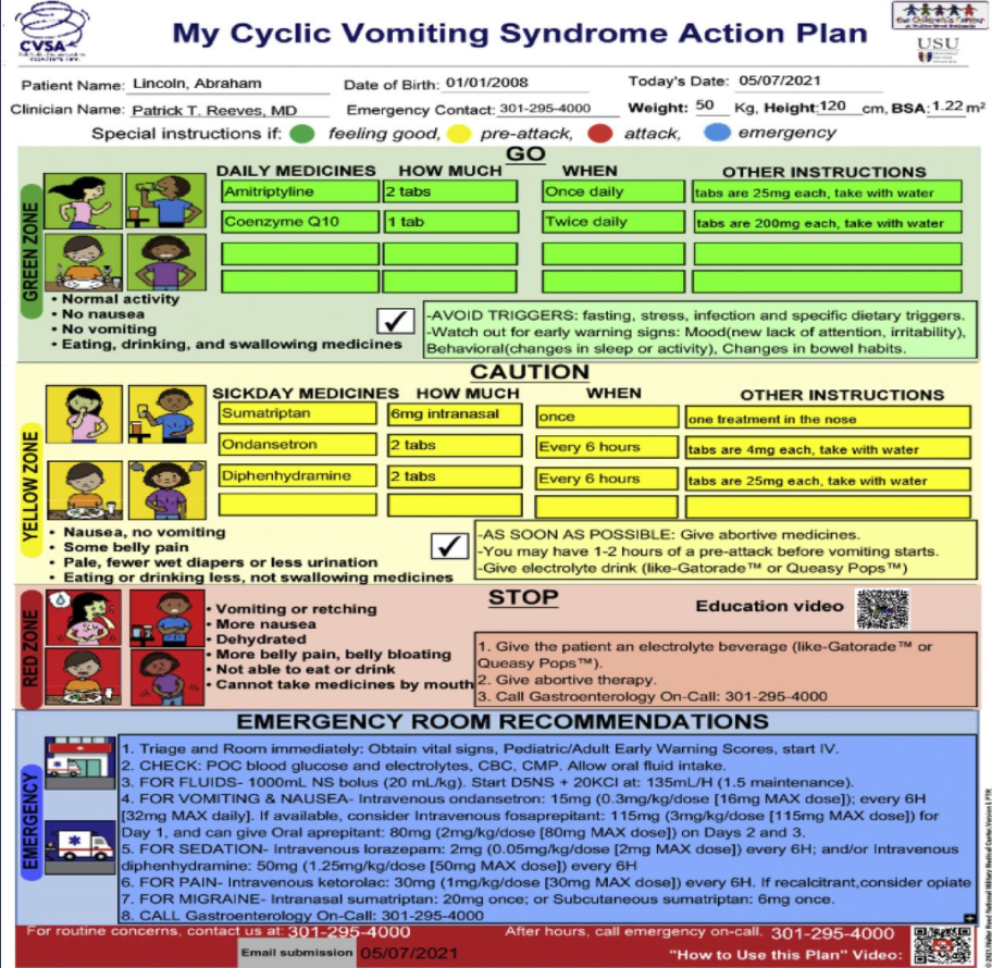
Similar to the constipation action plan (see blog link below), the authors have created a stepwise pictographic CVS action plan (CVSAP).
Image is from Pat Reeves twitter feed and corresponds to figure in study (pg 175)
Key points:
A composite readability score of 5.32 was consistent with a fifth-grade level.
Patients/caregivers (n = 70) judged the CVSAP to be of high quality with consumer information rating form rating of 84.2%
Six medical librarians rated the CVSAP to have 93% understandability and 100% actionability, and 33 clinicians completing the SAM generated a suitability rating of 87.5%
On the listed ED management, the authors note “consider fosaprepitant…and can give oral aprepitant on days 2 and 3.” It should be noted that oral dosing afterwards is generally not required as fosaprepitant can last 2-3 days after a single dose. In addition, many use a maximum dose of 150 mg rather than 115 mg. Also, the ED dosage of several agents need to be tailored to the individual based on weight and other medications. Lower doses of many of the medications in the protocol are often effective.
My take: Patients with cyclic vomiting syndrome, like those with constipation, are likely to benefit from clearly articulated plans for maintenance treatment, escalation approaches and for ED management. The need for ED management may lessen with more consistent treatment approaches.
Disclaimer: This blog, gutsandgrowth, assumes no responsibility for any use or operation of any method, product, instruction, concept or idea contained in the material herein or for any injury or damage to persons or property (whether products liability, negligence or otherwise) resulting from such use or operation. These blog posts are for educational purposes only. Specific dosing of medications (along with potential adverse effects) should be confirmed by prescribing physician. Because of rapid advances in the medical sciences, the gutsandgrowth blog cautions that independent verification should be made of diagnosis and drug dosages. The reader is solely responsible for the conduct of any suggested test or procedure. This content is not a substitute for medical advice, diagnosis or treatment provided by a qualified healthcare provider. Always seek the advice of your physician or other qualified health provider with any questions you may have regarding a condition
This retrospective study (n=45) shows that supplemental water added to blenderized tube feeds may have detrimental effects.
Key finding:
Patients receiving <20% thin liquids were less likely to undergo chest X-rays during follow-up than patients receiving larger amounts of thin liquids (10% in the minimal thin group versus 48% in the greater thin group, P = 0.03)
This relationship remained significant after controlling for underlying pulmonary disease, aspiration, method of feed administration (bolus or continuous feeds), fundoplication status, and oral intake status. CXRs likely indicate concern for pulmonary outcomes related to feedings.
From JPGN twitter feed
My take: Many thick formulas may be difficult to administer via GT. However, using too much water may hinder the benefits of a blenderized diet. Larger prospective studies are needed to determine optimal viscosity diets in these vulnerable populations.
The Boston group has several related articles (Thanks to Alison Miller for sharing these articles):
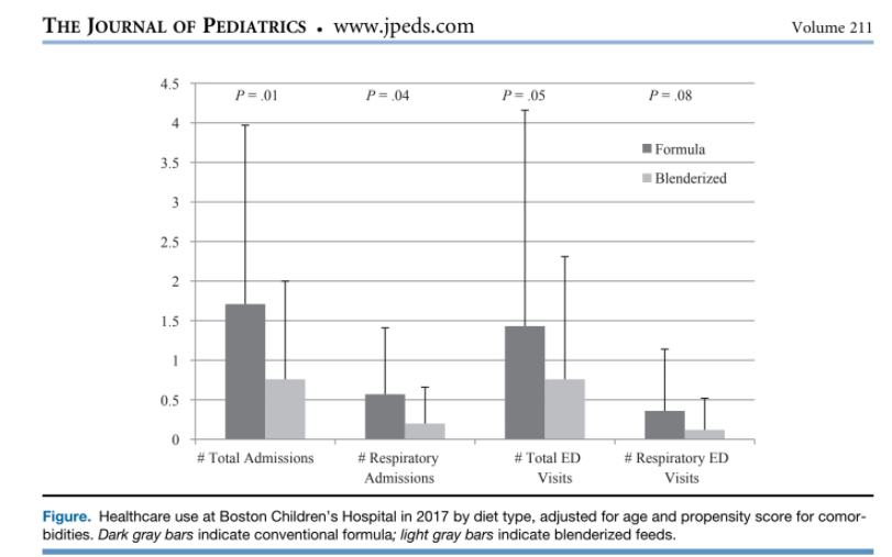
B Hron et al. J Pediatr 2019;211:139-45. Health Outcomes and Quality of Life Indices of Children Receiving Blenderized Feeds via Enteral Tube
Blenderized diets were associated with decreased healthcare use, improved symptom scores, and increased patient satisfaction compared with conventional formulas.
B Hron, R Rosen. JPGN 2020; 70: e124–e128. Viscosity of Commercial Food-based Formulas and Home-prepared Blenderized Feeds
This article shows that adding 90 mL of water can reduce viscosity of blenderized formula from >6000 cP to ~1000 cP. The authors suggest that those patients with significant reflux may benefit from higher viscosity formulas: “Low viscosity formulas such as Kate Farms and Compleat may not be ideal for patients fed via gastrostomy with significant reflux, in whom extremely thick or possibly moderately thick liquids may have a beneficial impact.”
Commercial food-based formulas vary even more widely, with some meeting criteria for thin liquids (Kate Farms Pediatric 1.2 and Compleat Pediatric), slightly thick (Harvest), mildly thick (Nourish), moderately thick (Compleat Organic Blends, Liquid Hope), and extremely thick (Real Food Blends).
Specific viscosity (cP) listed in Table 1 of this article:
Levine et al provide a good overview of the topic of emulsifiers. Key points:
Emulsifiers allow “the mixing of water and and water-soluble agents with fats and fat-soluble agents that is they possess both hydrophilic and lipophilic properties”
The FDA “has been responsible for approving the use of all direct food additives” (n=~3000) and “for regulatory purposes, [the FDA excluded] some substances that were generally regarded as safe (GRAS) (n=~450)…Precisely how some emulsifiers gained GRAS status is unclear.
“Lecithin” is derived from the Greek name for egg yolk (lekithos). “Over the years the use of the term “lecithin” has been taken to include various mixtures of different phospholipids” (not just phosphatidylcholine).
Lecithin can provide the substrate “for the production of trimethylamine N-oxide (TMAO)…linked to cardiac events and cardiovascular inflammation.”
“The list of emulsifiers that are widely used, but not considered GRAS, most notably include polysorbate 80 (p80), carboxymethylcellulose (CMC) and carrageenan…these emulsifiers have been linked to the disruption of the microbiota and gut mucosal lining…In addition, low-grade inflammation [has been] associated with consumption of emulsifying agents such as CMC and p80” [in mouse models].
The International Organization for the Study of Inflammatory Bowel Disease (IOIBD) has recommended that IBD patients “limit consumption of certain commonly encountered synthetic emulsifiers, specifically carboxymethylcellulose (E466/cellulose gum) and polysorbate 80 (E433) [which] are present in many processed foods, such as ice cream. The group also recommends a decrease in foods containing carrageenan”
In the second study by Chassaing et al with 16 healthy adults, the authors studied the effects of CMC in those with an emulsifier-free diet (n=9) or an identical diet enriched with CMC (n=7).
Key findings:
Relative to control subjects, CMC consumption modestly increased postprandial abdominal discomfort and perturbed gut microbiota composition in a way that reduced its diversity
CMC-fed subjects exhibited changes in the fecal metabolome, particularly reductions in short-chain fatty acids and free amino acids
2 subjects consuming CMC who exhibited increased microbiota encroachment into the normally sterile inner mucus layer, a central feature of gut inflammation, as well as stark alterations in microbiota composition
My take: The dramatic increase in the prevalence of IBD over the past 50 years indicates a strong influence of environment factors, particularly diet. Determining which of these factors are most important will be challenging. These articles indicate that some emulsifiers could be contributing to GI tract inflammation and non-GI tract inflammation as well.
The challenges with identifying dietary factors relate to difficulties with using randomized controlled trials (especially eliminating delicious foods) to assess the impact over a long period of follow-up.
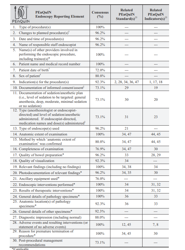
Several articles in a recent JPGN supplement issue describe the efforts to develop quality standards and indicators for pediatric endoscopy. All of these articles are open access.
My view: These detailed articles provide a good framework for improving pediatric endoscopy. After assuring that the facility and personnel are high quality, the pediatric endoscopist has the important responsibility of providing a high quality endoscopy. We need to strive to provide the best care for every single child entrusted in our care. In my view, the most important step is having an appropriate indication and despite guidelines, this remains highly subjective.
Growing up, I heard a number of Paul Harvey broadcasts on the radio. Often there would be an important twist at the end and he would conclude with ‘and that’s the rest of the story.’
This came to mind after reading a recent article on celiac disease and hepatitis B infection:
A cross-sectional study using the National Health and Nutrition Examination Survey (NHANES) database (2009–2014)
And a retrospective analysis of HBV infection in two cohorts: Mayo Clinic cohort (1998–2021) and the Rochester Epidemiology Project cohort (REP; 2010–2020)
Key findings:
Based on NHANES database, the rate of HBV infection in the United States was 0.33%
Of 93 patients with CD, 46 (49%) were vaccinated for HBV and of the remaining 19,422 without CD, 10,228 (53%) were vaccinated
Twenty-two (48%) vaccinated patients with CD had HBV immunity and 4405 (43.07%) vaccinated patients without CD had HBV immunity
In NHANES data, there were no cases of HBV infection in patients with CD. Among the 3568 patients with CD seen at Mayo Clinic and 3918 patients with CD in the REP database, only four (0.11%) at Mayo Clinic and nine (0.23%) of the REP patients had HBV infection.
This finding is probably applicable to other conditions in which HBV immunity is ascertained.
My take: In contrast to other small studies, this study showed that the “rate of HBV vaccination and immunity was similar in individuals with and without CD.” In addition, there was no increased risk of HBV infection detected in CD patients. Thus, testing for HBV is not necessary in patients with CD.
Methods: Prospective enrollment (n=146) included a ADA and those with a nonmedical switch from the ADA originator (n=98). Clinical remission and safety were assessed at baseline and at 3, 6, and 12 months
Key finding:
In the naïve cohort, the overall remission rate at 12 months was 60% (similar to originator adalimumab results); the remission rate in the switching cohort it was 75% with a treatment persistency of 82% at 12 months after the switch
No differences were found in terms of ADA serum trough levels at baseline, 3, and 6 months after switching. No patient developed antidrug antibodies after the switch
Fecal calprotectin (FC) values trended lower in both cohorts. In the naive cohort, the mean value of FC dropped from 665 (baseline) to 231 at 12 months. In the switch cohort, the mean value of FC dropped from 212 (baseline) to 84 at 12 months
My take: In this cohort, SB5 biosimilar for adalimumab was effective and safe.
Because our office is one of the centers participating in a mirikizumab study for adolescents, I was particularly interested in seeing the published results of a phase 2 study in 191 adults.
Background: “Mirikizumab (LY3074828) is a humanized immunoglobulin G4 (IgG4)–variant monoclonal antibody that binds specifically to the p19 subunit of IL23 and has demonstrated efficacy in psoriasis and ulcerative colitis, and is currently in phase 3 testing for psoriasis, ulcerative colitis, and CD. We evaluated the efficacy and safety of mirikizumab for the treatment of patients with moderately-to-severely active CD”
Methods: Patients (N = 191) were randomized (2:1:1:2) to receive placebo (PBO), 200, 600, or 1000 mg mirikizumab, administered intravenously (IV) every 4 weeks. Patients who received mirikizumab and achieved ≥1 point improvement in Simple Endoscopic Score-CD at Week 12 (rerandomized maintenance cohort) were rerandomized to continue their induction IV treatment (combined IV groups [IV-C]) or receive 300 mg mirikizumab subcutaneously (SC) every 4 weeks. Nonrandomized maintenance cohort included endoscopic nonimprovers (1000 mg) and PBO patients (PBO/1000 mg) who received 1000 mg mirikizumab IV from Week 12. The primary objective was to evaluate superiority of mirikizumab to PBO in inducing endoscopic response (50% reduction from baseline in Simple Endoscopic Score-CD) at Week 12.
**approximately two thirds of participants had received biologic therapy and approximately half of all patients in this trial having experienced at least 1 biologic failure
Key findings:
At Week 12, endoscopic response was significantly higher for all mirikizumab groups compared with placebo (PBO) (200 mg: 25.8%, P = .079; 600 mg: 37.5%, P = .003; 1000 mg: 43.8%, P < .001; PBO: 10.9 %).
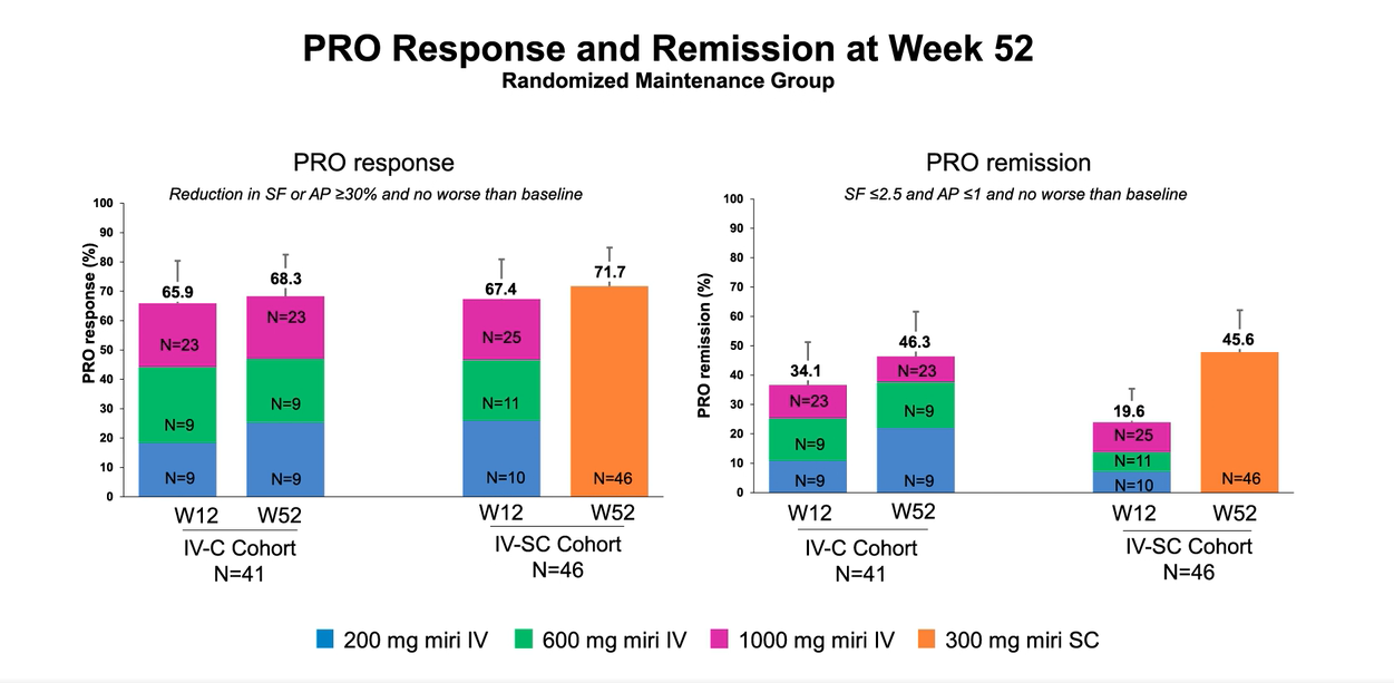
Endoscopic response at Week 52 was 58.5% (24/41) and 58.7% (27/46) in the IV-C (combined IV groups) and SC (subcutaneous) groups , respectively. See 4th and 6th slides below which show that those with response at 12 weeks continued with response at 52 weeks.
In the Non-Randomized group which included non-improvers and placebo, they received the highest dose, 1000 mg. A significant number of non-improvers responded at week 52.
My take: In this study of adults, with moderate to severe Crohn’s disease, Mirikizumab showed good efficacy and safety at both 12 weeks and 52 weeks. Because about half of the participants were biologic failures, this indicates that this agent shows promise in those with refractory disease.
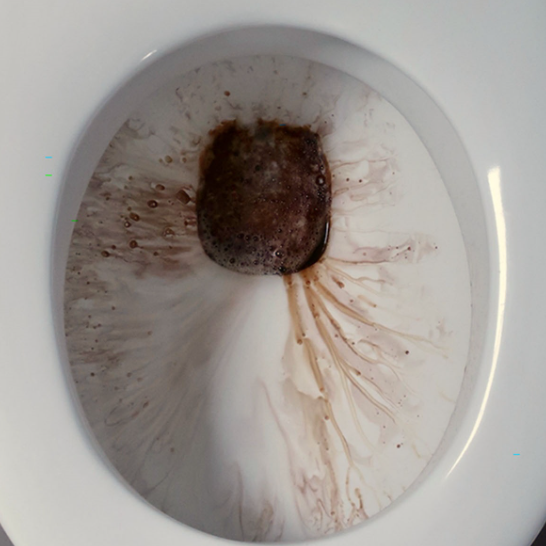
“The boy noticed the colorization only when he urinated in the toilet at home and this phenomenon was most pronounced after the toilet had been cleaned.” He had a normal urinalysis. “Discoloration of urine by a chemical reaction between mesalamine and sodium hypochlorite bleach has been widely reported in online patient forums, we only found 2 related case reports.”
“Mesalamine and its metabolite, N-acetyl-5-aminosalicylic acid, have structural similarity to methyldopa, which is metabolized to melanin-like compounds. In an alkaline environment (the pH of bleach is 11–13), polymerization of these melanin-like metabolites causes a brownish/red discoloration of urine after methyldopa ingestion.”
Chris Fritzen and Erica Riedesel passed along a case report: a 16 mo presented to our hospital system with coughing, gagging and hematemesis after an unwitnessed FB ingestion; she was emergently taken to the OR where a button battery was noted to be densely adherent to the posterior wall of the upper thoracic esophagus and removed with a flexible endoscope. Postoperatively, she received 7 days of ampicillin/sulbactam. 14 days after presentation, an MRI revealed esophageal injury was evident at T3 level as well as an abnormal signal was seen within the esophagus spanning C7-T4. In addition, there was edema and enhancement of the intervertebral disc space at T1-T2 and T2-T3 consistent with spondylodiscitis. Prior reports (see below) have emphasized the need for antimicrobial coverage for Staphylococcus and upper respiratory pathogens. Children with spondylodiscitis may present with refusal to walk or limping, back or leg pain, and local stiffness of the neck/spine.
My take: Spondylodiscitis is another rare complication following button battery ingestion.
References:
NEO Grey et al. Pediatr Radiol 2021; 51:1856–1866. In this report, 1 of 23 children had spondylodiscitis. Other complications: esophageal perforation (n=11), tracheoesophageal fistula (n=3)
V Kieu et al. J Pediatr 2014; 164: 1500-1501. Highlights increased risk from unwitnessed button battery ingestions.
H Eshaghi et al. Pediatr Emer Care 2013;29: 368Y370. Spondylodiscitis: A Rare Complication of Button Battery Ingestion in a 10-Month-Old Boy
Related blog posts:
Foreign Bodies in Children -Expert Guidance Even with “spent” batteries, there is enough residual charge to cause injury and all ingestions (even if asymptomatic) into the esophagus require emergent removal. If these batteries are in the stomach & asymptomatic, urgent removal is recommended if age < 5 years and BB ≥20 mm.
Disclaimer: This blog, gutsandgrowth, assumes no responsibility for any use or operation of any method, product, instruction, concept or idea contained in the material herein or for any injury or damage to persons or property (whether products liability, negligence or otherwise) resulting from such use or operation. These blog posts are for educational purposes only. Specific dosing of medications (along with potential adverse effects) should be confirmed by prescribing physician. Because of rapid advances in the medical sciences, the gutsandgrowth blog cautions that independent verification should be made of diagnosis and drug dosages. The reader is solely responsible for the conduct of any suggested test or procedure. This content is not a substitute for medical advice, diagnosis or treatment provided by a qualified healthcare provider. Always seek the advice of your physician or other qualified health provider with any questions you may have regarding a condition.