1000th Tweet: GI Symptoms Preceding IBD Diagnosis

Another milestone for this blog: since 2012, the blog has been publicized on twitter; this is the 1000th tweet. It is also 1314th blog post over nearly 4 years.

A recent study (H Singh et al. Clin Gastroenterol Hepatol 2015; 13: 1302-09) indicates that children with inflammatory bowel disease (IBD) were more likely to have gastrointestinal symptoms in each of the 4 years before the diagnosis of IBD than children without IBD.

In this study, the researchers identified all children with IBD from a population-based Manitoba database; Manitoba had a population of 1.27 million in 2012.  651 children were matched with 5950 controls without IBD.  The study’s Table 1 & 2 indicates that children with IBD had increased clinic visits prior to diagnosis:

  • 54-66 months prior: standardized rate ratio for number of ambulatory visits 1.15; & for ≥1 visit due to GI symptoms odds ratio 1.44
  • 42-54 months prior: standardized rate ratio for number of ambulatory visits  1.22; & for ≥1 visit due to GI symptoms odds ratio 2.05
  • 30-42 months prior: standardized rate ratiofor number of ambulatory visits 1.19; & for ≥1 visit due to GI symptoms odds ratio 2.16
  • 18-30 months prior: standardized rate ratio for number of ambulatory visits 1.23; & for ≥1 visit due to GI symptoms odds ratio 2.93
  • 6-18 months prior: standardized rate ratio for number of ambulatory visits  1.15; & for ≥1 visit due to GI symptoms odds ratio 5.23

There was not a clear trend in increased symptoms between those who developed Crohn’s disease compared with Ulcerative Colitis. In addition, the study noted a trend towards decreased colectomy and resective surgery in Crohn’s in the time period 2002-2010 compared with 1987-2001.  One limitation of this study is the few number of pediatric gastroenterologists in Manitoba (only 1 before 2003); the lack of pediatric gastroenterology availability could impact timely diagnosis.

My take: This data shows that GI symptoms still predate diagnosis in many children and indicate a potential for diagnosis delay. The authors note that noninvasive tools like stool calprotectin have not been widely adopted (at least in Manitoba) and could be helpful in reducing diagnostic delays.

Estes Park, Colorado
Estes Park, Colorado

Higher Stool Infliximab Correlates with Poor Response in Severe Ulcerative Colitis

A recent study (full text link: “Loss of Infliximab into feces is associated with lack of response to therapy in patients with severe ulcerative colitisGastroenterol 2015; 149; 350-55.e2) provides information about patients with ulcerative colitis who do not respond well to infliximab therapy.

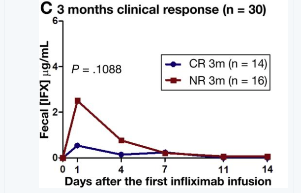
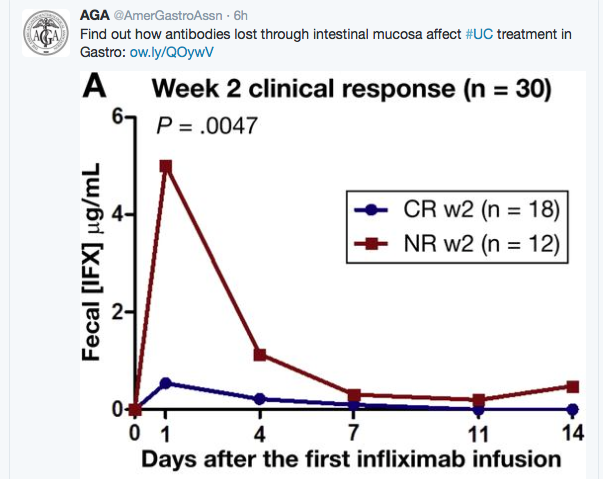
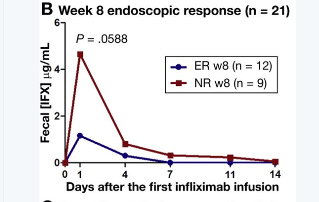
In this study, the authors obtained fecal samples from 30 consecutive patients with moderate to severe UC during the 1st 2 weeks of therapy.  In addition, they obtained serum infliximab levels as well as assessed clinical and endoscopic response at 2 weeks, 8 weeks, and 3 months after treatment began.

Key findings:

  • Fecal infliximab was detected in 129 of 195 (66%) samples.  The greatest loss was observed approximately 2 days after infusion. Low serum albumin was associated with greater infliximab levels in the stool.
  • Clinical nonresponders at week 2 had significantly higher fecal infliximab
  • The authors did not observe a correlation between fecal and serum infliximab concentrations.  However, it is possible that stool losses could indicate lower mucosal concentrations of infliximab.
From AGA twitter account

From AGA twitter account

From AGA twitter account

From AGA twitter account

From AGA twitter feed

From AGA twitter feed

Bottomline: It is not clear whether stool losses of infliximab directly contribute to drug failure or whether the loss is another biomarker of disease activity/high-risk patients.

The study authors note that “intestinal loss of IFX in moderate to severely active UC is associated with a diminished response to this treatment.  Patients with severe disease can, therefore, benefit from more intensive dosing regiments. This strategy warrants a prospective clinical trial.”

Related blog posts:

Head-to-Head: Nutritional Therapy versus Biological Therapy in Pediatric Crohn’s Disease

The best data to date: D Lee et al. Inflamm Bowel Dis 2015; 21: 1786-93. In this prospective study, the authors studied treatment initiation in children (N=90), comparing partial enteral nutrition (PEN, n=16), exclusive enteral nutrition (EEN, n=22), and anti-TNF therapy (n=52).

Results:

  • Clinical response, defined by PCDAI reduction ≤15 or final PCDAI ≤10, was achieved by 64% PEN, 88% EEN, and 84% anti-TNF.
  • Fecal calprotectin ≤250 noted in 14% PEN, 45% EEN, and 62% anti-TNF

Because of the discrepancy between EEN and PEN, the authors speculate that the “efficacy of EEN may be a consequence of elimination of table food rather than providing a uniquely therapeutic method of delivering nutrients.”  They note that “choice of formula has not impacted the efficacy of enteral nutrition.”

More extensive information on this subject: D Lee et al. Gastroenterol 2015; 148: 1087-1106.

Bottomline: Anti-TNF therapy was as effective or more effective than EEN. And, “for patients who prefer treatment with a nutrition-based therapy, EEN seems superior to PEN.”

Related blog posts:

Street Art, NYC

Street Art, NYC

Spice It Up? Curcumin for Ulcerative Colitis

This past week I’ve been on call and had not finished a few articles.  One article that was on the to do list: A Lang et al. Clinical Gastroenter Hepatol 2015; 13: 1444-9.

I’ve read it now.  However, even before finishing the article, I read a few good summaries of this article, including one from my colleague Stan Cohen/Nutrition4Kids: Curcumin Helps (A Lot) in Ulcerative Colitis

Here’s an excerpt:

The cover of a prestigious medical journal shows a pile of curcumin and over it, the announcement reads: Curcumin Helps Induce Remission in Mild-to-Moderate Ulcerative Colitis.  That’s big news for a lot of reasons: first, this Indian spice (derived from tumeric) is inexpensive and well-tolerated; second, in a well-designed scientific study, curcumin showed that it was more effective than some medicines; and third, it showed, again, that careful trials of long-used herbs can be done with important results being shown.  Again, because an earlier study (H Hanai, Clinical Gastroenterology 2006, pages 1502-6) had previously shown that curcumin can help keep ulcerative colitis (UC) patients from flaring for up to 12 months. 

This new study (A Lang, Clinical Gastroenterology 2015, pages 1444-9) compared curcumin to a placebo in patients who were not doing well on the standard therapy (mesalamine) for mild to moderate UC.  With a single daily dose of 3 grams of curcumin in capsule form, 65% responded (compared to 12% with a placebo) and 54% actually went into remission, having essentially no symptoms.  Perhaps even, more importantly, 38% of those taking the curcumin showed improvement in the intestinal tissue when a colonoscopy was performed.  That’s comparable or better than some of the medications that are being used.

A few other details: The researchers used a product called Cur-Cure from Bara Herbs Inc (Yokneam, Israel).  Also, the associated commentary in the same journal by CN Bernstein (pages 1450-52) suggests that the study may have targeted mild ulcerative colitis (rather than moderate ulcerative colitis). He comments that the increasing rates of ulcerative colitis among Indian immigrants could be related to including less curcumin in their now more westernized diets.  He also notes, as did Dr. Cohen, that there were previous promising studies dating back to 2006.  Why has it taken nine years for this report?

My Take: This is probably an article worth reading.  Although curcumin appears promising, I worry that a lack of financial incentive may hamper research efforts to better define its place as an agent for treatment of ulcerative colitis.

Related blog posts:

Curcumin

This has been a sad week in our office.  Here are links to two poems that come to mind:

Stress and IBD Flareups

Over the years, I’ve had several experiences in which some patients had flareups of their inflammatory bowel disease (IBD) in relation to specific stresses (eg. going to away camp).  This was not just stomach pain but instead bloody diarrhea.  While this is very infrequent, I’ve come to believe that there may be some individuals who develop IBD flareups in response to stress. A recent study (Targownik LE, et al. AJG 2015; 110, 1001-1012doi:10.1038/ajg.2015.147) suggests that most of the time when individuals report a flareup in response to stress, that there is not objective evidence of increased inflammation.

The Relationship Among Perceived Stress, Symptoms, and Inflammation in Persons With Inflammatory Bowel Disease

From Abstract:

METHODS:

Participants were recruited from a population-based registry of individuals with known IBD. Symptomatic disease activity was assessed using validated clinical indices: the Manitoba IBD Index (MIBDI) and Harvey Bradshaw Index (HBI) for Crohn’s disease (CD), and Powell Tuck Index (PTI) for ulcerative colitis (UC). Perceived stress was measured using Cohen’s Perceived Stress Scale (CPSS). Intestinal inflammation was determined through measurement of fecal calprotectin (FCAL), with a level exceeding 250μg/g indicating significant inflammation. Logistic regressions were used to evaluate the association between intestinal inflammation, perceived stress, and disease activity.

RESULTS:

Of the 478 participants with completed surveys and stool samples, perceived stress was associated with symptomatic activity (MIBDI) for both CD and UC (1.07 per 1-point increase on the CPSS, 95% confidence interval (CI) 1.03–1.10 and 1.03–1.11, respectively). There was no significant association between perceived stress and intestinal inflammation for either CD or UC. Active symptoms (MIBDI ≤3) were associated with intestinal inflammation in UC (odds ratio (OR) 3.94, 95% CI 1.65–9.43), but not in CD (OR 0.98, 95% CI 0.51–1.88).

CONCLUSIONS:

Symptomatic disease activity was unrelated to intestinal inflammation in CD and only weakly associated in UC. Although there was a strong relationship between perceived stress and gastrointestinal symptoms, perceived stress was unrelated to concurrent intestinal inflammation. Longitudinal investigation is required to determine the directionality of the relationship between perceived stress, inflammation, and symptoms in IBD.

Related blog posts:

For worried hikers in Yellowstone

For worried (“stressed”) hikers in Yellowstone

Is It Right? Anti-TNF Therapy Does Not Fix IBD-Related Anemia

A surprising study (Koutroubakis, IE et al. Inflamm Bowel Dis 2015; 21: 1587-93) of prospectively-collected data from 430 patients with inflammatory bowel disease (IBD) showed that the rate of anemia did not change after 1 year in patients treated with anti-tumor necrosis factor (anti-TNF) therapy and oral iron.

The data was derived from 2010-2012 and included 324 patients with Crohn’s disease (51.6% females) with a median age of 41 years.  Anemia was defined as hemoglobin (Hb) <13 g/dL in men and <12 g/dL in women.  Patients with Hb <10 g/dL were considered to have severe anemia. Key findings:

  • Prevalence of anemia in IBD patients treated with anti-TNF was 38.1% at baseline and then 36.6% at 1 year.
  • Severe anemia was identified in 10% at baseline and 9.9% at 1 year.
  • A hematopoietic response with a Hb ≥2 g/dL was observed in 33.6% (n=45 of 134 anemic patients) and 14 (40%) of those with severe anemia.
  • There were 45 new anemic patients at 1 year; 64.4% were nonresponders to anti-TNF treatment.
  • Using multivariate logistic regression analysis, the author noted that use of immunomodulators was associated with an odds ratio of 2.56 of improvement in hemoglobin levels.

The authors state that anemia is the most common extra intestinal manifestation of IBD and remains underappreciated.  Anemia in IBD correlates with the extent of intestinal disease and activity.

Bottomline: “Use of anti-TNF therapy had only a modest effect on patients’ Hb level.”

From related post: IBD Update January 2015 (Part 2)

Inflamm Bowel Dis 2014; 20: 2266-70.  This study with 749 patients from Sweden showed that a large number of inflammatory bowel disease patients did not receive with iron supplementation: “Only 46% of patients with anemia were treated with iron supplementation or blood transfusion.”  This study showed frequent persistence of anemia one year after diagnosis, especially in children. At time of diagnosis, 55% of children and 27% of adults had anemia and 28% and 16% at one year followup, respectively.

My take: Treatment of the underlying IBD, often helps anemia.  However, in some patients treating the anemia with iron may help improve symptoms as much or more than other aspects of treatment.

Related blog post: Microcytic Anemia Review | gutsandgrowth

Sandy Springs, Georgia

Sandy Springs, Georgia


Ulcerative Colitis with Questionable Response to Fecal Transplant

Fecal microbiota transplantation (FMT) is not going to be a “magic bullet” for most patients with inflammatory bowel disease. So far, it is unclear whether FMT works at all.  A recent study (full text link: Rossen NG et al. Gastroenterol 2015; 145: 110-8) with only 37 patients echo that experience. Here is the abstract:

Background & Aims

Several case series have reported the effects of fecal microbiota transplantation (FMT) for ulcerative colitis (UC). We assessed the efficacy and safety of FMT for patients with UC in a double-blind randomized trial.

Methods

Patients with mild to moderately active UC (n = 50) were assigned to groups that underwent FMT with feces from healthy donors or were given autologous fecal microbiota (control); each transplant was administered via nasoduodenal tube at the start of the study and 3 weeks later. The study was performed at the Academic Medical Center in Amsterdam from June 2011 through May 2014.

The composite primary end point was clinical remission (simple clinical colitis activity index scores ≤2) combined with ≥1-point decrease in the Mayo endoscopic score at week 12. Secondary end points were safety and microbiota composition by phylogenetic microarray in fecal samples.

Results

Thirty-seven patients completed the primary end point assessment. In the intention-to-treat analysis, 7 of 23 patients who received fecal transplants from healthy donors (30.4%) and 5 of 25 controls (20.0%) achieved the primary end point (P = .51). In the per-protocol analysis, 7 of 17 patients who received fecal transplants from healthy donors (41.2%) and 5 of 20 controls (25.0%) achieved the primary end point (P = .29). Serious adverse events occurred in 4 patients (2 in the FMT group), but these were not considered to be related to the FMT. At 12 weeks, the microbiota of responders in the FMT group was similar to that of their healthy donors; remission was associated with proportions of Clostridium clusters IV and XIVa.

Conclusions

In this phase 2 trial, there was no statistically significant difference in clinical and endoscopic remission between patients with UC who received fecal transplants from healthy donors and those who received their own fecal microbiota, which may be due to limited numbers. However, the microbiota of responders had distinct features from that of nonresponders, warranting further study. ClinicalTrials.gov Number: NCT01650038.

Related blog posts:

Cumberland Island

Cumberland Island


Will Infliximab Worsen Flare-ups Associated with Cytomegalovirus Infection?

Another look (Pillet S, et al. Inflamm Bowel Dis 2015; 21: 1580-86) at Cytomegalovirus (CMV) infection in patients with ulcerative colitis (UC) examines 109 flareups in 73 patients who were receiving maintenance therapy with anti-TNF therapy.

This was a single-center prospective observational study.  CMV load was determined with PCR based on a pair of biopsies. DNA load was either undetectable, mild (10-250 copies/mg of tissue) or high (>250 copies/mg of tissue). 69 patients with anti-TNF therapy were compared with 40 patients receiving azathioprine. Key findings:

  • CMV reactivation was noted in 35% of anti-TNF therapy patients and 38% in azathioprine patients.
  • Among 45 patients requiring infliximab optimization, clinical remission was not significantly impacted by the presence of CMV reactivation.
  • 17 of 20 who had repeat biopsies 8 weeks later had stable or decreased CMV load.

Bottomline: This prospective, small study shows that “in patients with moderate-to-severe UC, treatment with anti-TNF mab does not increase the risk of colonic CMV infection.”  In addition, “no adverse influence of CMV colonic infection was observed in patients with flare-up treated by anti-TNF mabs.”

Disclaimer: These blog posts are for educational purposes only. Specific dosing of medications/diets (along with potential adverse effects) should be confirmed by prescribing physician/nutritionist.  This content is not a substitute for medical advice, diagnosis or treatment provided by a qualified healthcare provider. Always seek the advice of your physician or other qualified health provider with any questions you may have regarding a condition.

Zoo Atlanta

Zoo Atlanta

Utility of Antiviral Therapy for Cytomegalovirus in the Setting of Inflammatory Bowel Disease

According to a recent study (Jones A et al. Clin Gastroenterol Hepatol 2015; 13: 949-55), the tissue density of cytomegalovirus (CMV) is an important determinant of antiviral response in patients with inflammatory bowel disease (IBD).

In this case-control study, the authors identified 68 samples from 1111 patients with IBD that were found to contain CMV.  Adequate data was available for 50, including 16 with high-grade CMV (all treated) and 34 with low-grade CMV (20 treated).  High-grade CMV was defined as biopsies with 5 or more inclusions.  Treatment included ganciclovir, valganciclovir or both; 33 of 36 treated patients received at least 21 days of therapy.

Key findings:

  • Patients with high-grade CMV showed significant benefit from treatment: they had the best outcomes with “only 33% undergoing surgery by 1 year after biopsy.”
  • All patients with low-grade CMV, treated or not, were more likely to undergo surgery than those with high-grade CMV, with HR of 2.13.  However, the treated low-grade CMV had a lower risk of surgery (HR 0.39) compared with the untreated group.  73% of the untreated low-grade CMV group had undergone resection by 1 year after biopsy.

The authors note the many limitations of the study.  Requests to rule out CMV were not done uniformly but “usually reflected refractoriness of steroids or failure to respond to escalation of therapy.”

Bottomline: In those with high-grade CMV, the likelihood of responding to antiviral therapy was much higher than in patients with low-grade CMV; however, treatment in all patients with CMV inclusions was associated with improved outcomes.

Disclaimer: These blog posts are for educational purposes only. Specific dosing of medications/diets (along with potential adverse effects) should be confirmed by prescribing physician/nutritionist.  This content is not a substitute for medical advice, diagnosis or treatment provided by a qualified healthcare provider. Always seek the advice of your physician or other qualified health provider with any questions you may have regarding a condition.

Bird in Flowers

Immune-Mediated Reactions to Anti-TNFs and What to Do About Them

Statue

A recent review article (Feuerstein JD et al. Inflamm Bowel Dis 2015; 21: 1176-86) serves as a useful reference regarding immune-mediated reactions to anti-tumor necrosis factor (anti-TNF) medications used in inflammatory bowel disease (IBD).

Background:

  • All anti-TNF agents induce antidrug antibodies (ADAs).
  • With regard to infliximab (IFX) which has the most literature, it is well-recognized that combination therapy with an immunomodulator reduces the risk of antibodies to infliximab (ATIs).  For example, in the SONIC study, ATIs were noted in 0.9% of those with combination therapy compared with 14.6% of those receiving monotherapy with IFX.  With the UC-SUCCESS, the rates were 19% and 3% respectively.

Acute Infusion Reactions -Key points:

  • Acute infusion reactions (IRs) are more common in patients with ADAs.  IRs can be categorized as acute (w/in 24h) and chronic (2-14 d after infusion).
  • Acute IRs can be mild (dizziness, flushing, nausea, palpitation), moderate (chest pain, hypertension [SBP increase of more than 20], hypotension, fevers urticaria, mild dyspnea, chills, rash) or severe (severe hypertensions [SBP increase of more than 40] , severe hypotension, significant dyspnea with brochospasm, stridor, and rigors)
  • The authors provide a treatment algorithm (Figure 1) based on severity of acute IR.  All reactions are initially treated by stopping infusion, but many can be restarted at a low rate after administration of acetaminophen (mild & moderate), normal saline (mild & moderate), diphenhydramine (moderate), and possibly hydrocortisone (if needed in moderate cases).  While the algorithm suggests the possibility of restarting infusion reaction in severe cases without anaphylaxis, if this is considered, it may be worthwhile to attempt in a hospital setting.
  • Typically if infusions are restarted, the rates are 10 mL/hr x 15 minutes –>20 mL/hr x 15 minutes–> 40 mL/hr x 15 minutes –>80 mL/hr x 15 minutes –>100 mL/hr x 15 minutes–>125 mL/hr until completion.
  • Following an IR, the authors recommend checking for ATIs and for IFX level.
  • Prophylaxis for mild IRs includes the use of acetaminophen and antihistamines (2nd generation antihistamine daily for 5 days prior or first generation antihistamine an hour prior to infusion).  In addition, the infusion should be started at 10 mL/hr
  • Prophylaxis for moderate IRs includes the use of acetaminophen and antihistamines and steroids (prednisone 50 mg q12 hr x 3 doses prior or hydrocortisone 100 mg (or equivalent) 20 minutes prior to infusion).  In addition, the infusion should be started at 10 mL/hr
  • The authors recommend against premedication in those who have not had IRs. Use of premedication may cause a paradoxical increase in IRs due to symptoms induced by the antihistamine.

Autoimmune Complications:

  • Autoantibodies: anti-nuclear antibody (ANA), anti-double-stranded DNA antibody (anti-dsDNA), anti-cardiolipin antibody, antihistone antibody
  • Drug-Induced Lupus Erythematosus (DILE) -“the most frequently presenting symptoms, seen in 90% of cases, is symmetric arthralgias.”  Systemic involvement of the kidneys or central nervous system is rare. Treatment is cessation of the offending medication.
  • Vasculitis -likely due to the development of circulating immune complexes that deposits into smaller capillaries–>result in a type III hypersensitivity reaction.  The most common manifestation would be palpable purpura due to a leukocytoclastic vasculitis.
  • While autoimmune complications can be a class effect, many patients have been able to switch to a different anti-TNF.

Dermatologic Complications:

The authors review both anti-TNF induced psoriasis and eczema.  Treatment should be in conjunction with dermatology.  For psoriasis that involves >5% of body surface area, this could require changing to a different anti-TNF or a different drug class.  For severe cases, “anti-TNF therapy should be discontinued.”

Related blog posts:

Disclaimer: These blog posts are for educational purposes only. Specific dosing of medications/diets (along with potential adverse effects) should be confirmed by prescribing physician/nutritionist.  This content is not a substitute for medical advice, diagnosis or treatment provided by a qualified healthcare provider. Always seek the advice of your physician or other qualified health provider with any questions you may have regarding a condition.

Sunflowers