A Better Budesonide for Eosinophilic Esophagitis (Part 2)

A recent study (ES Dellon et al Gastroenterol 2017; 152: 776-86) provides more data indicating that a premixed solution of budesonide improves eosinophilic esophagitis (EoE). This study complements a recent report highlighted in a blog post earlier this year:

A Better Budesonide for Eosinophilic Esophagitis

In the present study by Dellon et al, the authors performed a multicenter, randomized, double-blind, placebo-controlled, parallel-group trial of 93 EoE patients (ages 11-40).  All patients had dysphagia and active EoE. The active treatment group received 2 mg twice daily.

Key findings:

  • Dysphagia symptom questionnnaire (DSQ) scores improved more in the active treatment group compared to placebo.  At baseline, the DSQ scores were 29.3 and 29.0 respectively.  After 12 weeks, the the scores were 15.0 and 21.5 respectively.
  • Similarly, the active treatment group peak eosinophil counts improved more.  At baseline, the treatment group had a count (per hpf) of 156 and this dropped to 39; in contrast, the placebo group started at 130 and dropped to 113.
  • The overall histologic response (≤6 eos/hpf) was 39% for the treatment group and 3% for the placebo group.
  • No significant adverse effects could be attributed to budesonide.  There was 1 case of esohageal candidiasis.  “There were no notable differences between the groups in cortisol levels.”

My take: Budesonide suspension is useful for EoE but not effective in all patients. A reliable composition from a manufacturer, if not too expensive, would be a big improvement for many kids with EoE. Higher doses of budesonide may be warranted in some cases of EoE.

Related blog posts:

Statue outside the Louvre

Better to Do a Coin Toss than an ENT Examination to Determine Reflux

A recent study (R Rosen et al. J Pediatr 2017; 183: 127-31) adds additional data to the literature which has shown that ENT doctors are NOT able to tell if there is reflux by examining the airway.

Prior post on this topic: Accuracy of ENT diagnosis of Reflux Changes

This prospective, cross-sectional cohort study of 77 children correlated ENT examinations with “reflux finding score” (RFS) by three blinded otolaryngologists with objective measures of reflux: pH-metry and impedance.  All children had chronic cough and underwent bronchoscopy and esophagogastroduodenoscopy.

Key findings:

  • “There was no correlation between pH-MII variables and mean RFS”
  • The concordance correlation for RFS between ENT doctors was low (intraclass correlation coefficient =0.32)
  • Using pH-metry as a gold standard, the positive predictive value for the RFS was 29% whereas with MII as the gold standard, the positive predictive value for the RFS was 40%.

My take: ENT doctors are unable to tell if a patient has reflux.  The finding of a red or swollen airway has poor predictive value in determining the presence of reflux –a coin toss is more reliable.  Based on this study and others, starting a PPI because of an abnormal airway exam does not make sense.

Related blog posts:

Monet, Musee de l’Orangerie

Quick Take: New Rotavirus Vaccine Stable without Refridgeration.

A recent study (S Isanaka et al. NEJM 2017; 376: 1121-30) shows that a new low-cost oral rotavirus vaccine is effective and importantly, it does not require refridgeration.

Here is a link to a 1:35 min quick take video summary: Efficacy of a Low-Cost, Heat-Stable Oral Rotavirus Vaccine in Niger


Have you seen Medical Child Abuse?

“Medical Child Abuse” is the favored term for what has been called Munchausen Syndrome by Proxy (and “Factitious disorder imposed on another”).  A recent case series with 36 cases (AZ Ali-Panzarella, TJ Bryant, H Marcovitch, JD Lewis. Curr Gastroenterol Rep 2017; 19: 14) provides some of the most extensive information about this troubling condition.  Kudos to my colleague Jeff Lewis and his coauthors for this publication.

The authors conducted a retrospective review of all cases of medical child abuse that were confirmed by video.  During a 20-year period, covert video monitoring was used in suspected cases. This study is important because most of the literature on this topic are single case reports.  Furthermore, there is virtually no published follow-up outcome data available.

Key demographics:

  • All abusers were the mothers
  • Age at diagnosis: 2 months to 17 years, median 2 years. 61% were under 5 years.
  • 91% were Caucasian
  • 72% were Medicaid recipients
  • Only 5 patients were diagnosed on first admission. 13 of 36 (36%) were diagnosed before 1 year of age.

Clinical features:

  • 38% had 5 or more admissions prior to diagnosis (possibly more at other institutions)
  • Median time between first hospitalization and diagnosis was 15 months
  • Primary symptoms in those less than a year: reflux, feeding difficulty, apnea, and seizure-like movements.  These were often induced by partial suffocation, forced feeding and induced vomiting.
  • Older children were reported often as having a past history of leukemia, muscular dystrophy, food allergy, recurrent infections, and mitochondrial disorders.
  • Due to their presentations, the cohort underwent 24 procedures and 9 surgeries (3 Nissen fundoplications, 1 pacemaker, 5 gastrostomy tubes, 3 ENT surgeries)
  • Two-thirds had pediatric gastroenterology involved prior to diagnosis.

Multidisciplinary Process:

  • Setting up covert surveillance and undertaking monitoring requires a team of individuals.  This is well-described in this article -see Table 1.
  • Before monitoring is allowed, a core group has a meeting to determine if monitoring is warranted in each case.
  • During monitoring, “it is critical to have staff observing in real time to allow an opportunity for bedside staff to be notified immediately and intervene in life-threatening events.”

19 references are given with this report.  Two references that are highlighted:

  • Pediatrics 2015; 136 (5): e1361-5.  Describes how induced illness can look like a rare disease
  • Pediatrics 2013; 132 (3): 590-7.  Guides pediatrician in diagnostic evaluation of medical child abuse

My take: Making a diagnosis of medical child abuse is crucial for these children.  This report makes an important contribution to this end.  Institutions that do not have covert surveillance should consider collaboration with institutions that have set up this capability.

Related blog post: A Cautionary Tale–Is it Medical Child Abuse?

Monet, Musee L’Orangerie

Abdominal Pain -Placebo Effect and Celiac Effect

Briefly noted:

  • DR Hoekman et al. J Pediatr 2017; 182: 155-63.
  • M Saps et al. J Pediatr 2017; 182: 150-54.

In the first study, Hoekman et al identified 21 studies to determine the placebo response in pediatric abdominal pain-related functional GI disorders.  The authors found a pooled response to placebo of 41% (improvement) and resolution with placebo occurred in 17%.

The second study examined 289 children (55% U.S., 45% Italy) comparing the frequency of functional GI disorders in children with celiac disease on a gluten free diet compared with controls.  Overall, chronic abdominal pain was present in 30.9% of subjects with celiac disease compared with 22.7% of sibling controls and 21.6% of unrelated controls. This did not reach statistical significance.

Related post: Is functional pain more common with celiac disease?

Sawnee Mountain

USPSTF Takes Neutral Stance on Celiac Disease Screening in Asymptomatic Individuals

Full Text: JAMA. 2017;317(12):1252-1257. doi:10.1001/jama.2017.1462

From Abstract:

Objective  To issue a new US Preventive Services Task Force (USPSTF) recommendation on screening for celiac disease.

Evidence Review  The USPSTF reviewed the evidence on the accuracy of screening in asymptomatic adults, adolescents, and children; the potential benefits and harms of screening vs not screening and targeted vs universal screening; and the benefits and harms of treatment of screen-detected celiac disease. The USPSTF also reviewed contextual information on the prevalence of celiac disease among patients without obvious symptoms and the natural history of subclinical celiac disease.

Findings  The USPSTF found inadequate evidence on the accuracy of screening for celiac disease, the potential benefits and harms of screening vs not screening or targeted vs universal screening, and the potential benefits and harms of treatment of screen-detected celiac disease.

Conclusions and Recommendation  The USPSTF concludes that the current evidence is insufficient to assess the balance of benefits and harms of screening for celiac disease in asymptomatic persons. (I statement)

Winged Victory of Samothrace, 190 BC

Celiac Titers and Likelihood of Celiac Disease

A recent study (A Ermarth et al. Clin Gastroenterol Hepato 2017; 15: 396-402) retrospectively examined serology from 3555 pediatric patients who had intestinal biopsies, including 517 with celiac disease from a total of 30,413 with serologic testing These patients had their testing at a commercial lab (ARUP laboratiories). The authors note their regional prevalence of celiac disease was 1.7% of screened patients.

Table 1 lists the PPV, NPV, Sens, Spec based on the degree of elevation of the TTG IgA value.

Some examples:

  • If TTG IgA at 1-fold ULN, then PPV 61%, NPV 98%, Sens 90%, Spec 90%
  • If TTG IgA at 2-fold ULN, then PPV 79%, NPV 97%, Sens 82%, Spec 96%
  • If TTG IgA at 5-fold ULN, then PPV 93%, NPV 94%, Sens 62%, Spec 99%
  • If TTG IgA at 7-fold ULN, then PPV 96%, NPV 91%, Sens 41%, Spec 100%

Related blog links:

Sawnee Mountain

How Slow Do Objective Markers of Celiac Change After Treatment?

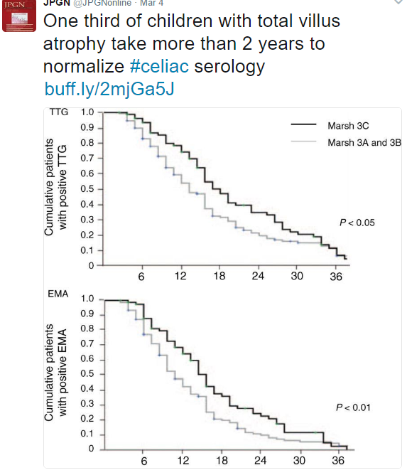
In a recent study (D Gidrewicz et al. JPGN 2017; 64: 362-7) with 228 consecutive, newly diagnosed children with biopsy-proven celiac disease, the authors followed improvement in serology.  Patients were divided into tertiles based on the degree of TTG IgA/EMA abnormalities. For example, Group A had the most abnormal serology: TTG >10 times ULN and EMA ≥1:80.

My take: This data confirms the fact that it takes a long time for resolution of celiac serology, particularly in those with the most severe disease.

Bowel Management Recommendations

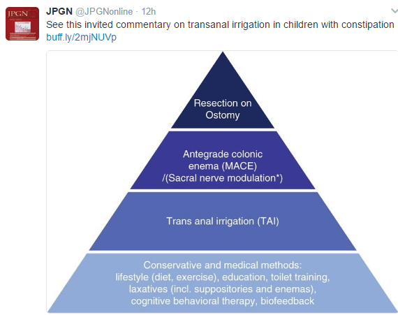
A recent “consensus” review on bowel management (G Mosiello et al. JPGN 2017; 64: 343-52) is available as an open access article –Link: Consensus Review of Best Practic of Transanal Irrigation in Children

The use of bowel management tube (or cone) for transanal irrigation has been around since ~1987 (B Shandling et. al. J Ped Surg 1987; 22: 271-3) and generally is considered in children older than 3 years of age with severe problems with defecation (organic and functional).

This particular review has a very good table on troubleshooting (Table 4) and a succinct summary of indications/contraindications (Table 2).

Related blog entries:


Why Do Canadians with Cystic Fibrosis Live Longer than Patients in U.S.?

NY Times Summarizes the reasons why Canadians with Cystic Fibrosis Live Longer: Link:  Canadians With Cystic Fibrosis Live 10 Years Longer Than Americans With the Disease

Cystic fibrosis is an inherited disease that causes recurrent lung infections and other problems. The average lifespan for an American with the illness is 37 years. In Canada, it is 49.

Researchers studied records of 5,941 Canadian and 45,448 American cystic fibrosis patients between 1990 and 2013. After controlling for severity of disease, age and other factors, they found that overall death rates were 34 percent lower in Canada than in the United States.

There was no difference in death rates between Canadians and Americans with private health insurance. But Canada provides universal health care coverage under a single-payer system, so every Canadian has some kind of health insurance. The Canadian death rate was 44 percent lower than that of Americans on Medicaid or Medicare, and 77 percent lower than Americans without insurance.

 AL Stephenson et al.  Ann Intern Med. 2017. DOI: 10.7326/M16-0858

Abstract

Background:In 2011, the median age of survival of patients with cystic fibrosis reported in the United States was 36.8 years, compared with 48.5 years in Canada. Direct comparison of survival estimates between national registries is challenging because of inherent differences in methodologies used, data processing techniques, and ascertainment bias.
Objective:To use a standardized approach to calculate cystic fibrosis survival estimates and to explore differences between Canada and the United States.
Design:Population-based study.
Setting:42 Canadian cystic fibrosis clinics and 110 U.S. cystic fibrosis care centers.
Patients:Patients followed in the Canadian Cystic Fibrosis Registry (CCFR) and U.S. Cystic Fibrosis Foundation Patient Registry (CFFPR) between 1990 and 2013.
Measurements:Cox proportional hazards models were used to compare survival between patients followed in the CCFR (n = 5941) and those in the CFFPR (n = 45 448). Multivariable models were used to adjust for factors known to be associated with survival.
Results:Median age of survival in patients with cystic fibrosis increased in both countries between 1990 and 2013; however, in 1995 and 2005, survival in Canada increased at a faster rate than in the United States (P < 0.001). On the basis of contemporary data from 2009 to 2013, the median age of survival in Canada was 10 years greater than in the United States (50.9 vs. 40.6 years, respectively). The adjusted risk for death was 34% lower in Canada than the United States (hazard ratio, 0.66 [95% CI, 0.54 to 0.81]). A greater proportion of patients in Canada received transplants (10.3% vs. 6.5%, respectively [standardized difference, 13.7]). Differences in survival between U.S. and Canadian patients varied according to U.S. patients’ insurance status.
Limitation:Ascertainment bias due to missing data or nonrandom loss to follow-up might affect the results.
Conclusion:Differences in cystic fibrosis survival between Canada and the United States persisted after adjustment for risk factors associated with survival, except for private-insurance status among U.S. patients. Differential access to transplantation, increased posttransplant survival, and differences in health care systems may, in part, explain the Canadian survival advantage.
Primary Funding Source:U.S. Cystic Fibrosis Foundation.

Sainte-Chapelle, Paris