We had a brilliant lecture given to our group by Dr. Benjamin Gold. I have had the good fortune of getting to know Ben and working alongside Ben for more than 15 years. Most readers of this blog are very familiar with Dr. Gold who is a leader in our field.
My notes below may contain errors in transcription and in omission.
.Key points:
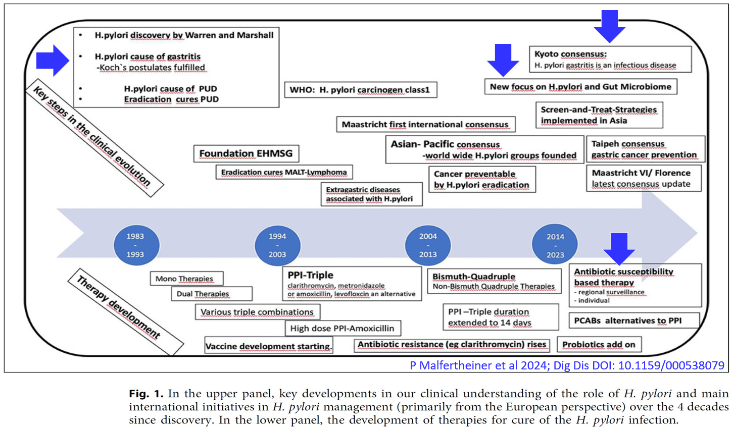
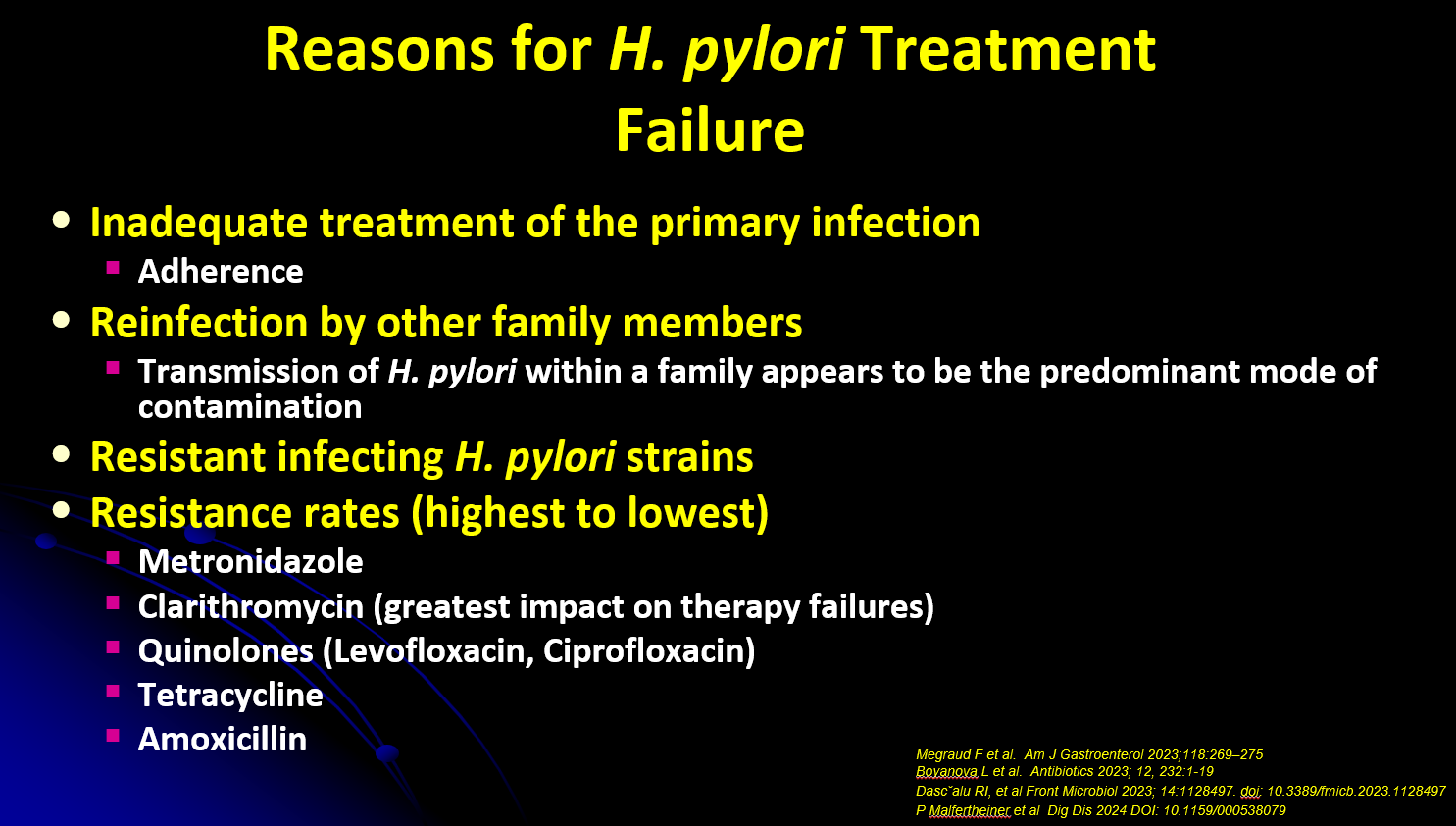
- While H pylori prevalence has decreased, it is becoming more difficult to treat
- Knowing if there is clarithromycin resistance in individuals with H pylori infection is most likely to impact treatment success. Metronidazole resistance can often be overcome with adequate dosing


- H pylori is an infectious disease with GI manifestations (rather than a GI disease). It needs to be treated as such, using tools like antimicrobial sensitivity


- Improving water supply in endemic areas reduces reacquisition of infection

- Transmission can occur from one generation to the next. Dr. Gold (& coauthors) has published a study showing transmission from grandfather to mother to child using DNA fingerprinting


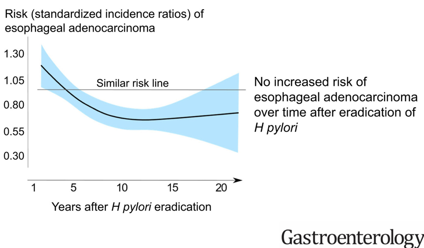
- Eradication of H pylori lowers the risk of developing gastric cancer
- Vonoprazan has been an effective part of treatment in adults. Pediatric studies are underway
Related blog posts:
- Give the Right Dose (for H pylori) -It Works Better!
- Helicobacter Pylori Stool Susceptibility in Children –How Good Is It?
- Understanding FDA Approval of Vonoprazan-Based Therapies for Helicobacter Pylori
- Synergistic Dangers: Helicobacter Pylori and Cancer Genes
- AGA: Best Practice Advice for Refractory H pylori
- Treating Helicobacter Pylori Lowers The Risk of Gastric Cancer
Disclaimer: This blog, gutsandgrowth, assumes no responsibility for any use or operation of any method, product, instruction, concept or idea contained in the material herein or for any injury or damage to persons or property (whether products liability, negligence or otherwise) resulting from such use or operation. These blog posts are for educational purposes only. Specific dosing of medications (along with potential adverse effects) should be confirmed by prescribing physician. Because of rapid advances in the medical sciences, the gutsandgrowth blog cautions that independent verification should be made of diagnosis and drug dosages. The reader is solely responsible for the conduct of any suggested test or procedure. This content is not a substitute for medical advice, diagnosis or treatment provided by a qualified healthcare provider. Always seek the advice of your physician or other qualified health provider with any questions you may have regarding a condition.