Timing of Cleft Palate Surgery

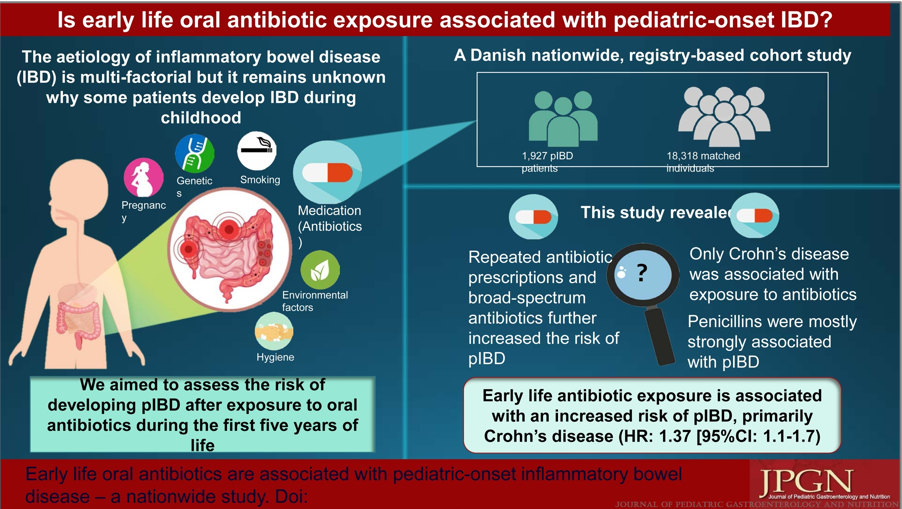
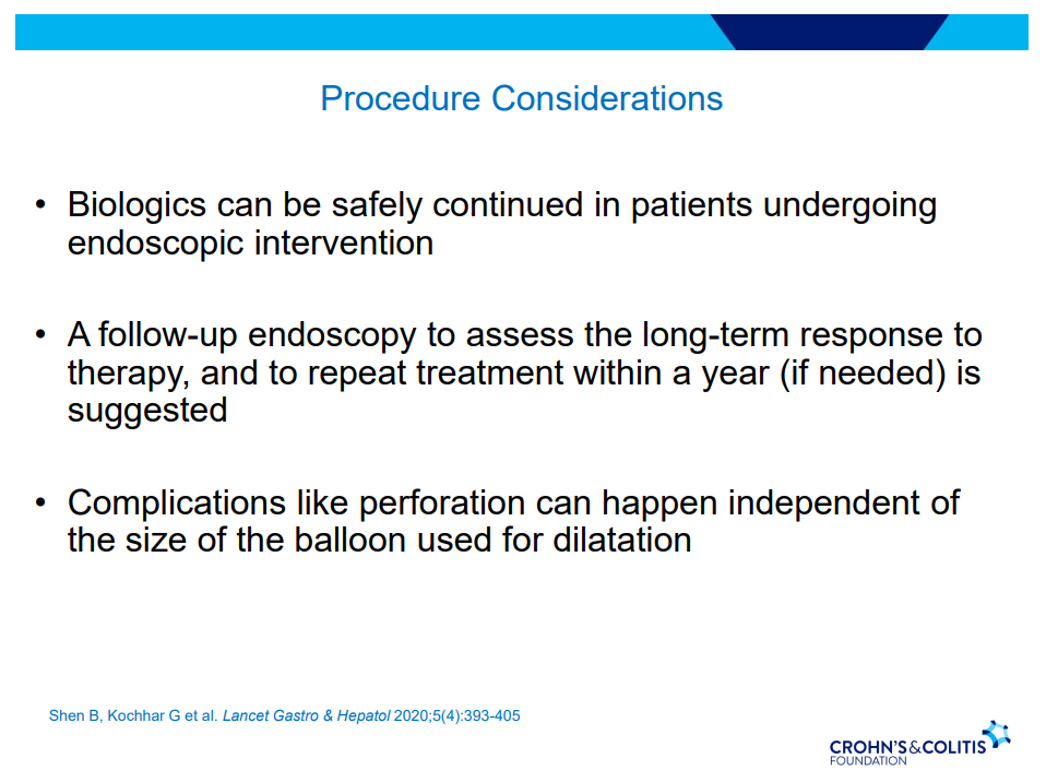
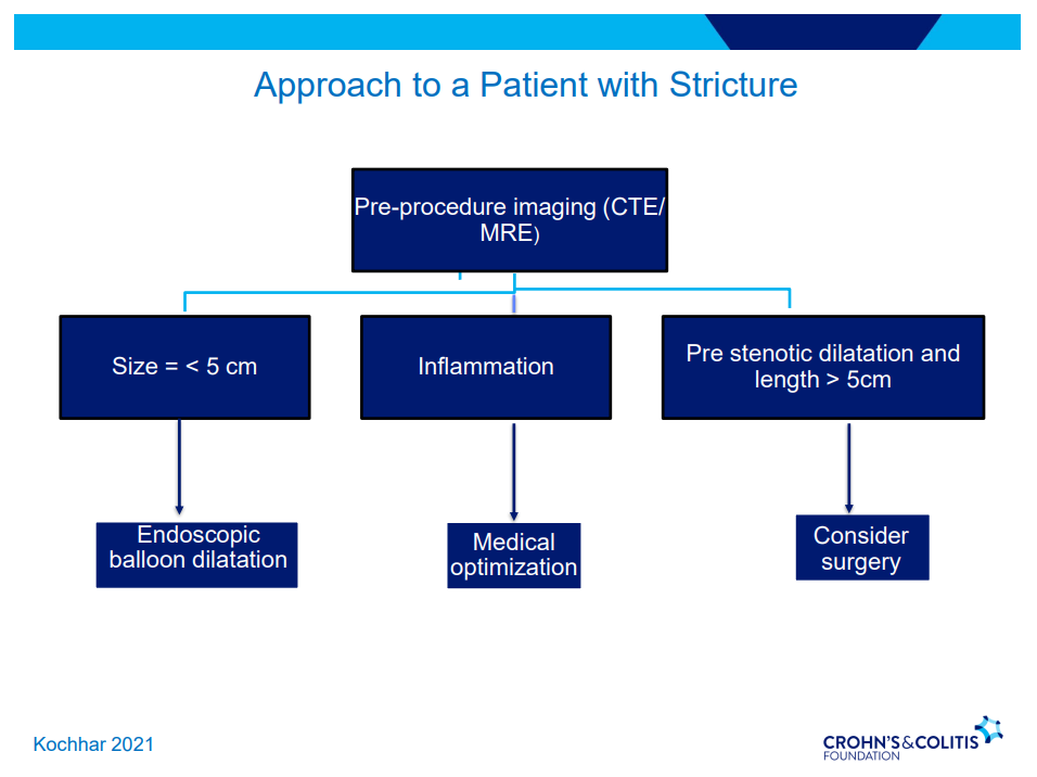
C Gamble et al. NEJM 2023; 389:795-807. Timing of Primary Surgery for Cleft Palate

Conclusion from study authors: Medically fit infants who underwent primary surgery for isolated cleft palate in adequately resourced settings at 6 months of age were less likely to have velopharyngeal insufficiency at the age of 5 years than those who had surgery at 12 months of age.

The associated editorial (R Tse, O Jackson, N Engl J Med 2023; 389:857-858. Mind the Gap) notes that “over two thirds of infants who were screened were excluded because their cleft was part of a syndrome, they were deemed to be medically unfit for early surgery, or they had a cleft that was too wide.” Also, “the incidence of additional surgery to treat velopharyngeal insufficiency was greater in the 6-month group than in the 12-month group (30 procedures in 27 children vs. 17 procedures in 16 children), and speech assessments performed after the secondary procedures were used in the analysis. Thus, the trial evaluated the mixed effects of primary surgery and secondary surgery in some patients.” Finally, “the considerations with regard to early surgery include the greater technical complexity of the procedure and the greater risks associated with anesthesia (airway complications and potential neurodevelopmental sequelae). Early surgery may also contribute to midfacial growth restriction, which becomes apparent only later, in adolescence, and may require complex corrective jaw surgery.”

My take: Early cleft palate surgery is not best for all infants with this defect.

Related blog post: Skinny Babies with Cleft Lips and/or Cleft Palates

Risk Factors for Inflammatory Bowel Disease: Antibiotics (Part 2)

AB Jawad et al. JPGN 2023; 77: 366-372.Early Life Oral Antibiotics Are Associated With Pediatric-Onset Inflammatory Bowel Disease-A Nationwide Study

Key findings:

  • Oral antibiotic exposure during the first 5 years of life was associated with a higher risk of developing pIBD (HR = 1.33,  P <0.0001). The risk was also increased if patients had ≥4 antibiotic prescriptions compared to no antibiotics (HR = 1.33, P <0.0001).
  • Broad-spectrum antibiotics increased the risk of pIBD compared to narrow-spectrum antibiotics (HR = 1.29, P < 0.0001).
  • When stratified by IBD subtypes, only Crohn disease was significantly associated with exposure to antibiotics (HR = 1.37, P = 0.002).

My take: This study indicates that antibiotics (and/or serious infections) are associated with an increased the risk of pediatric Crohn’s disease but the absolute risk is very low. We still have a lot to learn about how environmental exposures, including diet, infections, antibiotics, and pollution, contribute to the increasing prevalence of inflammatory bowel disease.

Related blog posts:

Risk Factors for Inflammatory Bowel Disease: Ultra-Processed Food (Part 1)

N Narula et al. Clin Gastroenterol Hepatol 2023; 21: 2483-2495. Open Access! Food Processing and Risk of Inflammatory Bowel Disease: A Systematic Review and Meta-Analysis

JA Fitzpatrick et al. Clin Gastroenterol Hepatol 2023; 21: 2478-2480 (editorial). Open Access! Ultra-processed Foods and Risk of Crohn’s Disease: How Much is Too Much?

Figure 1 from editorial: The NOVA classification of food

A total of 1,068,425 participants were included (13,594,422 person-years) among 5 cohort studies published between 2020 and 2022. The average age of participants ranged from 43 to 56 years. Key findings:

  • Crohn’s disease:  During follow-up, 916 participants developed CD, and 1934 developed UC. There was an increased risk for development of CD for participants with higher consumption of ultra-processed foods compared with those with lower consumption (HR, 1.71; 95% CI, 1.37–2.14; I2 = 0%) and a lower risk of CD for participants with higher consumption of unprocessed/minimally processed foods compared with those with lower consumption (HR, 0.71; 95% CI, 0.53–0.94; I2 = 11%). 
  • Ulcerative colitis: There was no significant association between risk of UC and ultra-processed foods (HR, 1.17; 95% CI, 0.86–1.61; I2 = 74%) or unprocessed/minimally processed foods (HR, 0.84; 95% CI, 0.68–1.02; I2 = 0%).

The associated editorial by Fitzpatrick et al, notes that “there are plausible mechanisms that explain the associations of higher UPFs and development of CD, such as: (1) displacing the intake of minimally processed foods and subsequently reducing exposure to beneficial micronutrients, antioxidants, and phytochemicals; (2) driving overconsumption of total calories7; and (3) increasing exposure to non-nutritive food substances that have been implicated in the development of CD in pre-clinical studies…The notion is that a lower UPF intake is better, but a cutoff value remains elusive.”8

My take (borrowed from editorial): “the population studies have indicated that the extremes of UPF intake are related to risk of CD and that such associations are underpinned by plausible biological mechanisms, suggesting causality.”

Related blog posts:

How to Handle Gastric Intestinal Metaplasia

I Mansuri et al. JPGN 2023; 77: 332-338. Gastric Intestinal Metaplasia in Children: Natural History and Clinicopathological Correlation

This retrospective single-center study (2013-2019) identified 38 patients with gastric intestinal metaplasia (GM) with a mean age of 12.5 years.

Background: “Gastric intestinal metaplasia (GIM) is defined as the replacement of the normal gastric epithelium by intestinal-type epithelium. GIM is considered a preneoplastic lesion for gastric adenocarcinoma in adults and is found in 25% of Helicobacter pylori ( H pylori ) exposed adults.”

Key findings:

  • The prevalence was 0.53% based on 7104 patients who were 18 or younger who underwent EGDs
  • 2 cases of H pylori were identified; chronic gastritis was noted in 47%
  • Only a third of patients had f/u EGDs; none of these had progression to dysplasia. In fact, GIM was NOT identified in the majority at followup

In their discussion, the authors note that the AGA’s clinical practice guidelines for GIM in adults provides the following recommendation: “Routine surveillance endoscopy in patients with incidental detection of gastric intestinal metaplasia (GIM) is discouraged.”

Also, it is worthwhile to consider that many cases of GIM are likely overlooked given the often patchy distribution.

My take (borrowed from authors): GIM appears to be an incidental gastric biopsy finding of uncertain significance. Unless there are high risk factors for gastric cancer, routine followup is unlikely to be beneficial.

Pictures from Villa Ephrussi de Rothschild:

Disclaimer: This blog, gutsandgrowth, assumes no responsibility for any use or operation of any method, product, instruction, concept or idea contained in the material herein or for any injury or damage to persons or property (whether products liability, negligence or otherwise) resulting from such use or operation. These blog posts are for educational purposes only. Specific dosing of medications (along with potential adverse effects) should be confirmed by prescribing physician.  Because of rapid advances in the medical sciences, the gutsandgrowth blog cautions that independent verification should be made of diagnosis and drug dosages. The reader is solely responsible for the conduct of any suggested test or procedure.  This content is not a substitute for medical advice, diagnosis or treatment provided by a qualified healthcare provider. Always seek the advice of your physician or other qualified health provider with any questions you may have regarding a condition.

Customized Postoperative Therapy for Biliary Atresia -Does It Help?

S Pandurangi et al. J Pediatr Surg 2023; 58: 1483-1488. Customized Postoperative Therapy Improves Bile Drainage in Biliary Atresia: A Single Center Preliminary Report

This single center retrospective study compared  20 consecutive infants underwent hepatoportoenterostomy (HPE) (beginning in 2017) for biliary atresia (BA) to a historical cohort. Analysis of successful biliary drainage 3 months after HPE (defined as serum total bilirubin (TB) <2 mg/dL) was the primary endpoint; survival with native liver at 2 years was the secondary endpoint.

Protocol:

  • Cefoxitin was administered to all infants following HPE for 3-4 days.
  • Standard protocol: If the stool color normalized (pigmented), the infant received “conventional” treatment with trimethoprim-sulfamethoxazole cholangitis prophylaxis, fat-soluble vitamin supplementation with DEKAsPlus or AquaADEKs (1 mL daily), and ursodeoxycholic acid (5 mg/kg twice daily).
  • Customized protocol: If the stools were acholic (or not consistently pigmented) and </=45 days, the infants received intravenous cefoxitin or piperacillin-tazobactam and methylprednisolone, initial dose 5 mg/kg/day and decreased by 1 mg/kg/day each day thru day 5; then orally treated with dose dropped 0.25 mg/kg weekly. When switched to oral steroids, IV antibiotics were stopped and infant was placed on amoxicillin-clavulanate which was continued until TB <2 mg/dL or discontinuation of corticosteroids (whichever came first).
  • If stools were acholic and infant was >45 days, then the same treatment was given if there was liver inflammation on histology.

Key findings:

  • 8 had pigmented stools after HPE and received standard protocol.
  • 12 had acholic/inconsistent stools. All of those >45 days had liver inflammation; thus, all 12 received the customized protocol. Two infants had two cycles of steroids/antibiotics who had initial response to treatment and then worsened.
  • Sixteen of 20 (80%) infants had successful bile drainage, compared to 8 of 20 (40%) infants in the historical cohort (P = 0.0225)
  • Among the sixteen who have reached two years of age, 11 (68.8%) are alive with native livers versus 10 of 20 (50%) in the historical cohort (P = 0.0970).  This did not achieve statistical significance.

The authors established their protocol based on data from Kings College in 2016 suggesting that steroids appeared effective in younger patients who underwent HPE prior to 45 days (Peg Surg Int 2016; 32: 193-200). The START study showed no significant improvement in biliary drainage between patients receiving corticosteroids and placebo. However, in the group <70 days, 72% of infants receiving corticosteroids achieved biliary drainage compared with 57% of the placebo group (P=0.36).

My take: This is a small sample size. Perhaps, this protocol will help improve outcomes. If so, we still don’t know which factor is more important —the IV antibiotics or the high dose steroids. If these agents are helpful, are there other predictive factors –microbiome? MMP-&?

Related blog posts:

Pictures from the Villa Ephrussi de Rothschild

According to the study which you would never qualify for… (2023)

In 2012, this blog highlighted a study which showed that “only 31.1% of 206 patients with IBD would have been eligible to participate in any of the selected RCTs.” (Post: According to the study which you would never qualify for…).

A recent study shows the same phenomenon in pediatric IBD studies: O Atia et al. Aliment Pharmacol Ther 2022; 56 (5): 794-801. Open Access! Children included in randomised controlled trials of biologics in inflammatory bowel diseases do not represent the real-world patient mix

This study utilized data for children initiating biologics from two prospective real‐world cohorts and one retrospective cohort.

Key findings:

  • Only 62 of 164 (38%) children with moderate–to‐severe disease would have been eligible for inclusion in the original RCTs.
  • The steroid-free remission rate was higher in the eligible children (51%) than in the ineligible children (31%; OR 2.3 [95%CI 1.2–4.5]; p = 0.01)
  • The main exclusion criterion was prohibited previous therapies (47%)

My take (borrowed from authors): “Remission rates were higher among eligible children raising the concern that results presented in regulatory RCTs in paediatric IBD do not necessarily reflect the patient‐mix in the real‐world and should be interpreted with caution when applied to clinical practice.”

Pictures from VilleFranche-Sur-Mer:

Expect Costs of Liquid Omeprazole to Increase Due to FDA Approval

Last fall, the FDA approved a liquid version of omeprazole.

9/3/22 Drugs.com:FDA Approves Konvomep. FDA Approves Konvomep (omeprazole and sodium bicarbonate for oral suspension) for Gastric Ulcer and Reduction of Risk of Gastrointestinal Bleeding in Critically Ill Patients

WOBURN, Mass. (September 2, 2022) – Azurity Pharmaceuticals, Inc., a pharmaceutical company focused on developing innovative dose-forms and formulations of products to serve the needs of overlooked patients, announced today that the U.S. Food and Drug Administration (FDA) has approved Konvomep (omeprazole and sodium bicarbonate for oral suspension).

Now that this FDA approved product is available, it may be that it will be more difficult to receive a compound version. The cost of this new formulation is much higher. Here are some of the costs from GoodRx.com.

My take: The high cost of this liquid preparation is another good reason to avoid using a PPI in patients with low likelihood of benefit.

Related blog posts:

Don’t Put the Cart Before the Horse: Biliary Atresia Screening

R Lerer et al. JPGN Reports2023; 4(4):p e345. Open Access! Evaluation of Newborn Direct Bilirubin As Screening for Cholestatic Liver Disease

This retrospective study analyzed data from 11,965 infants who had fractionated bilirubin obtained in the nursery (2016-2019). Key findings:

  • DB of 0.6 mg/dL was chosen as the cut-off based on a high sensitivity (100%) and specificity (99%) for screening newborns for CLD
  • Out of 60 infants who met criteria for DB ≥0.6 mg/dL, only 15 (25%) had a repeat level drawn after nursery discharge; 3 (5%) were eventually diagnosed with CLD (2 with BA and 1 with Alagille syndrome)

It is fairly easy to get fractionated bilirubins on infants. Many need to get a bilirubin check and in many centers, a fractionated bilirubin is automatically generated at no additional costs. The hard part is making sure that those with abnormal values receive timely followup.

My take: It is a mistake to get fractionated bilirubins in newborns unless one has developed a plan/infrastructure to make sure those with abnormal values receive appropriate followup.

Related blog posts:

Off the coast of Southern France (near Juan-Les-Pines)

Gene Therapy for Crigler-Najjar

L D’Antiga et al. NEJM; 2023; 389: 620-631. Gene Therapy in Patients with the Crigler–Najjar Syndrome

Methods: Five patients received a single infusion of the gene construct (GNT0003): two received 2×10¹² vector genomes (vg) per kilogram of body weight, and three received 5×10¹² vg per kilogram. The primary end points were measures of safety and efficacy; efficacy was defined as a serum bilirubin level of 300 μmol per liter or lower measured at 17 weeks, 1 week after discontinuation of phototherapy. The infusion protocol included administration of sirolimus adjusted for a trough of 4-12 mcg/L (starting 1 week prior to infusion) and steroids (IV day prior then oral for 8 weeks). .

Key findings

  •  By week 16, serum bilirubin levels in patients who received the lower dose of GNT0003 exceeded 300 μmol per liter.
  • The patients who received the higher dose had bilirubin levels below 300 μmol per liter in the absence of phototherapy at the end of follow-up; mean level at the final follow-up visit [week 78 in two patients and week 80 in the other], was149±33 μmol per liter.
  • No serious adverse events were reported. Mild increase in ALT levels were seen in 4 of 5 patients; this was “potentially related to an immune response against the infused vector; these patients were treated with a course of glucocorticoids.”
This figure shows the response of the serum bilirubin in patients receiving the higher dose of the infusion.

My take: This study shows that the GNT0003 increased UGT1A1 activity to levels that permitted cessation of phototherapy; this persisted for 18 months after treatment. Further studies are needed.

Related blog posts:

CCFA 2023 (Atlanta) Part 5

Another very good review by David Schwartz on The Daunting Duo: Management of Stictures and Fistula. Below are some of my notes and some slides; my notes may contain errors in transcription or omission. Can get access to all 49 slides here: IBD Pro CCFA 2023 Atlanta

  • Fistula healing rates are poor
  • 78% have complex fistulas
  • Long-term healing: 67% with simple fistulas but only 37% with complex fistulas
  • Seton placement prior to biologic therapy increases likelihood of better outcomes
  • Antibiotic therapy recommended until Seton removed (not short-term treatment) –improved healing rates along with medical therapy
  • Anti-TNF therapy effective in ~40% long-term; higher rates of healing with higher anti-TNF levels
  • MRI and/or EUS helpful in improving fistula healing rates
  • Ustekinumab and Vedolizumab both had fistula healing rates ~40%
  • Adipose derived stem cells with ~50% healing rates (study with high placebo healing rate too ~37%); changes local cytokine profile, cells gone in about 2 weeks but goal for changing trajectory

Strictures

  • 5-24% with stricturing phenotype
  • No effective medical treatments
  • Endoscopic balloon dilatation in shorter strictures (<5 cm) without associated abscess or fistula (needs imaging prior)
  • One-third of balloon dilated strictures will still wind up needing surgery despite dilatation. 5-10% risk of complication. Goal is 15 mm or more (Dr. Schwartz typically does not increase by more than 3 sizes from baseline in one session)

Disclaimer: This blog, gutsandgrowth, assumes no responsibility for any use or operation of any method, product, instruction, concept or idea contained in the material herein or for any injury or damage to persons or property (whether products liability, negligence or otherwise) resulting from such use or operation. These blog posts are for educational purposes only. Specific dosing of medications (along with potential adverse effects) should be confirmed by prescribing physician.  Because of rapid advances in the medical sciences, the gutsandgrowth blog cautions that independent verification should be made of diagnosis and drug dosages. The reader is solely responsible for the conduct of any suggested test or procedure.  This content is not a substitute for medical advice, diagnosis or treatment provided by a qualified healthcare provider. Always seek the advice of your physician or other qualified health provider with any questions you may have regarding a condition.