Non-Invasive Studies Often Fail to Detect Advanced Liver Fibrosis in Steatotic Liver Disease

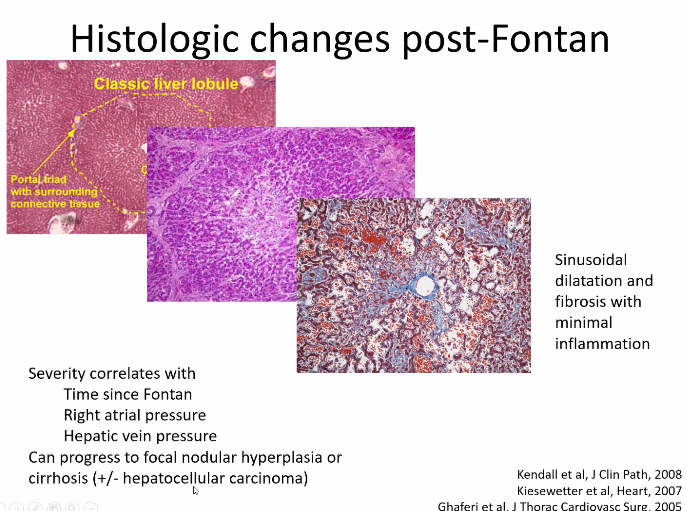
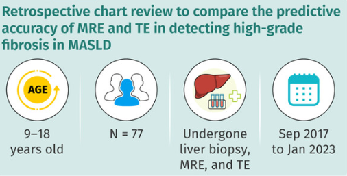
N Ravanbakhsh et al. JGPN 2024; https://doi.org/10.1002/jpn3.12368. Comparing imaging modalities in the assessment of fibrosis in metabolic dysfunction-associated steatotic liver disease

Key findings:

  • TE and MRE did not have high correlation with liver biopsy in the detection of high-grade fibrosis
  • Fibrosis was identified in 90% of liver biopsies with bridging fibrosis in 15 (19%) and cirrhosis in 1 (1%)
  • AUROC curves of MRE and TE for detection of high-grade fibrosis were 0.817 and 0.750, respectively, and not significantly different.

The authors note that previous adults studies suggest that MRE is more accurate in the identification of liver fibrosis than TE (MRE detected ≥ F1 fibrosis with an AUROC of 0.82, while TE detected fibrosis with an AUROC of 0.67).20 

My take: Trying to identify accurate non-invasive testing is crucial to help identify patients most in need of treatment and for limiting costs.

Related blog posts:

Isle of Palms, SC

Blog Case Report: A Persistent Elevated AST in Teen with IBD and ADHD

A recent case reminded me of the quote by Helena Ravenclaw in Harry Potter: “”If you have to ask, you’ll never know. If you know, you need only ask.”

One of my colleagues recently diagnosed a teenage boy with ulcerative colitis. His past medical history was notable for ADHD. At the time of his evaluation, he was noted to have an elevated AST.

Labs:

  • June: AST 143, ALT 8, Hepatitis B immune
  • August: AST 190, ALT 10, Albumin 4.7, T protein 7.3, T bili 0.4, D bili 0.1, Alk phos 168; GGT 10, CPK 93

The concern at the time was whether his elevated AST should preclude using his ADHD medicine and whether there was an underlying liver disease. Based on the pattern of liver enzyme abnormalities, it was suspected that the patient had macro AST. A blood test was sent to the Mayo clinic and confirmed this diagnosis:

“”The sample was investigated for the presence of macro AST by polyethylene glycol (PEG) precipitation. Serum AST activity = 316 U/L. The AST result post-PEG precipitation = 22 U/L. The results obtained are positive for the presence of macro AST (93% of activity precipitated with PEG). Based on validation studies performed at the Mayo Clinic, a cut-off of >80% AST activity precipitated by PEG indicates the presence of macro AST.” This test is rarely ordered at the Mayo Clinic and is ordered as a miscellaneous test; it is not on the Mayo Clinic’s regular test menu.

Internet description of macro AST: Macro-aspartate aminotransferase (macro AST) is a rare, benign condition that causes a persistent elevation of aspartate aminotransferase (AST) levels in the blood. It’s caused by the binding of AST to immunoglobulins, which results in a high molecular weight macroenzyme that’s excreted from the serum more slowly than normal.

My take: Macro AST diagnosis is useful –it helps eliminate the concern for other conditions. Since it is quite uncommon, it is easier to think of this problem once you have seen it before.

Related blog posts:

Grand Point View Overlook at Canyonlands National Park

Treats (Tips) and Tricks for Using IBD Drugs in Patients with Hepatobiliary Comorbidities

S Massironi et al. Inflamm Bowel Dis 2023; 29: 1477-1487. Use of IBD Drugs in Patients With Hepatobiliary Comorbidities: Tips and Tricks

This review article makes a number of useful points:

  • Almost all of the IBD medications (Anti-TNF, Vedolizumab, Ustekinumab, Tofacitinib, Ozanimod) should not be used in patients with untreated hepatitis B surface antigen positivity. Antiviral prophylaxis is recommended even patients with inactive disease (HBV DNA <2000 IU/mL/normal ALT) starting 2 weeks prior to immunosuppression up to 12 weeks after immunosuppression discontinuation.
  • Anti-TNF, Vedolizumab, and Ustekinumab can be safely used in patients with cirrhosis, tofacitinib requires a dose reduction, and ozanimod is NOT recommended
  • Autoimmune hepatitis may be triggered (rarely) by use of anti-TNFs; however, these agents have been uses off-label as a rescue therapy for AIH as well.
  • Anti-TNFs do not appear worsen liver function in PSC and may be associated with improvement in MASH (NASH)
  • Drug-induced liver injury (DILI) is common with anti-TNFs, often seen between the second and fifth doses. It is “generally transient and asymptomatic.” DILI may occur with ustekinumab, tofacitinib and ozanimod.
  • Tofacitinib may have a favorable effect on PSC. A retrospective study with 5 patients reported a 28% and 39% decrease respectively in total bilirubin and alkaline phosphatase within 6 months of starting therapy, A separate study with 42 patients showed a non-significant drop in alkaline phosphatase from 150 U/L to 132 U/L at 12 month followup.
  • The authors note that in patients with cirrhosis and posttransplantation, “vedolizumab and ustekinumab should be preferred due to their safer profile linked to infectious risk.”
  • “Patients with hepatobiliary disorders are often excluded from pivotal trials.” This contributes to a significant knowledge gap for patients with these comorbid conditions which are frequent in patients with IBD

My take: I don’t think I will be too popular if I hand out copies of this article to the kids I see later today –despite the useful advice. (This post was meant to be published on Halloween)

Related blog posts:

Disclaimer: This blog, gutsandgrowth, assumes no responsibility for any use or operation of any method, product, instruction, concept or idea contained in the material herein or for any injury or damage to persons or property (whether products liability, negligence or otherwise) resulting from such use or operation. These blog posts are for educational purposes only. Specific dosing of medications (along with potential adverse effects) should be confirmed by prescribing physician.  Because of rapid advances in the medical sciences, the gutsandgrowth blog cautions that independent verification should be made of diagnosis and drug dosages. The reader is solely responsible for the conduct of any suggested test or procedure.  This content is not a substitute for medical advice, diagnosis or treatment provided by a qualified healthcare provider. Always seek the advice of your physician or other qualified health provider with any questions you may have regarding a condition.

Sickle Cell Related Liver Disease

A terrific review of sickle cell disease (SCD) associated liver problems: F Lacaille et al. JPGN 2021; 72: 5-10. The Liver in Sickle Cell Disease

While the most frequent liver-related problem in individuals is cholelithiasis (>25% after age 5 yrs), a host of other problems can develop –this article is a good reference.

Key points:

  • Acute Sickle Cell Hepatic Crisis, Intrahepatic cholestasis, and acute hepatic sequestration
    • ~6% of children and 10% of adults develop severe liver complications of SCD
    • With sequestration, indications include pain with acute drop in hemoglobin (>2 g/dL)
    • Acute hepatic crisis is often signaled by elevated conjugated bilirubin
    • With severe liver disease/liver ischemia, authors advocated for exchange transfusion which “more efficiently decreases HbS percentage, faster restoring the blood flow than simple transfusion.” Consider after excluding biliary complication if INR is >1.4 with increased conjugated bilirubin (>3 mg/L). “Simple transfusion should be discussed in other cases.”
  • Cholangiopathy and autoimmune liver disease
    • Although autoimmune sclerosing cholangitis/autoimmune hepatitis are rare, it may account for 8% of children with SCD referred for hepatic dysfunction
    • Liver biopsy, needed for diagnosis, “is a dangerous procedure in SCD, which cannot be performed without at least a transfusion”
    • “Steroids can induce sickle crisis”
    • Look for ANA, SMA, LKM, and ANCA
  • Iron Overload
    • “It is not usually a significant concern in children…In our patients, the median ferritin level was about 3000 ng/mL, and none had a severe overload on MRI”
  • Infections/Drug toxicity
    • Need to consider hepatitis B, hepatitis C, and hepatitis E in particular
    • Inquire about herbal medicines and recreative drugs
  • Liver transplantation
    • Results are often poor.
    • Problems include sickle cell crisis in the transplanted liver, and drug toxicity which can add to the neurological and renal morbidities of SCD
  • Stem cell transplantation
    • Consider for severe complications of SCD including hepatic complications

Related blog posts: 

From NY Times

Online Aspen Webinar (Part 9) -Liver Disease After Fontan, Acute on Chronic Liver Disease and Immunosuppression Withdrawal Strategies

Below I’ve included a few more slides form recent Aspen Webinars

Fontan Associated Liver Disease  Greg Tiao

Related blog posts:

Acute on Chronic Liver Failure  Estella Alonso

Immunosuppression strategies ..and is withdrawal possible  Kathleen Campbell

Online Aspen Webinar -COVID-19, Autoimmune Hepatitis (Part 8)

For those who want to view the actual lectures, you can sign up and view the recordings:  Aspen Webinar Lecture Series


Below I’ve included a few slides and some notes; my notes may have errors of omission or transcription.

COVID-19 and the Liver — Fred Suchy

Key Points:

  • The extent and severity of liver disease related to COVID-19  is still being determined.  Many individuals have mild liver test abnormalities (5-60%)
  • Avoid imaging unless it will change your management (eg. thrombus)
  • In those with worsening/significant liver abnormalities, look for other etiologies of elevated liver tests (eg. other viral hepatitis, drugs, myositis, coinfection, clots, multi-system inflammatory disorder)
  • Currently, no change in immunosuppression is recommended in the post-transplant population WITHOUT COVID-19. In those with severe COVID-19 infection, reduction in immunosuppression is recommended


How I Manage Patients with Autoimmune Hepatitis -Diagnosis and Treatment   Amy Taylor.

Key points:


Stratifying Cystic Fibrosis Liver Disease with Ultrasonography

A recent multicenter case-controlled study (MJ Siegel, J Freeman, W Ye et al. J Pediatr 2020; 219: 62-9) show that ultrasonography can identify children with cystic fibrosis (CF) at increased risk for developing advanced CF liver disease; it is well-recognized that currently advanced CF liver disease occurs predominantly in children.

Among 722 participants who underwent ultrasonography as part of a planned 9 year research study, 65 had heterogeneous liver pattern and 592 had a normal liver pattern at baseline.

Key findings at planned 4-year interim analysis in a subgroup of 55 with (baseline) heterogeneous liver pattern and 116 with (baseline) normal liver pattern:

  • Those with heterogeneous liver pattern had higher median biochemical markers of liver disease: ALT 42 vs 32, GGT 36 vs 15, AST to platelet ratio index: 0.7 vs 0.4
  • Those with heterogeneous liver pattern were at ~9-fold increased risk of developing a nodular liver pattern (23% vs. 2.6%).
  • The authors did not identify specific clinical risk factors that predict the development of nodular liver ultrasound pattern

My take: Even among those with a heterogeneous pattern, the majority will not develop a ultrasonography (nodular) pattern of advanced liver disease.  Much more insight in this area is needed with the advancement in medical treatments and with the more availability of elastography.

Related blog posts:

Island Ford Nat’l Recreation Area/Chattahoochee River

“When the Cause of Liver Disease Is the Heart”

A recent review (S Ofei, C Gariepy. JPGN 2017; 64: 3-7) provides a good review of “congestive hepatopathy.”

Key points:

  • Overall, the liver receives 25% of cardiac output; though, 70% of blood flow to the liver is partially deoxygenated blood.  Cardiac disease can lead to liver disease due to hypoxic injury.
  • “Congestive hepatopathy (CH) results from chronic right heart dysfunction with decreased hepatic blood flow, arterial saturation, and increased central venous pressure.”  Ultimately, CH can lead to hepatic cirrhosis, termed ‘cardiac cirrhosis’ by the authors.
  • “Symptoms of CH are vague.” These symptoms could include abdominal pain nausea, and early satiety.
  • Treatment is uncertain.  “Guidelines and expert consensus..favor use of loop diuretics in patients with jaundice, hepatic congestion, and ascites.”
  • With regard to patients with Fontan-associated liver disease (FALD), “there is no consensus.” Patients should be treated for complications like varices, coagulopathy, and nutritional deficiencies.”  Some patients will need liver transplantation, though liver disease may be reversible with cardiac transplantation.  The article provides many references that provide more in-depth review of this topic.

My take: Overall, this article provides a succinct review of congestive hepatopathy.  There are many other cardiac conditions associated with liver dysfunction including heart disease associated with NAFLD, Alagille syndrome, and Kawasaki’s.

Cozumel

Cozumel

Related blog posts:

PSC 2013 Review

A recent review of PSC was published (Gastroenterology 2013; 145: 521-36).  This review is a little more detailed than a previous review noted in this blog less than 6 months ago (Staying current with PSC | gutsandgrowth).

A couple of useful comments from the review:

  • “An increased serum level of alkaline phosphatase is the most common biochemical abnormality detected in patients with PSC.  In some cases, it is the only biochemical alteration observed, such as in patients with intrahepatic involvement.”
  • “Typically, a liver biopsy is not required to diagnose PSC unless small duct PSC is suspected or if there are concerns that a patient also has AIH.”  Cholangiography is the best way to identify PSC.
  • “Patients diagnosed with PSC should undergo colonoscopy… to determine if they have IBD, even when there are no symptoms.”
  • Autoimmune hepatitis-PSC overlap is thought to occur in ≤6% of cases.  AIH-PSC should be suspected if there are biochemical features of AIH (positive serology, increased transaminases), histology suggestive of AIH, or in AIH patients that become refractory to treatment.
  • No controlled trials have identified effective medical treatments.  Studied medications have included corticosteroids, etanercept, tacrolimus, cyclosporine, azathioprine, methotrexate, infliximab, and ursodeoxycholic acid.  The latter may increase disease progression, particularly at higher doses.

Also noted:

Hepatology 2013; 58: 1392-1400. “Primary Sclerosing Cholangitis, Autoimmune Hepatitis, and Overlap in Utah Children: Epidemiology and Natural History”

In this study the authors identified 607 cases of IBD, 29 cases of PSC, 12 cases of ASC (overlap), and 44 cases of AIH.  “Cholangiocarcinoma developed in 2 of the 29 PSC patients (6.9%).  PSC occurred in 9.9% of patients with ulcerative colitis (UC) and 0.6% of patients with Crohn’s disease.”

Related blog posts:

Disclaimer: These blog posts are for educational purposes only. Specific dosing of medications (along with potential adverse effects) and specific medical management interventions should be confirmed by prescribing physician.  Application of the information in a particular situation remains the professional responsibility of the practitioner.

Mitochondrial Liver Disease

A recent review provides advice for the evaluation of the child with suspected mitochondrial liver disease (JPGN 2013; 57: 269-79).

Clinical presentations:

  • Acute in a child with no history of hepatic dysfunction
  • Chronic liver and CNS dysfunction
  • Onset of liver disease in patient with known CNS disorder

Suggested Tiered Diagnostic Evaluation: Table 1 provides extensive suggestions.

  • 1st tier: CMP, INR, AFP, CPK, Phos, CBC/d, ammonia, Lactate/pyruvate (preferably 1 hour after feeding), serum ketone bodies, serum acylcarnitine profile, carnitine profile, urine organic acids, serum amino acids, urine acylglycines and 2-ethylmalonic acid quantification, plasma thymidine (especially if intestinal dysmotility), quantitative serum methylmalonic acid, CSF analysis (lactate and pyruvate, amino acids, protein)
  • 2nd tier: genotyping for more common genes (eg. panel with POLG1, DGUOK, MPV17), other genetic tests based on tier 1 testing (see table for details)
  • 3rd tier: liver biopsy, skin biopsy, and muscle biopsy (see table for details)
  • 4th tier: additional genetic tests based on 1st three tiers

Table 2 describes potential evaluations in other organs.  For example, for brain, MRI, EEG and CSF.

Table 3 lists ~27 mutation/syndromes and clinical features.

The last three words from the conclusion of the publication are not supported by the review.  The authors state that this systematic approach “can aid in making a timely, accurate, and cost-effective diagnosis.”  While the authors do not provide estimates of the expense of these tests, they are probably very expensive, though less costly than a failed liver transplantation.

Bottomline: “Available technology to aid in diagnosis has improved substantially.  Nonetheless, diagnosis of suspected mitochondrial disease in children is complicated.”

Related blog post:

Proven treatments for mitochondrial disorders | gutsandgrowth