Big Study of Primary Sclerosing Cholangitis -Pediatrics 2017

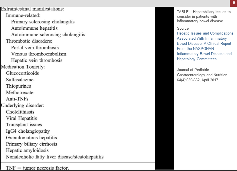
In July, this blog reviewed a recent big study of primary sclerosing cholangitis (PSC) in adults with more than 7000 patients. A recent study in the pediatric age group enrolled 781 children from 36 institutions: MR Deneau et al. Hepatology 2017; 66: 518-27 (Congratulations to Nitika Gupta and Miriam Vos -in-town colleagues and contributing authors to this study.)

This retrospective study’s key findings:

  • Median age 12 years at diagnosis; 39% were female
  • Autoimmune hepatitis (overlap) was present in 33%
  • Small-duct PSC was present in 13%
  • Inflammatory bowel disease (IBD) was present in 76%
  • PSC-IBD and Small-duct PSC (normal cholangiograms) had more favorable prognosis with hazard ratio of 0.6 of developing complications
  • Portal hypertensive and biliary complications were noted in 38% and 25% respectively. After developing these complications, the median survival with native liver was 2.8 years and 3.5 years respectively
  • Survival with native liver was noted in 70% at 5 years and 53% at 10 years
  • Elevations in bilirubin, GGT, and AST-to-platelet count ratio were associated with highest risk of progressive disease.
  • Cholangiocarcinoma (CCA) developed in 1% (median, 6 years after diagnosis)

The discussion notes that while pediatric PSC is a progressive disease, complications were slower to develop compared with adult-onset PSC. 10-year survival with native liver is typically lower in adults ~60% (vs 70% in this study).  “Up to one third of adults with PSC may have esophageal varices within a year of diagnosis” compared with only 13% in this cohort.  Dominant strictures are more common in adults, occurring in the majority within 5 years whereas this occurred in 16% of this cohort.

With regard to CCA, the authors note that current recommendations suggest starting to screen for CCA in patients over age 18 years with ultrasound and CA 19-9 at 6-12 month intervals. These studies “could reasonably be extended to PSC patients aged 15 and above and [for those requiring dilatation] of biliary strictures.”

My take: This large pediatric PSC study provides more clarity on the outcomes of patient’s with PSC and the associated conditions.

Related blog posts:

Steps at the High Museum


Big Study of Primary Sclerosing Cholangitis

This blog has reviewed multiple publications on primary sclerosing cholangitis (see blog posts below). Now, a study from 37 centers with 7121 patients with PSC has been published: TJ Weismuller et al. Gastroenterol 2017; 152: 1975-84. Given the relative infrequency of PSC, this retrospective report offers more insight into the predictors of the clinical course of PSC.

Key points:

  • Most of the patients in the study had large duct PSC (89.8%); 3.6% had small duct disease and 6.6% had overlapping PSC/autoimmune hepatitis.
  • Mean age of cohort at diagnosis was 38.5 yrs.
  • 70% of PSC patients developed IBD with ulcerative colitis (UC) about 5-times more common than Crohn’s disease.
  • 37% of patients met the primary endpoint of either liver transplantation or death
  • Individuals with small duct PSC had a favorable outcome; only one of 254 (0.4%) developed cholangiocarcinoma (CCA). Risk of primary endpoint was much lower in small duct PSC compared with classical PSC with an adjusted hazard ratio of 0.23.
  • Individuals with PSC/AIH variant also had a reduced risk of primary endpoint compared with classical PSC with an adjusted hazard ratio of 0.73.
  • Overall, CCA occurred in 594 patients (8.3%); the incidence of CCA changed markedly with the age of the patient.  In the youngest group (<20 years), the rate was 1.2 per 100 patient-years, it was 6.0 in 21-30 yr-olds, 9.0 for 31-40 yr-olds, 14.0 fr 41-50 year olds, 15.2 for 51-60 yr-olds, and 21.0 per 100 patient-years in those older than 60 years.
  • The absence of IBD, particularly UC, was associated with a lower risk PSC clinical course. Patients with UC had increased liver disease progression compared with patient’s with Crohn’s disease, with a HR of 1.56.
  • The median transplant-free survival time was 14.5 years; the estimated survival was approximately 21 years in the entire cohort

It is noted that an important limitation is that the cohort is from specialist centers and may not reflect a more typical population-based cohort; that is, this patient population is likely to be severely affected.

My take: Patients with small-duct PSC have a much lower risk of disease progression.

Related blog posts:

Piedmont Park Arts Festival

Liver Problems with Inflammatory Bowel Disease

A recent review (Full text: LJ Saubermann et al. JPGN 2017; 64: 639-52)  discusses the hepatic issues and complications associated with inflammatory bowel disease.

Key topics:

  • Primary Sclerosing Cholangitis (PSC)
  • Autoimmune Hepatitis (AIH)
  • Autoimmune Sclerosing Cholangitis (ASC)
  • Portal Venous Thrombosis/hypercoagulability
  • Cholelithiasis (more common in Crohn’s disease if diseased terminal ileum)
  • Viral hepatitis
  • Drug-Induced Liver Disease
  • Fatty Liver disease

Many of these topics have been discussed previously on this blog.  A couple of pointers in this review:

PSC:

  • Greater risk of colorectal carcinoma
  • IBD-PSC patients are at higher risk for pouchitis
  • GGT of >252 U/L “was highly sensitive (99%) and had good specificity (71%) for PSC” [or ASC]
  • The authors recommend “screening all newly diagnosed patients with IBD with ALT and GGT
  • Immunosuppressive therapy is NOT effective
  • Vancomycin therapy is currently being tested (clinical trials: NCT02137668 & NCT01802073)

AIH:

  • Less frequent in IBD patients than PSC
  • Most common treatment is prednisone/azathioprine
  • 40-80% of children have cirrhosis at AIH diagnosis, but “progression to end-stage liver disease is rare and …with appropriate treatment, 80% of patients achieve remission.”

ASC:

  • ASC is an overlap syndrome between AIH and PSC
  • “It is important that children with IBD and apparent AIH are routinely investigated for evidence of biliary disease with MRCP”
  • “ASC responds to the same immunosuppressive combination therapy used for AIH”

HAV/HBV Immunization:

  • HAV vaccination is effective in patients with IBD…although the rate [seroconversion] was significantly lower” in patients receiving anti-TNF therapy (92.4% vs 99.1% in one study).
  • In those needing HBV immunization: “One strategy evaluated to improve HBV immunity in adults with IBD is an accelerated course with double vaccine doses at 0, 1, and 2 months.”

Methotrexate (MTX):

  • “The extent of histological features of hepatotoxicity secondary to long-term MTX use in IBD has been infrequently described; however, the inicdence of significant abnormal histological findings appears to be rather low.”

My take: This article is a good starting point for liver-related issues in IBD.  For concerns regarding medications, the NIH livertox website is more useful and much more comprehensive.

Related blog entries:

DILI:

PSC:

AIH:


Primary Sclerosing Cholangitis (PSC) –Natural History Study

Last week, I posted an blog referencing new guidelines for peanut introduction.  A more detailed explanation of these guidelines: New Guidelines: Early Introduction of Peanut to Prevent Peanut Allergy from David Stukus (Thanks to Kipp Ellsworth for sharing this information)

_________________

A recent study (PL Valentino et al. JPGN 2016; 63: 603-09) follows “the largest reported pediatric PSC cohort” to determine the natural history.

Study characteristics:

This retrospective study followed 120 children (1-21 yrs) with a median age of 14 years.  27% had autoimmune sclerosing cholangitis (ASC), 63% had PSC; 24% (n=29) of entire cohort had exclusive small duct PSC. Median followup was 3.7 years.

Key findings:

  • 81% of PSC patients had inflammatory bowel disease; most (72/97) had ulcerative/indeterminant coliits. 40/72 had pancolitis.
  • PSC-IBD was more common than ASC-IBD (85% vs 68%).
  • 10-year transplant-free survival in this cohort was 89%; there were 6 liver transplants.
  • The rate of cirrhosis was lower in the group who had IBD preceding PSC (15% vs 31%,P=0.05).
  • PSC is clinically silent in the majority of patients; 64% presented with abnormal chemistries and no other symptoms.
  • ERCP therapeutic intervention was low, 3% for stenting and 7% for balloon dilatation.

The authors speculate that one reason for milder PSC-IBD disease could relate to the fact that IBD patients undergo frequent chemistries.  In those without IBD, subacute PSC could be present for a much longer period before detection.  The authors note that PSC in children presents as a milder disease with only 10% having cirrhossi compared with 30% in studies with adult patients.

My take: We have a lot to learn about PSC including which patients are likely to develop clinically significant liver disease and whether most patients benefit from treatment.

Related blog post: Should we care about subclinical PSC? (This post has links to others related to PSC)

Thunder Hole, Acadia Nat'l Park

Thunder Hole, Acadia Nat’l Park

Should We Care About Subclinical Primary Sclerosing Cholangitis with Inflammatory Bowel Disease?

A recent study (AK Lunder et al. Gastroenterol 2016; 151: 660-69, editorial 590-3) provides more information about the prevalence of subclinical primary sclerosing cholangitis (PSC) in the setting of long-term inflammatory bowel disease. From a cohort of 756 Norwegian patients in the “IBSEN” cohort of patients with inflammatory bowel disease, the authors analyzed 327 patients with magnetic resonance cholangiography (MRC).

Key findings:

  • 24 (7.5%) of 327 patients who had been followed for 20 years were found to have PSC lesions.  Only 7 (2.2%) were known to have PSC based on biochemical or clinical features. Subsequently, a missed case of small-duct PSC was recognized increasing the rate to 8.1%.
  • Subclinical PSC, interestingly, was detected more often in Crohn’s patients (9.0%) compared with ulcerative colitis (6.8%)
  • Extensive colitis, high prevalence of colectomy, and refractory IBD symptoms were more common in patients with suspected PSC compared with those without PSC features (P= .029, P= .002, and P= .012 respectively)

The natural history of these subclinical cases of PSC is unclear.  Studies have shown that patients with PSC with normal alkaline phosphatase values have an excellent outlook.  Yet, there should be some concern.  PSC has been associated with 400-fold higher chance of cholangiocarcinoma and 5-fold increased risk of developing colorectal cancer.  This could indicate the need for more intensive surveillance in these patients –though the exact risks in those with subclinical disease is unknown.

My take: Until we know more, I doubt looking for subclinical PSC makes sense outside research protocols.

Related blog posts:

insurancecoverage

Primary Sclerosing Cholangitis 2016

Though this blog has reviewed primary sclerosing cholangitis (PSC), it has been a while since I’ve posted much.  As such, I thought I would place a post of a recent review (KN Lazaridis, NF LaRusso. NEJM 2016; 375; 1161-70).

Key points:

Epidemiology:

  • Strongly associated with inflammatory bowel disease with 70-80% of PSC patients having IBD
  • Median age at diagnosis 41 years with ~6-% male

Clinical manifestations:

  • Insidious disease in most.  “About half the patients with this condition do not have symptoms but receive a diagnosis after liver-function tests are found to be abnormal.”
  • Diagnostic criteria include increased alkaline phosphatase for more than 6 months
  • In adults, a liver biopsy is not need for diagnosis
  • Tends to be slowly progressive
  • Bacterial cholangitis is reported as initial presentation in ~6% and can be recurrent and intractable
  • Colon cancer is more frequent in patients with PSC.  “Colonoscopy is warranted in all patients who have received a new diagnosis”

Subtypes:

  • Classic subtype (90%) involves the entire biliary tree
  • ~5% have only small intrahepatic bile duct involvement
  • ~5% of adults have overlap syndrome with autoimmune hepatitis.  In children, overlap syndrome is present in ~35%.
  • There are numerous “secondary” PSC causes including AIDS-related cholangiopathy, amyloidoiss, eosinophilic cholangiopathy, histiocytosis X, IgG4-associated cholangitis, and sarcoidosis (most extensive list -see Table 1)

Pathogenesis:

  • The exact reasons remain unclear.  There are associations with environmental triggers but these have not been proven to be causally related.  For example, patients with PSC are more likely to consume steak or hamburger compared with controls and less likely to consume similar amounts of fish.
  • Due to its association with IBD, there are “microbiota hypothesis” to account for the aberrant cholangiocytic response.

psc-nejm

Treatment:

  • “As of this writing, no effective medical therapy exists.”
  • The authors detail eight potential treatments that are being studied: obeticholic acid, simtuzumab, 24-nor-ursodeoxycholic acid, an apical sodium-dependent bile acid transporter inhibitor (LUM001), a human monoclonal antibody that targets vascular adhesion protein 1 (BTT1023), oral vancomycin (NCT01802073 -pediatric trial), and fecal microbiota transplantation.
  • Management: includes managing varices in those with cirrhosis, following for benign and malignant biliary strictures, following for gallbladder disease (eg. polyps or masses), colon cancer surveillance (typically yearly screening), and managing metabolic bone disease.

Briefly noted: M Bramuzzo et al. JPGN 2016; 63: 259-64.  Using an Italian Pediatric IBD registry, the authors noted 6.8% of 677 patients had autoimmune liver disease: 61% with PSC and 33% with overlap syndrome.

Related blog posts:


Vedolizumab for Primary Sclerosing Cholangitis (with IBD)?

“The monoclonal antibody vedolizumab may reduce biliary inflammation in patients with primary sclerosing cholangitis and comorbid inflammatory bowel disease, according to early, open-label study findings reported at the meeting sponsored by the European Association for the Study of the Liver”  –according to GIHepNews: Biliary inflammation reduced by IBD drug

“Vedolizumab given to 27 patients with primary sclerosing cholangitis (PSC) and inflammatory bowel disease (IBD) resulted in a 50% reduction or normalization of serum alkaline phosphatase levels in 17 cases (63%).”

This was an open-label, proof-of-concept study involving 27 patients aged 25-30 years with PSC and comorbid IBD.

My take: This is interesting but needs a lot more study.

Atlanta Zoo 2016

Atlanta Zoo 2016

Why the Genetics of Inflammatory Bowel Diseases Matter Now

A terrific update on the genetics of inflammatory bowel diseases (DPB McGovern, S Kugathasan, JH Cho. Gastroenterol 2015; 149: 1163-76) explains why and how this information matters right now.  The article is a little difficult to read due to its review of highly technical material.

Here’s what I think were the key points:

  • Big advances in understanding the genetics started with the first genome-wide association studies (GWAS) using genome-wide single nucleotide polymorphisms (SNP) chips in 2005.  “The conceptual basis of GWAS is that most complex (ie, not single-gene Mendelian) genetic disorders are polygenic, being driven by multiple common genetic polymorphisms.”
  • “Early GWAS identified the most significant loci.”  Now, more than 200 loci associated with IBD have been identified with GWAS and Immunochip data. Table 1 lists these loci over 4 pages.  About 2/3rds of these are associated with Crohn’s disease (CD) and ulcerative colitis (UC) whereas the remaining 1/3rd are unique to either CD or UC.
  • These loci provide insight into disease mechanisms. NOD2 mutations result in “impaired activation of NF-κB” This supported “the general concept that deficiencies of innate immune cell function represent a central factor in Crohn’s disease, distinguishing it from ulcerative colitis.”
  • ATG16L1 gene mutation “establish the fact that the CD risk allele is correlated with impaired autophagy.”  This is leading directly into treatment efforts.
  • IL23R.  “The most significant association is Arg381Gln…confers a 2- to 3-fold protection against development of IBD.”  The protective effect is thought to be due to “decreased numbers of interleukin (IL)-23 dependent CD4+ Th17 and CD8+ Tc17 cells…decreasing IL-23 signaling, such as through monoclonal antibody blockade of anti-p40 or anit-p19 may be beneficial.”
  • FUT2 mutations.  These mutations affect the mucus layer in Crohn’s disease.
  • Studies in non-Caucasians highlight other susceptibility regions.
  • “Currently, sequencing of the whole exome has become not only a practical method but also a cost-effective option to identify functionally relevant variants in the protein encoding regions of the genome.”

Very Early Onset IBD:

  • Whole exome sequencing (WES) identified XIAP (X-linked inhibitor of apoptosis) in a case of boy with very early onset (VEO) IBD.  XIAP is a positive regulator of NOD2 function.  WES has also identified FOXP3, and IL10RB genes.
  • “The VEO group experiences a more severe disease course and more frequently shows a positive family history for IBD in support of higher genetic load.”  Table 2 lists ~40 genes associated with VEO.  These genes are involved in epithelial barrier function, neutropenia/defects in phagocyte function, hype-and autoinflammation, and  regulatory T cells and immune regulation.

Genetic Testing Will Impact Current Therapies and Help Explain Extraintestinal Manifestations:

  • Currently testing for TPMT variations is recommended prior to use of thiopurines due to concerns of toxicity in individuals with decreased metabolism of these medications.  However, genetic testing can identify other individuals with propensity to leukopenia (eg. NUDT15 polymorphism) and those with increase risk for pancreatitis (eg. HAL-DQA1-HLA-DRB1)
  • Primary Sclerosing Cholangitis (PSC) is associated with numerous genetic loci as well. PSC “genetically is more similar to UC than to CD.” Most other extraintestinal manifestation studies have been underpowered.
  • IBD share more genetic similarity to spondyloarthropathy (SpA) than any other immune-mediated diseases.  “The vast majority of shared susceptibility loci are concordant between IBD and SpA.”
  • With regard to psoriasis, the genetic relationship to IBD is complex.  Anti-TNF agents can cause psoriaform lesions in IBD patients.  In addition, anti-IL17a therapy, “so successful in psoriasis, appears to worsen Crohn’s disease” but not in those with a TNFSF15 variant.  Specific genotyping may help identify which patients with CD are susceptible to psoriaform lesions and those who may improve with therapy typically given for psoriasis.

My take: This article shows how understanding genetics of IBD is providing insight into pathophysiology and more personalized treatment approaches.

Briefly noted: EM Stoffel, CR Boland. Gastroenterol 2015; 149: 1191-1203. Excellent review of the genetics and genetic testing for Hereditary Colorectal Cancer.  The review includes polyposis syndromes and Lynch syndrome.

Atlanta Botanical Gardens

Atlanta Botanical Gardens

Understanding the Reasons for Abnormal Liver Enzymes in Pediatric Inflammatory Bowel Disease

A recent large single center study (Pusateri AJ et al. JPGN 2015; 60: 592-97) provides some very practical information regarding elevated liver enzymes in the setting of inflammatory bowel disease (IBD).  Because there are some serious liver diseases associated with IBD and due to the potential for liver toxicity from many of the medications, bumps in liver enzymes need to be carefully considered.

This retrospective study with 514 patients indicates that 77% of these elevations are transient. Table 1 lists the definitions (chronicity, severity) and patterns that were analyzed.  Transient elevations were broken down into brief (<30 days), prolonged <180 days, chronic >180 days and either intermittent or continuously abnormal. The three types were the following:

  • Hepatic: elevated ALT and/or AST; normal alkaline phosphatase (AP), GGT, and direct bilirubin (DB)
  • Cholestatic: elevated AP, GGT, and/or DB; normal ALT and AST
  • Mixed

Severity or degree was classified as follows:

  • 1 –peak liver enzyme 0-1 x ULN
  • 2 –peak liver enzyme >1-2 x ULN
  • 3 –peak liver enzyme >2-4 x ULN
  • 4 –peak liver enzyme >4 x ULN

Key findings:

  • 219 of 514 patients had 1 or more episode of abnormal liver enzymes; five patients with preexisting liver disease were excluded from the analysis.
  • Of 214 patients (152 with Crohn’s disease [CD], 62 with Ulcerative colitis [UC]) with abnormalities, 69% had a hepatitic pattern, 8% had a cholestatic pattern, and 23% had a mixed pattern. There was no association between the pattern and the final diagnosis (eg. idiopathic vs defined etiology)
  • Only 128 had adequate data to assess chronicity.  In this group, 77% had transient elevations (CD 75%, UC 80%)
  • 87% of elevations were considered idiopathic.  65% of patients with idiopathic elevation had levels < 2 times ULN.
  • Among patients with levels <2 times ULN, 95.3% had an idiopathic etiology.
  • Among patients with levels >4 times ULN, 63% had a benign idiopathic etiology
  • Figure 1 provides a pie chart of diagnoses.  Among the 12.6% with a specific etiology for elevated liver tests, drug toxicity was the most common reason: 51.9% were considered due to 6-MP therapy, 3.7% due to methotrexate, 3.7% due to acetaminophen.
  • Other identified causes among the 12.6% with a defined etiology included NAFLD in 11.1%, infections (CMV,EBV, Histoplasmosis) in 14.8%, cholelithiasis in 3.7%, autoimmune hepatitis in 3.7%, primary sclerosing cholangitis/overlap in 3.7%, and vascular malformation in 3.7%.

As with any retrospective study, there are a number of limitations, especially underdiagnosis given a lack of uniform approach to evaluation.  That being said, all patients had a minimum follow-up of at least nine months and most patients with prolonged liver enzyme elevation would have been examined closely.

Bottomline: This study provides reassurance that liver enzyme elevations are common in children with IBD, occurring in >40% of patients over 3 years at this center; most often these elevations are benign and transient.

Related blog posts:

What is the Role of Ursodeoxycholic Acid in Primary Sclerosing Cholangitis?

While previous studies of ursodeoxycholic acid (UDCA) at high doses (28-30 mg/kg/day) have been shown to have detrimental effects, a number of randomized controlled trials (RCTs) have shown that low-dose UDCA has been associated with biochemical improvements but no differences in endpoints like death, liver transplantation or cholangiocarcinoma.  Given this conflicting information, a new study (Hepatology 2014; 60: 931-40, editorial 785-88) has examined the effects of withdrawal of low-dose UDCA.

In this cohort, the median age was 34 years, “62% were male, 69% had IBD, 19% had cirrhosis, and the baseline UDCA dose was 10-15 mg/kg/day.”

Key findings:

  • “At 3 months, discontinuation of UDCA in patients with PSC causes significant deterioration in liver biochemistry and influences concentrations of bile acid metabolites.”
  • Alkaline phosphatase increased 75.6%, GGT increased 117.9%, bilirubin increased by 50%, aspartate aminotransferase increased by 45.0%, and alanine amiontransferase increased by 63.9%
  • The Mayo Risk Score for PSC (associated with PSC prognosis) also increased 0.5 points from baseline.

Conclusion (from editorial): “there may still be a role for judicious use of UDCA in patients with well-compensated disease.”  A suggested “yet unproven” algorithm for use of UDCA is noted in Figure 1 pg 787 and considers UDCA for patients with alkaline phosphatase >1.5x ULN and/or PSC-associated symptoms like pruritus.  If no clinical improvement within 6 months, then stopping UDCA is recommended.

Related blog posts: