Unknown's avatar

About gutsandgrowth

I am a pediatric gastroenterologist at GI Care for Kids (previously called CCDHC) in Atlanta, Georgia. The goal of my blog is to share some of my reading in my field more broadly. In addition, I wanted to provide my voice to a wide range of topics that often have inaccurate or incomplete information. Before starting this blog in 2011, I would tear out articles from journals and/or keep notes in a palm pilot. This blog helps provide an updated source of information that is easy to access and search, along with links to useful multimedia sources. I was born and raised in Chattanooga. After graduating from the University of Virginia, I attended Baylor College of Medicine. I completed residency and fellowship training at the University of Cincinnati at the Children’s Hospital Medical Center. I received funding from the National Institutes of Health for molecular biology research of the gastrointestinal tract. During my fellowship, I had the opportunity to work with some of the most amazing pediatric gastroenterologists and mentors. Some of these individuals included Mitchell Cohen, William Balistreri, James Heubi, Jorge Bezerra, Colin Rudolph, John Bucuvalas, and Michael Farrell. I am grateful for their teaching and their friendship. During my training with their help, I received a nationwide award for the best research by a GI fellow. I have authored numerous publications/presentations including original research, case reports, review articles, and textbook chapters on various pediatric gastrointestinal problems. In addition, I have been recognized by Atlanta Magazine as a "Top Doctor" in my field multiple times. Currently, I am the vice chair of the section of nutrition for the Georgia Chapter of the American Academy of Pediatrics. In addition, I am an adjunct Associate Clinical Professor of Pediatrics at Emory University School of Medicine. Other society memberships have included the North American Society for Pediatric Gastroenterology Hepatology and Nutrition (NASPGHAN), American Academy of Pediatrics, the Food Allergy Network, the American Gastroenterology Association, the American Association for the Study of Liver Diseases, and the Crohn’s and Colitis Foundation. As part of a national pediatric GI organization called NASPGHAN (and its affiliated website GIKids), I have helped develop educational materials on a wide-range of gastrointestinal and liver diseases which are used across the country. Also, I have been an invited speaker for national campaigns to improve the evaluation and treatment of gastroesophageal reflux disease, celiac disease, eosinophilic esophagitis, hepatitis C, and inflammatory bowel disease (IBD). Some information on these topics has been posted at my work website, www.gicareforkids.com, which has links to multiple other useful resources. I am fortunate to work at GI Care For Kids. Our group has 17 terrific physicians with a wide range of subspecialization, including liver diseases, feeding disorders, eosinophilic diseases, inflammatory bowel disease, cystic fibrosis, DiGeorge/22q, celiac disease, and motility disorders. Many of our physicians are recognized nationally for their achievements. Our group of physicians have worked closely together for many years. None of the physicians in our group have ever left to join other groups. I have also worked with the same nurse (Bernadette) since I moved to Atlanta in 1997. For many families, more practical matters about our office include the following: – 14 office/satellite locations – physicians who speak Spanish – cutting edge research – on-site nutritionists – on-site psychology support for abdominal pain and feeding disorders – participation in ImproveCareNow to better the outcomes for children with inflammatory bowel disease – office endoscopy suite (lower costs and easier scheduling) – office infusion center (lower costs and easier for families) – easy access to nursing advice (each physician has at least one nurse) I am married and have two sons (both adults). I like to read, walk/hike, bike, swim, and play tennis with my free time. I do not have any financial relationships with pharmaceutical companies or other financial relationships to disclose. I have helped enroll patients in industry-sponsored research studies.

Do antibiotics contribute to obesity? Not in recent study

There have been studies suggesting that antibiotics at a young age promote obesity and other studies that have NOT found an association. A recent study (JAMA Pediatr. 2017;171(2):150-156. doi:10.1001/jamapediatrics.2016.3349could not find an effect of chronic prophylactic antibiotics.

Link: Weight Gain and Obesity in Infants and Young Children Exposed to Prolonged Antibiotic Prophylaxis

From Abstract:

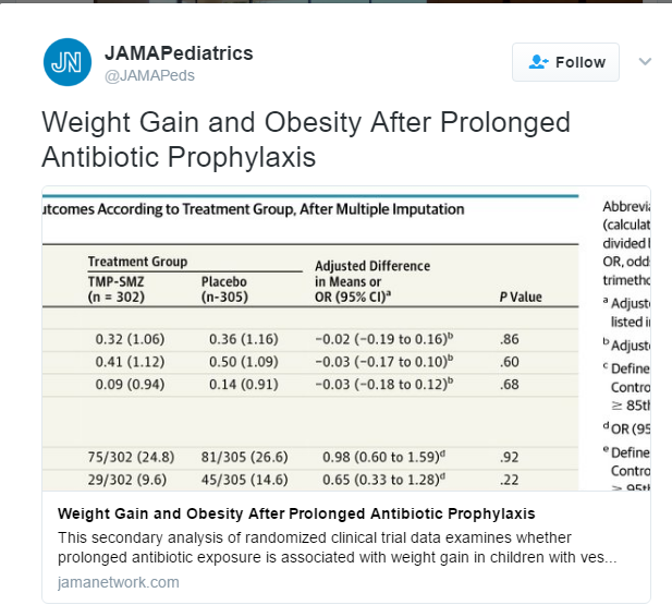
Design, Setting, and Participants  Secondary analysis of data from the Randomized Intervention for Children With Vesicoureteral Reflux Study, a 2-year randomized clinical trial that enrolled participants from 2007 to 2011. All 607 children who were randomized to receive antibiotic (n = 302) or placebo (n = 305) were included. Children with urinary tract anomalies, premature birth, or major comorbidities were excluded from participation.

Interventions  Trimethoprim-sulfamethoxazole or placebo taken orally, once daily, for 2 years.

Results  Participants had a median age of 12 months (range, 2-71 months) and 558 of 607 (91.9%) were female. Anthropometric data were complete at the 24-month visit for 428 children (214 in the trimethoprim-sulfamethoxazole group and 214 in the placebo group). Weight gain in the trimethoprim-sulfamethoxazole group and the placebo group was similar (mean [SD] change in weight-for-age z score: +0.14 [0.83] and +0.18 [0.85], respectively; difference, −0.04 [95% CI, −0.19 to 0.12]; P = .65). There was no significant difference in weight gain at 6, 12, or 18 months or in the prevalence of overweight or obesity at 24 months (24.8% vs 25.7%; P = .82). Subgroup analyses showed no significant interaction between weight gain effect and age, sex, history of breastfeeding, prior antibiotic use, adherence to study medication, or development of urinary tract infection during the study.

My take: Whether antibiotics could contribute to obesity is not entirely clear –even the possibility could encourage better stewardship of antimicrobials.

Related blog posts:

screenshot-143

screenshot-144

Triglyceride Levels and Pancreatitis

A recent study (JAMA Intern Med 2016; 176: 1834-42) suggests that even mild to moderate hypertriglyceridemia may increase risk of pancreatitis.

Among two large cohorts that were followed prospectively for a median of 6.7 years, 434 out of 116,550 patients developed acute pancreatitis.

Key finding (which persisted after adjustment of multiple potential confounding factors):

screenshot-140

More complete summary at GI & Hepatology News: “Mild, moderate hypertriglceridemia tied to pancreatitis”


What’s Happening on the Edge of Viability

A study (N Younge et al. NEJM 2017; 376: 617-28) provides some data on the slowly changing survival and neurodevelopmental outcomes among periviable infants (22-24 weeks gestation).

From epoch 1 (2000-2003), to epoch 3 (2008-2011), there has been some improvements. Overall survival increased from 30% to 36% and the percentage without neurodevelopmental impairment increased from 16% to 20%.

Mortality and Neurodevelopmental Outcomes at ~18 months of age (combined data and 11 centers)

Mortality and Neurodevelopmental Outcomes at ~18 months of age (combined data and 11 centers)

The insightful commentary (pgs 694-6) notes that there has not been improvement in survival in infants born at 22 weeks.  Furthermore, in reviewing multiple studies on outcomes, neurodevelopmental impairment was >94% in patients born at 22 weeks and between 80-90% for infants born at 23 weeks.  At 24 weeks, neurodevelopmental impairment was present between 51-72%

screenshot-137

Related blog posts:

Will Bariatric Surgery Become an Endoscopic Procedure?

A recent study (BK A Dayyeh et al. Clin Gastroenterol Hepatol 2017; 15: 37-43) provides evidence that endoscopic sleeve gastoplasty can be an effective treatment for obesity.

AGA Website Summary Endoscopic Sleeve Gastroplasty: A Promising New Weight Loss Procedure

An excerpt:

In the fight against obesity, bariatric surgery is currently the most effective treatment; however, only 1 to 2 percent of qualified patients receive this surgery due to limited access, patient choice, associated risks and the high costs. A novel treatment method — endoscopic sleeve gastroplasty — might offer a new solution for obese patients. Endoscopic sleeve gastroplasty is a minimally invasive, safe and cost-effective weight loss intervention, according to a study1 published online in Clinical Gastroenterology and Hepatology, the official clinical practice journal of the American Gastroenterological Association…

In this study of 25 patients with obesity who underwent the procedure at the Mayo Clinic in Rochester, MN, endoscopic sleeve gastroplasty reduced excess body weight by 54 percent at one year. Further, the procedure delayed solid food emptying from the stomach and created an earlier feeling of fullness during a meal, which resulted in a more significant and long-lasting weight loss.

Endoscopic sleeve gastroplasty was well tolerated as an outpatient treatment, requiring less than two hours of procedure time. Patients resumed their normal lifestyle within one to three days. The treatment was performed using standard “off-the-shelf” endoscopic tools as opposed to specific weight loss devices or platforms. The cost of endoscopic sleeve gastroplasty is roughly one-third that of bariatric surgery.

4 minute YouTube description from Johns Hopkins: What is Endoscopic Sleeve Gastroplasty and How Does it Work?

screenshot-73

screenshot-74 screenshot-75

Consensus Pancreatitis Recommendations

The INSPPIRE Group (CE Gariepy et al. JPGN 2017; 64: 95-103) has published consensus recommendations for acute recurrent pancreatitis (ARP) and chronic pancreatitis (CP).

While the authors acknowledge the need for high-level evidence/further research, they provide a large number of consensus recommendations.  These recommendations are succinctly summarized in Table 1 and Table 2.  From a reader’s perspective, my preference would have been to separate the recommendations for ARP and CP rather than to intermix them (though many of the recommendations are the same for both conditions).

ARP specific recommendations:

  • “Initial evaluation should include AST,ALT, GGT, Total bilirubin (fractionate if elevated), fasting lipids, and total serum calcium.”
  • Evaluate for fat-soluble vitamin deficiency, and pancreatic exocrine insufficiency at least annually

ARP and CP recommendations:

  • Consider ammonia and urine organic acids if there is a concern for undiagnosed metabolic disease.
  • Check for celiac disease.
  • Check for O&P if immunosuppressed, travel to endemic areas of Ascaris, or if peripheral eosinophilia.
  • Evaluation of genetic causes: should include sweat chloride test and PRSS1 gene testing. Consider SPINK1, CFTR, and CTRC evaluation.
  • Evaluate with MRCP (not ultrasound) acutely if GGT >2 x ULN or if direct bilirubin is elevated.
  • Non-acutely, MRCP recommended to evaluate pancreatic ductal abnormalities.  “When available, secretin-enhanced MRCP …should be obtained.” sMRCP can provide dynamic images of the pancreatic duct allowing differentiation of fixed from nonfixed lesions; this technique has not been widely adopted by pediatric radiologists compared with adult radiologists.

CP specific recommendations:

  • Evaluate for fat-soluble vitamin deficiency, pancreatic exocrine insufficiency, and pancreatic endocrine insufficiency at least annually

The authors did not recommend checking serum IgG4 in the absence of associated systemic disease or suggestive imaging for autoimmune pancreatitis.

Briefly noted: J-H Choi et al. Clin Gastroenterol Hepatol; 2017: 15: 86-92.  This study indicated that vigorous hydration with lactated ringer’s (LR) reduces risk of pancreatitis after ERCP.  A potential inference would be that LR would be an optimal fluid for pancreatitis more broadly. (Related: Why an ERCP Study Matters to Pediatric Care | gutsandgrowth)

Related blog posts:

Dragon Point, Labadee

Dragon Point, Labadee

“Addressing Physician Burnout”

In the last few years, there have been increasing reports of physician burnout.  A recent report (JAMA. Published online February 9, 2017. doi:10.1001/jama.2017.0076) (thanks to Ben Gold for this reference) provides a concise review of the reasons and potential mitigating strategies.

Full Text Link: “Addressing Physician Burnout”

An excerpt:

Physicians also have to navigate a rapidly expanding medical knowledge base, more onerous maintenance of certification requirements, increased clerical burden associated with the introduction of electronic health records (EHRs) and patient portals, new regulatory requirements (meaningful use, e-prescribing, medication reconciliation), and an unprecedented level of scrutiny (quality metrics, patient satisfaction scores, measures of cost).

These challenges have taken a toll on US physicians. Burnout is a syndrome of exhaustion, cynicism, and decreased effectiveness at work…The first large, national study of burnout among US physicians across all specialties did not occur until 2011. That study of 7288 participating physicians documented that approximately 45% reported at least 1 symptom of burnout and that burnout was more common among physicians than US workers in other fields…

The first large, national study of burnout among US physicians across all specialties did not occur until 2011. That study of 7288 participating physicians documented that approximately 45% reported at least 1 symptom of burnout and that burnout was more common among physicians than US workers in other fields…

Physician burnout has been linked to self-reported errors, turnover, and higher mortality ratios in hospitalized patients…

The current burden of documentation related to the clinical encounter required to meet billing requirements, quality reporting, and separate justification for each test ordered individually is unsustainable…

Individual physicians must also do their part…Individual physicians have a professional responsibility to take care of themselves. Adequate sleep, exercise, and attending to personal medical needs should be considered a minimal standard for self-care. Physicians must also proactively identify personal and professional priorities and take deliberate steps to integrate their personal and professional lives.

Related blog posts:

birthoutcomesaftertnt

Ileocecal Resection in Pediatric Crohn’s Disease

A recent retrospective study (K Diederen et al. Inflamm Bowel Dis 2017; 23: 272-82) provides data on the likelihood of complications and recurrence following ileocecal resection in pediatric Crohn’s disease (n=122).

Key findings:

  • Severe postoperative complications were noted in 9.8%.  Risk factors included colonic disease (Odds ratio 5.6), microscopically positive resection margins (OR 10.4), and emergency surgery (OR 6.8)
  • Overall complication rate was reported as 29.5% which is similar to rates reported in adults
  • Clinical recurrence rates after 1, 5, and 10 years: 19%, 49%, and 71%
  • Surgical recurrence rates after 1, 5, and 10 years: 2%, 12%, and 22%
  • Immediate postoperative therapy reduced the risk of clinical recurrence (HR 0.3) and surgical recurrence (HR 0.5)
  • “In this study, postoperative catch-up growth was found in patients younger than 16 years in the year after surgery.” Thus, surgery could be an important to reverse growth retardation.

Complications within 30 days of surgery were categorized with the Clavien-Dindo classification. Those with grade ≥III which required either surgical, endoscopic or radiologic intervention were considered severe.  In this population, the complications included intraabdominal septic complications and/or anastomotic leakage.

My take: In some patients, ileocecal resection should NOT be a last resort.  Waiting too late, increases the risk of complications.  The task at hand is prospectively identifying those who merit surgery sooner and then convincing the family to proceed.

screenshot-118

Related blog posts:

Clostridium difficile Infection in Inflammatory Bowel Disease: Expert Updates

A recent clinical practice update (S Khanna et al. Clin Gastroenterol Hepatol; 2017; 15: 166-74) provides some succinct recommendations regarding Clostridium difficile infection (CDI) in Inflammatory Bowel Disease (IBD).

Background: In 2011, the authors note that CDI was associated with 29,000 deaths and is now the most lethal enteric pathogen in the U.S.

Differences in pathogenesis of C diff in IBD compared to those without IBD:

  • Younger age
  • Less frequent antibiotic exposure
  • More often community onset (rather than hospital onset)
  • Higher recurrence (may be related to dysbiosis)

Key recommendations:

  • In patients with IBD flare, test for CDI
  • In patients with CDI and IBD, clinicians should consider “using vancomycin instead of metronidazole.”
  • In patients with recurrent CDI and IBD, consider fecal microbiota transplantation

Figure 4 proposes a management algorithm (for adults).  If uncomplicated CDI, recommended dose of vancomycin was 125 mg q6h. If no improvement in 3-4 days, then “consider escalation of immunosuppression.” For complicated CDI, consider oral vancomycin at 500 mg q6h and IV metronidazole 500 mg q8.  In addition, consider rectal vancomycin and surgery consult.

Complicated CDI includes ICU admission, hypotension, T >38.5, ileus/megacolon, mental status changes, leukocyte count >35,000  or < 2000, or lactate >2.2 mmol/L

Another review article (Y Chen et al. Inflamm Bowel Dis 2017; 23: 200-07) is a meta-analysis that identified six studies.  One of these studies was a case-control study with nearly 400,000 patients (and about 7000 cases of C diff). Key finding: CDI results in nearly a doubling of the risk of colectomy (OR 1.90), mainly in patients with ulcerative colitis.

Related blog posts

Disclaimer: These blog posts are for educational purposes only. Specific dosing of medications (along with potential adverse effects) should be confirmed by prescribing physician.  This content is not a substitute for medical advice, diagnosis or treatment provided by a qualified healthcare provider. Always seek the advice of your physician or other qualified health provider with any questions you may have regarding a condition.


Rare Tragic Reaction to Infliximab

A recent post on the pediatric GI Listserv pointed out a troubling case report: “Fatal Central Nervous System Disease Following First Infliximab Infusion in a Child With Inflammatory Bowel Disease,” FM. Baumer et al; Pediatric Neurology 2016; 57: 91-94.

“A seven-year-old boy diagnosed with ulcerative colitis and primary sclerosing cholangitis received infliximab. Six hours following his uneventful infusion, he awoke with headache and emesis and rapidly became obtunded…Cranial computed tomography revealed hypodense lesions in the cerebral hemispheres, cerebellum, and pons accompanied by hemorrhage…Within four days he met criteria for brain death.”

The authors note that “the close temporal association between our patient’s presentation and the infliximab infusion raises concern for a drug-related cause for his cerebral injury.” While this case report is terribly sad, severe and fatal reactions can unfortunately be encountered with a wide range of medications, including commonly used antibiotics.

My take: Thus, while the vast majority of pediatric patients with inflammatory bowel disease will benefit from infliximab therapy, there are rare tragic outcomes.  img_3954


Gastrojejunostomy Complications Frequent

Gastrojejnostomy (GJ) placement allows enteral feeds to bypass the stomach.  When a gastrostomy is already in place, GJ placement may allow patients to avoid surgery (eg. fundoplication).  Most practitioners would consider the risk of GJ placement to be low, but a recent report (J Moorse et al. JPS 2017; http://dx.doi.org/10.1016/j.jpedsurg.2017.01.026) suggests that it is higher than expected.  The abstract and link are below.

Link: Gastrojejunostomy tube complications — A single center experience and systematic review

Abstract

Purpose

Gastrojejunostomy tubes (GJTs) enable enteral nutrition in infants/children with feeding intolerance. However, complications may be increased in small infants. We evaluated our single-institution GJT complication rate and systematically reviewed existing literature.

Methods

With REB approval, a retrospective single-institution analysis of GJT placements between 2009 and 2015 was performed. For the systematic review, MOOSE guidelines were followed.

Results

At our institution, 48 children underwent 154/159 successful insertions primarily for gastroesophageal reflux (n = 27; 55%) and aspiration (n = 11; 23%). Median age at first GJT insertion was 2.2 years (0.2–18). Thirty-five (73%) had an index insertion when ≤10 kg. GJTs caused 2 perforations and 1 death. The systematic review assessed 48 articles representing 2726 procedures. Overall perforation rate was estimated as 2.1% (n = 36 studies, 23/1092, 95% CI: 1.0–3.2). Perforation rates in children <10 kg versus ≥10 kg were estimated as 3.1%/procedure (95% CI: 1.1%–5.0%) and 0.1%/procedure (95% CI: 0%–0.3%), respectively. The relative risk of perforation was 9.4 (95% CI: 2.8–31.3). Overall mortality was estimated as 0.9%/patient (n = 39 studies; 95% CI: 0.2–1.6%). Most perforations (19/23; 83%) occurred ≤30 days of attempted tube placement.

Conclusion

Gastrojejunostomy tubes are associated with significant complications and frequently require revision/replacement. Insertion in patients <10 kg is associated with increased perforation risk. Caution is warranted in this subgroup.

With regard to the methodology

  • ~90% of the procedures were performed by interventional radiology and the interventionist had a median of 6.6 years of experience
  • Most GJs were 16 French in width and most were either 15 cm or 22 cm in length

My take: This report highlights the significant risks associated with GJ placement, particularly in smaller patients (<10 kg).  Despite these risks, GJ placement is often the safest option.

Costa Maya, Mexico

Costa Maya, Mexico