Eosinophilic Esophagitis: Once vs Twice Daily Steroid Treatment

CC Reed et al. Clin Gastroenterol Hepatol 2025; 23: 946-953. Open Access! Daily or Twice Daily Treatment With Topical Steroids Results in Similar Responses in Eosinophilic Esophagitis

Methods: This was a retrospective cohort study using the UNC EoE Clinicopathologic Database of newly diagnosed patients with EoE treated with a tCS who had a follow-up endoscopy with biopsy. In total, there were 522 patients, including 195 pediatric patients (<18 yr). 122 patients received once daily dosing and 400 patients received twice daily dosing.

At our center, patients are typically treated on a clinical basis with either oral viscous budesonide or fluticasone from a multidose inhaler, with daily doses ranging from 1–2 mg for budesonide and 440–1760 μg for fluticasone based on patient size and at the discretion of the provider.

Key findings:

  • Global symptomatic response (78% vs 76%; P = .82), posttreatment eosinophil count (20.8 vs 25.6; P = .21), posttreatment EoE Endoscopic Reference Score (2.2 vs 2.2; P = .92), and histologic response (<15 eos/hpf; 56% vs 58%; P = .66) did not differ by dosing frequency
  • Candida was less frequent with daily dosing (2% vs 8%; P = .04)

My take: This study suggests that once daily dosing can be as effective as twice daily dosing. It may be that the total dose administered may be more important than the frequency. More studies are needed to confirm these results.

Related blog posts:

How HLA DQA1*05 and Combination Therapy Modulate Treatment Outcomes in Children with Crohn’s Disease

J Adler et al. Am J Gastroenterol 2025; 120: 1076-1086. HLA DQA1*05 and Risk of Antitumor Necrosis Factor Treatment Failure and Anti-Drug Antibody Development in Children’s with Crohn’s Disease.

This was a prospective, double-blind, placebo-controlled trial with 204 patients examining the clinical outcomes of anti-TNF with or without methotrexate (COMBINE).

Key findings:

  • Treatment failure in HLA DQA1*O5: A trend toward increased treatment failure among HLA DQA1*05-positive participants was not statistically-significant (hazard ratio 1.58; P = 0.08).
  • HLA DQA1*05 and Treatment Failure Rate: During the followup period, HLA DQA1*05-positive patients had a 35% failure rate compared to 26% for those who were HLA DQA1*05-negative (P=0.098). The overall failure rate was 30%
  • Methotrexate Combined with HLA DQA1*05 Effect: Patients who were HLA DQA1*05 negative and assigned to methotrexate experienced less treatment failures than HLA DQA1*05-positive patients on placebo (hazard ratio 0.31, 95% CI 0.13-0.70; P = 0.005).
  • Anti-TNF Medication Comparison: Treatment failure was similar between infliximab and adalimumab, 29% and 33% respectively
  • Antidrug antibodies (ADA): A trend toward increased ADA development among HLA DQA1*05-positive participants was not significant (odds ratio 1.96, P = 0.09). The addition of methotrexate to the treatment regimen mitigated the risk of treatment failure among individuals positive for HLA DQA1*05 and reduced the odds of developing ADA by 90%.
  • Rate of ADA: “After further stratification, HLA DQA1*05-negative participants assigned to methotrexate were less likely to develop ADA relative to HLA DQA1*05-positive patients on placebo (odds ratio 0.12; P = 0.008).”

Discussion Points:

  • “A retrospective by Fuentes-Valenzuela et al …found that if patients underwent proactive TDM, there was no increase in the rate of treatment discontinuation among those who were HLA DQ-A105 positive compared with HLA DQ-A105 negative”
  • “The totality of evidence suggests that HLA DQ-A1*05 seems to confer a risk of both immunogenicity and treatment failure, particularly among infliximab-treated patients. Furthermore, this risk may be mitigated by the use of proactive TDM and/or concomitant immunomodulators.”

My take (borrowed in part from authors):  “40% of patients were HLA DQ-A1*05 positive, which was associated with a trend toward increased risk of both treatment failure and ADA. These risks were mitigated, but not eliminated, by adding oral methotrexate.” The use of combination therapy (methotrexate with anti-TNF) was associated with the lowest failure rates.

Also, unrelated article: DA Carlson et al. Gastroenterology 2025; 168: 1114-1127. A Standardized Approach to Performing and Interpreting Functional Lumen Imaging Probe Panometry for Esophageal Motility Disorders: The Dallas Consensus. Congratulations to Dr. Garza from our group who was one of the authors

Related blog posts:

Dr. Arun Singh: Tips and Tricks to Managing Celiac Disease

Recently Dr. Arun Singh gave our group a terrific update on Celiac Disease. I have taken some notes and shared some slides. There may be inadvertent omissions and mistakes in my notes.

Key points:

  • Celiac disease (CD) global prevalence is about 1.4%, though there are ‘pockets’ with much higher rates (~3% prevalence in Colorado). This equates to more than 3 million Americans with CD
  • There is a huge gluten free diet market of ~$7 billion. This market includes CD, nonceliac gluten sensitivity (NCGS) and those with wheat allergies
  • Many patients have atypical symptoms which can include ADHD, brain fog, headaches, and elevated LFTs. Many have silent CD with no symptoms
  • Higher risk groups include family members (~10% risk for 1st degree, ~80% risk for identical twin), autoimmune diseases (thyroid, diabetes, others), and genetic disorders (Down syndrome, Williams syndrome, Turner syndrome)
  • Transient elevation of TTG IgA is common. In TEDDY study, 19% had TTG IgA spontaneously normalize. Thus, a single abnormal lab is not reliable
  • Genetic testing can be a useful adjunct in a few specific situations, including prior to instituting a gluten challenge
  • 1-3% of those with celiac disease may be negative for HLA-DQ2 and HLA-DQ8
  • If a gluten challenge is needed, 12 weeks is ideal. However, if poorly tolerated, then consider endoscopy earlier and Dr. Singh recommends checking in with family 4-6 weeks into the challenge
  • Endoscopy recommendations: Taking a single biopsy per pass can improve orientation when obtaining duodenal biopsies (bulb and distal portion, 5-6 in total)
  • NASPGHAN has not updated comprehensive guidelines for CD in 20 years
  • A survey of NASPGHAN members indicated that ~40% utilize a “no-biopsy” approach in patients. Dr. Singh noted that the accuracy of this approach, based on data from North America, may be about 96%
  • Drug trials for CD require a biopsy-confirmed diagnosis
  • Surveillance practice is quite variable. Important to follow growth and serology. CHOP approach includes surveillance for type 1 DM
  • Followup endoscopy to assess mucosal healing is not the current standard of care but could be helpful in some patients
  • Among patients with potential CD, about 30% develop CD over time. Thus, these patients should be monitored (yearly labs, f/u scope after 2 years)
  • Nonresponsive/refractory CD: start with nutrition assessment, often needs a f/u scope before consideration of budesonide therapy (9 mg x 12 weeks) or gluten contamination elimination diet

Related blog posts:

The Guardian: UnitedHealth Secretly Paid Nursing Homes to Reduce Hospital Transfers

The Guardian 5/21/25:
Revealed: UnitedHealth secretly paid nursing homes to reduce hospital transfers

An excerpt:

UnitedHealth Group, the nation’s largest healthcare conglomerate, has secretly paid nursing homes thousands in bonuses to help slash hospital transfers for ailing residents – part of a series of cost-cutting tactics that has saved the company millions, but at times risked residents’ health, a Guardian investigation has found…

Those secret bonuses have been paid out as part of a UnitedHealth program that stations the company’s own medical teams in nursing homes and pushes them to cut care expenses for residents covered by the insurance giant.

In several cases identified by the Guardian, nursing home residents who needed immediate hospital care under the program failed to receive it, after interventions from UnitedHealth staffer…UnitedHealth said the suggestion that its employees have prevented hospital transfers “is verifiably false”. It said its bonus payments to nursing homes help prevent unnecessary hospitalizations that are costly and dangerous to patients and that its partnerships with nursing homes improve health outcomes…

To reduce residents’ hospital visits, UnitedHealth has offered nursing homes an array of financial sweeteners…

Two current and three former UnitedHealth nurse practitioners told the Guardian that UnitedHealth managers pressed nurse practitioners to persuade Medicare Advantage members to change their “code status” to DNR even when patients had clearly expressed a desire that all available treatments be used to keep them alive...

UnitedHealth’s insurance arm covers millions more Medicare Advantage seniors than any of its rivals

Commentary from Dr. Glaucomflecken account on X: https://x.com/i/status/1925348490680340979

My take: Healthcare insurance companies are incentivized to act as financial institutions and gatekeepers/third party administrators. This often compromises and delays patient care.

Related blog posts:

Treating Pediatric Metastatic Crohn’s Disease

E Chang et al. JPGN Reports. 2025;1–8. DOI: 10.1002/jpr3.70022. Open Access! Genital Crohn’s disease in pediatrics and genetic associations

This case report of four patients provides a good review of metastatic Crohn’s disease (MCD). MCD indicates that there is noncontiguous dermatological spread of CD involving the genitalia and perineum.

Key points:

  • “Less than 100 cases of pediatric MCD have been reported in the literature to date. These lesions are characterized by swelling, plaques, nodules, fissures, ulcerations, or crusts. In children, MCD typically presents as genital swelling with or without erythema in approximately 85% of cases.”
  • “Prior studies have shown that MCD co-occurs with CD in 50.8% of children, while others may develop GI symptoms after MCD diagnosis (15.3%) or even lack signs of CD (11.9%).”
  • “Scrotal histopathology revealed granulomatous inflammation, and genetic testing identified pathogenic variants in NOD2, COL7A1, and Chek2, as well as additional variants of uncertain significance.”
  • The optimal treatment is not clear. “Prior case reports and case series have shown positive responses to TNF-α inhibitors, but relapses may be common. Similarly, only partial improvement was noted in our patients treated with infliximab and adalimumab.”

Discussion: “Many patients do not demonstrate GI symptoms and may experience significant delays in diagnosis.”

My take: This article provides a good review of metastatic Crohn’s disease which is a rare problem. I have had two patients with this disorder. This problem fits the adage of “the more you see, the more you know; and, the more you know, the more you see.”

Related blog posts:
Springway Trail in Sandy Springs, GA

Prospective Study: Safety of Live Rotavirus Vaccine in Infants with In Utero Exposure to Biologics

K Ernest-Suarez et al. Clin Gastroenterol Hepatol 2025; 23: 835-845. Open Access! Live Rotavirus Vaccination Appears Low-risk in Infants Born to Mothers With Inflammatory Bowel Disease on Biologics

Background: “Caution regarding live vaccine administration emerged following reports of 5 fatal outcomes following the administration of the Bacille Calmette-Guérin vaccine in biologic-exposed infants.9 This has resulted in gastroenterology guidelines recommending that biologic-exposed infants should avoid live vaccines within the first 6 to 12 months of life or until drug concentrations are no longer detectable in the infant’s blood to reduce potential risks.2,10 [J Crohns Colitis. 2023; 17:1-27, Gastroenterology. 2021; 161:669-680.e0]. Contrary to this, inadvertent administration of the live oral rotavirus vaccine in biologic-exposed individuals has not been associated with significant adverse effects.7,11 Withholding the rotavirus vaccine has implications, given that rotavirus infection in infants is a leading cause of severe, dehydrating diarrhea in children under the age of 5 years globally.12,13

This prospective cohort study enrolled 57 biologic-exposed infants, including infliximab (n = 21), adalimumab (n = 19), vedolizumab (n = 10), and ustekinumab (n = 7), in the third trimester.

Key findings:

  • Immunologic assessments validated for age were normal in all infants despite median infliximab concentrations of 6.1 ug/mL, adalimumab concentrations of 1.7 ug/mL, ustekinumab concentrations of 0.6 ug/mL, and undetectable for vedolizumab at median of 10.7 weeks of age
  • The live oral rotavirus vaccine series was provided to 50 infants with the first dose given at a median of 13 weeks of age. No adverse effects following immunization were reported

Discussion:

  1. “Administration of the live rotavirus vaccine appeared low-risk in biologic-exposed infants born to mothers with IBD”
  2. “Routine drug concentration testing in the infant should not be utilized to determine the safety of live rotavirus vaccination”
  3. “Physicians should advise patients to ‘be more concerned about active disease rather than active medications’ and to continue effective therapy through pregnancy and lactation”

My take: Given the difficulty in excluding rare adverse outcomes, it is unlikely that formal vaccine recommendations will change in infants exposed to biologics; however, inadvertent administration of a live oral rotavirus vaccine poses a very lowl risk based on current studies.

Related blog posts:
Floating flowers -seen in several places in Thailand

Disclaimer: This blog, gutsandgrowth, assumes no responsibility for any use or operation of any method, product, instruction, concept or idea contained in the material herein or for any injury or damage to persons or property (whether products liability, negligence or otherwise) resulting from such use or operation. These blog posts are for educational purposes only. Specific dosing of medications (along with potential adverse effects) should be confirmed by prescribing physician.  Because of rapid advances in the medical sciences, the gutsandgrowth blog cautions that independent verification should be made of diagnosis and drug dosages. The reader is solely responsible for the conduct of any suggested test or procedure.  This content is not a substitute for medical advice, diagnosis or treatment provided by a qualified healthcare provider. Always seek the advice of your physician or other qualified health provider with any questions you may have regarding a condition.

Rapid Gut Microbiome Recovery: Diet Outperforms Microbial Transplant

Briefly noted: MS Kennedy et al. Nature (2025). https://doi.org/10.1038/s41586-025-08937-9. Diet outperforms microbial transplant to drive microbiome recovery in mice

Methods:  Here we characterize the trajectory by which the gut microbiome recovers its taxonomic and functional profile after antibiotic treatment in mice on regular chow (RC) or Western Diet (WD).

Key findings: “Only mice on RC undergo a rapid successional process of recovery. Metabolic modelling indicates that a RC diet promotes the development of syntrophic cross-feeding interactions, whereas in mice on WD, a dominant taxon monopolizes readily available resources without releasing syntrophic byproducts. Intervention experiments reveal that an appropriate dietary resource environment is both necessary and sufficient for rapid and robust microbiome recovery, whereas microbial transplant is neither.”

Conclusion (from authors): “Our data challenge widespread enthusiasm for faecal microbiota transplant (FMT) as a strategy to address dysbiosis, and demonstrate that specific dietary interventions are, at a minimum, an essential prerequisite for effective FMT, and may afford a safer, more natural and less invasive alternative.”

My take: This study suggests that the best way to get a “healthy” microbiome is to eat a healthy diet rather than to try to alter with FMT. This finding likely would be the same for probiotics as well.

Related blog posts:
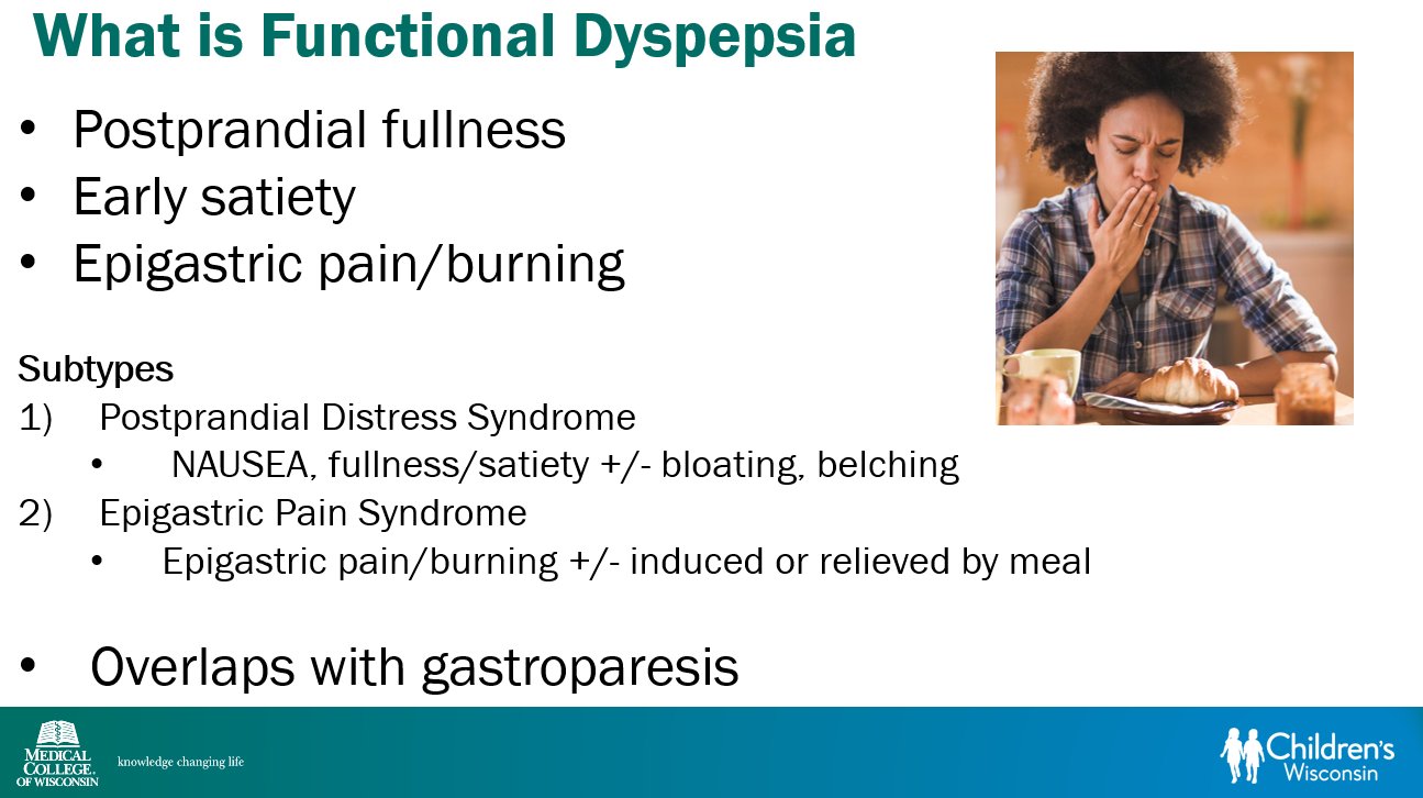
Andaman Sea (Thailand)

When an Inflammatory Bowel Disease Diagnosis is Far Away

JF McLaughin et al. Clin Gastroenterol Hepatol 2025; 23: 825-834. Travel Time to Treating Center Is Associated With Diagnostic Delay in Pediatric Inflammatory Bowel Disease

This was a cross-sectional study of newly diagnosed pediatric patients (n=869) with IBD at 22 United States sites from 2019 to 2022. 57% were diagnosed with CD, 34% with UC, and 4% with IBD-U.

Key findings:

  • Overall, the mean time from symptom onset to diagnosis was 265.9 days
  • Factors associated with longer diagnosis time included CD vs UC (odds ratio [OR], 2.6), and longer travel time to clinic (>1 hour [OR, 1.7], >2 hours [OR, 1.8] each vs <30 minutes)
  • There was no association with race, ethnicity, birth country, gender, parent education, household income, insurance type, health literacy, and health system distrust

The finding that there is a longer diagnostic delay with CD than UC is consistent with prior studies. The longer travel time has not been widely recognized as a factor associated with delayed diagnosis, though it has been associated with other negative outcomes like higher mortality with chronic liver disease.

Regarding the lack of a negative impact from factors like race/ethnicity and income, my suspicion is that this is probably related to several factors:

  • Overall, the pediatric age group has a very high rate of being insured as most children without commercial insurance currently qualify for Medicaid. This helps improve access to needed/timely health care
  • A recent study showed that pediatric GI specialists do not have disparities in treatment compared to pediatric GI providers with an IBD focus; thus, pediatric specialists are more likely to minimize treatment delay (Treatment Disparities in Adult vs. Pediatric IBD Care Related to Provider Specialization)
  • Parents help limit diagnostic delay in their children

My take: There are many places that are far away from pediatric specialists. This results in diagnostic delays.

Related blog posts:
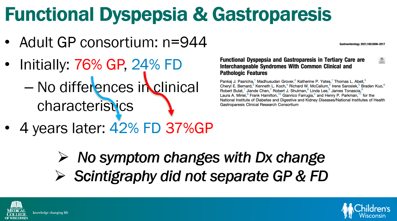
Mai Khao Beach, Phuket, Thailand

Impact of Disease Severity on Eosinophilic Esophagitis Treatment Responses

CC Reed et al. Clinical Gastroenterology and Hepatology 2025; Worsening Disease Severity as Measured by I-SEE Associates With Decreased Treatment Response to Topical Steroids in Eosinophilic Esophagitis Patients

This was a retrospective study with children and adults with eosinophilic esophagitis (EoE). Among 1312 patients, there were 657 (50%), 461 (35%), and 194 (15%) with mild, moderate, and severe disease by I-SEE, respectively. Disease activity was categorized as mild (I-SEE 1–6), moderate (I-SEE 7–14), or severe (I-SEE ≥15).

Key findings:

  • Patients with severe disease were less likely to have histologic response (49% (severe) vs 55% (moderate) vs 64% (mild); P = .03 for <15 eosinophils per high-power field) 
  • Patients with severe EoE also had lower global symptom response rates (53% vs 79% vs 83%, respectively) and higher post-treatment EoE Endoscopic Reference Scores (EREFS; 3.6 ± 2.3 vs 2.4 ± 1.8 vs 1.6 ± 1.6, respectively)

My take: More severe disease is harder to treat with EoE (and most everything else too).

Related blog posts:
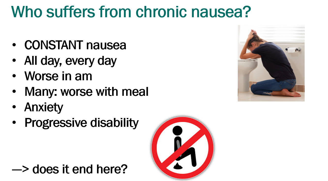
Link: I-SEE Tool Scoring Table

Crazy wiring in Chiang Mai, Thailand (but prevalent in many areas of Thailand)

How Gut Bacteria Might Increase Colon Cancer Risk in Young Adults

Will Stone, NPR 4/25/25: Damage from gut bacteria may play a role in the rise in colon cancer in young adults

An excerpt:

It’s unclear why colon cancer cases have doubled in people under 55 over the past two decades, a staggering rise that has alarmed doctors and cancer researchers.

But part of the story could be colibactin, a toxin made by certain strains of E. coli and other bacteria. In a study out this week, researchers have identified a strong link between this DNA-damaging toxin and colon cancer among younger patients.

The team, based at the University of California, San Diego, analyzed tissue samples from close to 1,000 colorectal cancer patients across four continents. They found the majority had cancers bearing mutations that signaled a past encounter with colibactin.

“You can think of it as the weapon system of a bacteria to fight other bacteria and to defend themselves,” says Ludmil Alexandrov, the lead author of the study, which was published in Nature this week.

Strikingly, those under the age of 40 with early-onset colon cancer were three to five times more likely to have these mutations than those in their 70s and older.

The thinking goes that in some people, this bacterial weaponry — technically called a “genotoxin” — can get directed at their gut cells, seeding mutations that put them at increased risk of developing colorectal cancer.

According to their data, this exposure isn’t ongoing when the cancer is diagnosed. Instead, it appears to have happened during childhood.

“Our estimate is that it happens within the first 10 years of life,” Alexandrov says. “So if you get that mutation at age 5, that puts you 20 to 30 years ahead of schedule for getting colorectal cancer.”

While the study shows a strong association, the data can’t prove colibactin caused these patients to develop cancer at a younger age. And researchers in the field don’t expect E. coli, or any single microbe for that matter, to be the skeleton key for the surge in colorectal cancer.

Related article: M Diaz-Gay, et al. Nature https://doi.org/10.1038/s41586-025-09025-8 (2025). Geographic and age variations in mutational processes in colorectal cancer

Related blog posts:
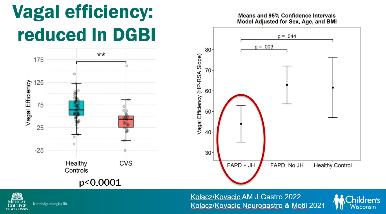
View from plane over Thailand