IPAA (Pouch) for Crohn’s Disease and Indeterminate Colitis

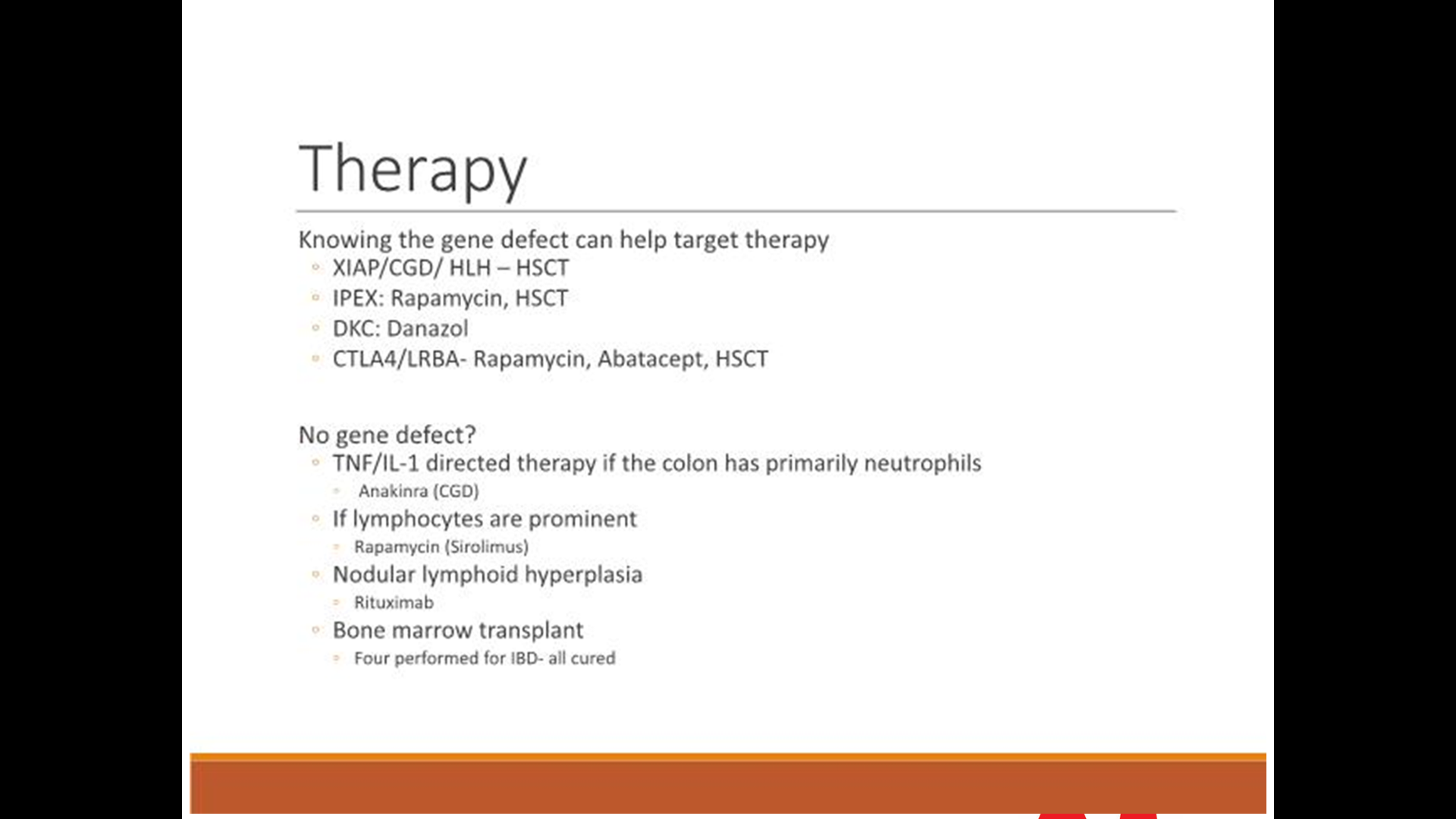
A recent review (S Chang, B Shen, F Remzi. Gastroenterology & Hepatology 2017; 13: 466-75 Full text link: When Not to Pouch: Important Considerations for Patient Selection for Ileal Pouch-Anal Anastomosis) makes recommendations regarding Ileal pouch-anal anastomosis (IPAA) for Crohn’s disease and indeterminate colitis. Key points:

  • In CD patients with isolated colitis and without perianal disease, “there were no differences in the rates of postoperative complications, pelvic sepsis, or pouch failure compared with UC patients” (GE Reese et al. Dis Colon Rectum 2007; 50: 239-50).
  • Rates of pouch retention for CD (Table 2) ranged from 43% to 94% in 19 studies. Most of these studies had small numbers (less than 40 patients). In the two largest studies with 97 patients and 150 patients, both with ~10 year followup, pouch retention rates were 74% and 87% respectively.
  • “Patients carrying the diagnosis of IC have pouch function on par with patients with UC, with no significant difference in the number of bowel movements…However, ..are more likely to develop CD of the pouch. Nevertheless, pouch failure rates among IC, IBD-unclassified, and UC are similar in multiple cohorts.”
  • Rates of pouch retention for IC ranged from 73%-100% among the 13 cited studies, though only 2 studies reported rates less than ~90%. The two largest studies with ~340 patients had retention rates of ~95% and followup of 3.4 yrs and 10.2 years.

This review also discusses IPAA and other issues including obesity (which increases the likelihood of complications), sphincter dysfunction, elderly patients, and radiation therapy.

Of note, recent ESPGHAN IBD Porto Group guideline for surgical Crohn’s disease management in children (J Amil-Dias et al JPGN 2017; 64: 818-35) at first glance seems to be at odds with Chang et al recommendations:

  • “Statement 8. Ileal pouch-anal anastomosis is not recommended when a patient has CD. (Agreement 100%)”
  • The body of the report is more nuanced: “There is, however, recent growing evidence that supports highly selective use of restorative proctocolectomy with ileal pouch-anal anastomosis for CD. These patients have isolated colonic CD and no evidence of ileal or perianal involvement.”

To me, statement 8 should have been worded to include “except in limited circumstances.”  As it stands now, it misleads those who do not carefully review the entire report.

My take: The report by Chang et al makes a strong case for its conclusion: “Although it is true that the diagnosis of CD is a potential contraindication to IPAA, patients with isolated Crohn’s colitis may thrive after pouch surgery.  At this time, patients with isolated Crohn’s colitis (without perianal disease or small bowel involvement) have good pouch retention rates.”  Their review prompted me to look more closely at the ESPGHAN IBD Porto Group guideline; their Statement 8 recommendation is, in fact, quite misleading.

Disclaimer: These blog posts are for educational purposes only. Specific dosing of medications (along with potential adverse effects) should be confirmed by prescribing physician.  This content is not a substitute for medical advice, diagnosis or treatment provided by a qualified healthcare provider. Always seek the advice of your physician or other qualified health provider with any questions you may have regarding a condition.

Keyhole view , looking into the Rotunda UVa, of Thomas Jefferson (or TJ for those in the know)

Programming for Fatty Liver Disease May Occur Prior to Birth

A recent study (KP Newton et al. J Pediatr 2017; 187: 141-6; associated editorial pg 13-15)) in a multicenter retrospective cross-sectional study of children (n=538) with biopsy-proven nonalcoholic fatty liver disease (NAFLD) showed that birth weight influenced the development of NAFLD.  The participants were enrolled in the Nonalcoholic Steatohepatitis Clinical Research Network (NASH CRN).

Key findings:

  • There was increased NAFLD among both low birth weight (LBW) and high birth weight (HBW).
  • LBW occurred more commonly in the NAFLD cohort 9.3% compared with the general population prevalence 6.1%.
  • HBW occurred more commonly in the NAFLD cohort 14.9% compared with the general population prevalence 10.5%

The authors speculate that the explanation/mechanisms for increase in both LBW and HBW are likely to differ. It has been recognized that LBW is associated with higher cardiovascular disease and type 2 diabetes.  HBW start bigger and often stay bigger; that is, there are increased risks of more severe obesity.

There are numerous limitations to this study -there is a lot of data that is not available, including gestational age, maternal weight, breastfeeding exposure, and antibiotic administration.

My take: These findings add to the literature that risks for NAFLD along with other metabolic problems may be present at birth.  Is there a way to modify this risk?

Related study: ET Jensen et al. J Pediatr 2017; 187: 50-7, editorial pg 10-12.  In this study of 535 ten-year-old children, enrolled in a prospective multicenter extremely low gestational age newborn cohort study, the authors found that maternal overnutrition and undernutrition affected the brain health of these children. The authors used neurocognitive assessment tools.

  • Children born to women with a pregravid BMI >30 scored “lower on measures of general cognitive ability, executive functioning, fine motor function, and academic achievement.”
  • Children born to women with inadequate maternal weight gain during pregnancy had “lower language and academic achievement.”

Hidden Falls, Highlands NC


Likelihood of Genetic Disease with Early-Onset Pancreatitis

Another study (MJ Giefer et al. J Pediatr 2017; 186: 95-100) from the INSPPIRE group provides data on early-onset pancreatitis.  Specifically, the group presents clinical information on 342 children with acute recurrent pancreatitis or chronic pancreatitis regarding disease burden and associations with genetic mutations.

Key findings:

Genetic disease is much more common in younger ages:

  • In subset younger than 6 years, 72 of 102 had genetic diseases identified:  PRSS1 in 42, CFTR in 27, SPINK1 in 12, and CTRC in 8.
  • In subset 6-11 years of age, 52 of 90 had genetic diseases identified: PRSS1 in 20, CFTR in 23, SPINK1 in 22, and CTRC in 1.
  • In subset greater than 12 years of age, 39 of 72 had genetic diseases identified: PRSS1 in 13, CFTR in 24, SPINK1 in 6, and CTRC in 1.
  • Testing for newer susceptibility genes were not evaluated as they had not become commercially available: carboxypeptidase 1, claudin 2, carboxylesterlipase, and carboxyesterlipase-hybrid
  • SPINK1 is noted to be present in 1-3% of general population; CTRC mutation occur in 0.7% of healthy controls.

Obstructive causes were common.  Pancreas divisum was identified in 38 patients but there was not a great deal of difference among the age groups; similarly, other obstructive causes were identified in about one-third of patients and included sphincter of Oddi dysfunction (n=9), gallstones (n=17), pancreaticobiliary malunion (n=12), biliary cyst (n=11), pancreatic stricture (n=2), annular pancreas (n=3), and duodenal diverticulum.

Disease burden:

  • Exocrine insufficiency noted in 52 (no strong age predilection)
  • Diabetes in 18 (11 of the cases occurred in those >12 years)
  • Constant moderate pain noted in 82 (25 in group <6, 27 in 6-12 group, and 30 in group >12 years)
  • Constant severe pain noted in 18  (4 in group <6, 5 in 6-12 group, and 9 in group >12 years)
  • Average number of hospitalizations in past year was 1 in those ≤12 and 2 in those older than 12.

My take: PRSS1 and CTRC mutations are associated with early onset pancreatitis.

Related blog posts:

Rotunda dome at the University of Virginia


Clostridium difficile Risk Factors in Children

From J Pediatr -full text: Risk Factors for Community-Associated Clostridium difficile Infection in Children  (DJ Adams J Pediatr 2017; 186: 105-9)

Methods: We performed a case-control study using billing records from the US military health system database

Results (from abstract):

A total of 1331 children with CA-CDI were identified and 3993 controls were matched successfully. Recent exposure to fluoroquinolones, clindamycin (OR 73.00; 95% CI 13.85-384.68), third-generation cephalosporins (OR 16.32; 95% CI 9.11-29.26), proton pump inhibitors (OR 8.17; 95% CI 2.35-28.38), and to multiple classes of antibiotics, each was associated strongly the subsequent diagnosis of CA-CDI. Recent exposure to outpatient healthcare clinics (OR 1.35; 95% CI 1.31-1.39) or to a family member with CDI also was associated with CA-CDI.

Table 2 lists other medications and their risks; for example, corticosteroids had adjusted OR of 1.22 and H2-receptor antagonists had adjusted OR of 3.33.  The OR of fluoroquinolone could not be calculated as 51 cases were exposed compared with 0 controls

In their discussion, the authors note the following:

Our study supports the occurrence of CDI among a population of children who were never hospitalized previously and provides a broad characterization of the medication and epidemiologic exposures associated with pediatric CA-CDI cases. Recent exposure to fluoroquinolones, clindamycin, third-generation cephalosporins, and to multiple classes of antibiotics was associated strongly with the subsequent diagnosis of CA-CDI in children; however, a sizeable minority had no preceding antibiotic exposure.

My take: This large study shows that CDI is more frequent after antibiotic usage and after usage of acid suppression (particularly with proton pump inhibitor) therapy.

Related blog posts:

Calprotectin in Triaging Potential Pediatric IBD Cases

Thanks to KT Park’s Twitter feed for this reference: GA Holtman et al. JAMA Pediatr. Published online August 14, 2017. doi:10.1001/jamapediatrics.2017.1736

An excerpt from abstract:

Results  Of the 16 eligible studies, authors of 8 studies (n = 1120 patients) provided their data sets. All blood markers and fecal calprotectin individually significantly improved the discrimination between pediatric patients with and those without IBD, when added to evaluation of symptoms. The best marker—fecal calprotectin—improved the area under the curve of symptoms by 0.26 (95% CI, 0.21-0.31). The second best marker—erythrocyte sedimentation rate—improved the area under the curve of symptoms by 0.16 (95% CI, 0.11-0.21). When fecal calprotectin was added to the model, the proportion of patients without IBD correctly classified as low risk of IBD increased from 33% to 91%. The proportion of patients with IBD incorrectly classified as low risk of IBD decreased from 16% to 9%. The proportion of the total number of patients assigned to the intermediate-risk category decreased from 55% to 6%.

Conclusions and Relevance  In a hospital setting, fecal calprotectin added the most diagnostic value to symptoms compared with blood markers. Adding fecal calprotectin to the diagnostic workup of pediatric patients with symptoms suggestive of IBD considerably decreased the number of patients in the group in whom challenges in clinical decision making are most prevalent.

From: Inflamm Bowel Dis. 2017 Aug 16. doi: 10.1097/MIB.0000000000001202. [Epub ahead of print]

Vaccination and Inflammatory Bowel Disease -Resources Targeted for Adult Patients

From a recent Gastroenterology & Hepatology –Full Link:

Gastroenterology & Hepatology  July 2017 – Volume 13, Issue 7; Vaccination of Patients With Inflammatory Bowel Disease.  Francis A. Farraye, MD, MSc

Thanks to John Pohl’s twitter feed for this link that provides recommendations for Adults with IBD.

An excerpt:

G&H  What specific resources for vaccinations are available to help gastroenterologists?

FF  It is helpful for providers to keep a copy of the Crohn’s and Colitis Foundation’s health maintenance recommendations posted in their office. This 1-page checklist (available at http://www.crohnscolitisfoundation.org/science-and-professionals/programs-materials/ccfa-health-maintenance.pdf) includes all recommended vaccines and also comments on other important health maintenance items, such as screening for cervical and skin cancer, depression, and osteoporosis. In addition, Cornerstones Health has a vaccination checklist (available at http://www.cornerstoneshealth.org/wp-content/uploads/2017/06/Monitoring-and-Prevention-3.10.2017.pdf) that can be downloaded, printed, and placed in each examination room to reinforce the importance of vaccination. Primary care providers as well as gastroenterologists can use these checklists as reminders in their busy practices.

Related blog post:

Disclaimer: These blog posts are for educational purposes only. Specific dosing of medications (along with potential adverse effects) should be confirmed by prescribing physician.  This content is not a substitute for medical advice, diagnosis or treatment provided by a qualified healthcare provider. Always seek the advice of your physician or other qualified health provider with any questions you may have regarding a condition.

Two Viewpoints: Anti-TNF Therapy Shortly After Crohn’s Disease Surgery

A recent AGA perspectives issue provides two viewpoints on when to start/resume anti-TNF therapy after Crohn’s disease surgery:

Dr. Bressler states that he considers anti-TNF therapy for patients with ongoing immune dysfunction after surgery who are at high risk for recurrence.  Attributes of high risk disease include the following:

  • younger age (<30 years)
  • smoker
  • two or more surgeries for penetrating disease.

His commentary indicates that a “‘wait and see’ approach is appropriate for most patients. He frequently will measure a calprotectin three months postoperatively and every three months and perform a colonoscopy typically 6-9 months postoperatively. Those with endoscopic recurrence will be placed on anti-TNF therapy.

Dr. Requiero states:

  • The most effective way to prevent recurrence is to initiate an anti-TNF within four weeks of surgery. It has been my practice that patients at high risk for postoperative Crohn’s disease recurrence initiate anti-TNF shortly after they are discharged from the hospital.
  • If a patient had been on an anti-TNF prior to the surgery, I will usually resume the same anti-TNF after the surgery. In these patients, I do not give a re-induction course unless they had not received the anti-TNF for more than three months prior to surgery.
  • Concomitant therapy: “In the majority of patients, I treat with an anti-TNF, I will use a concomitant immunomodulator…One year after surgery, if there is no disease recurrence, I will decrease and often stop the immunomodulator. With the advent of therapeutic drug monitoring, I have a number of postoperative anti-TNF patients on monotherapy without an immunomodulator.
  • [In] patients at moderate risk for postoperative recurrence… I perform an ileocolonoscopy six months postoperatively and, if there is evidence of endoscopic recurrence, I add an anti-TNF agent. After finding a high rate of recurrence in these patients, I am beginning to shift my practice to initiating anti-TNFs in this moderate-risk group as well.

My take: I tend to favor Dr. Reguieiro’s approach in my patient population.

Related blog posts: