Vedolizumab for Chronic Pouchitis

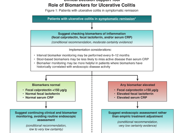
S Travis et al. NEJM 2023; 388: 1191-1200. Vedolizumab for the Treatment of Chronic Pouchitis

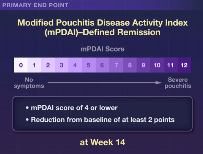
Methods: This was a a phase 4, double-blind, randomized trial (n=102 adults,EARNEST trial) to evaluate vedolizumab in adult patients in whom chronic pouchitis had developed after undergoing IPAA for ulcerative colitis.All patients received 4 weeks of ciprofloxacin and the treatment group received standard vedolizumab dosing. The primary end point was modified Pouchitis Disease Activity Index (mPDAI)–defined remission (an mPDAI score of ≤4 and a reduction from baseline of ≥2 points in the mPDAI total score; scores range from 0 to 12, with higher scores indicating more severe pouchitis) at week 14. The mPDAI is based on clinical symptoms and endoscopic findings.

Key findings:

  • The incidence of mPDAI-defined remission at week 14 was 31% (16 of 51 patients) with vedolizumab and 10% (5 of 51 patients) with placebo
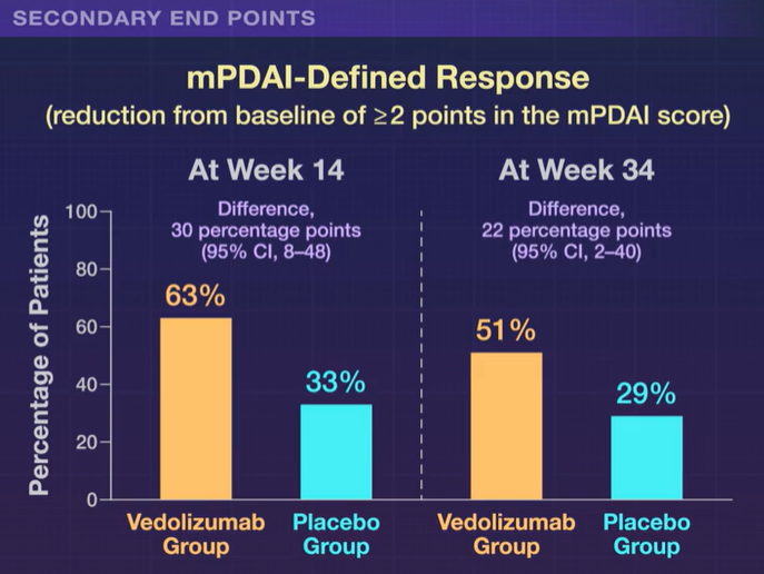
  • Differences in favor of vedolizumab over placebo were also seen with respect to mPDAI-defined remission at week 34 (difference, 17 percentage points, 35% vs 18%)

My take: Vedolizumab is an effective treatment for chronic pouchitis.

This figure is from NEJM Twitter Feed

Related blog posts:

Risk of Recurrent Surgery in Pediatric Crohn’s Patients

MD Edberg et al. Crohn’s and Colitis 360, 2023; https://doi.org/10.1093/crocol/otad003 Open Access! Trends in Surgical Recurrence Among Pediatric Crohn’s Disease Patients Using Administrative Claims Data

Methods: This study analyzed postresection pediatric (≤18 years) CD patients (n=434) identified in the 2007–2018 IQVIA Legacy PharMetrics administrative claims database

Key findings:

  • Risk of surgical recurrence was 3.5%, 4.6%, and 5.3% at 1, 3, and 5 years, respectively
  • Postoperatively, patients were most commonly prescribed an immune modulator (33%), anti-tumor necrosis factor agent (32%), or antibiotic (27%)
  • 24% underwent colonoscopy 6–15 months postoperatively

My take: Current recommendations include postoperative endoscopic surveillance 6-12 months after surgery. Would the surgical recurrence rate have been lower if there had been higher postoperatively endoscopic evaluation?

Related blog posts:

Sendero-Esperanza to Hugh Norris Trail, Saguaro National Park (Tucson, AZ)

Isolated Ileitis in Children

A Alper et al. JPGN 2023; 76: 338-342. Isolated Terminal Ileitis in Children

This single center retrospective study reviewed 640 colonoscopies in symptomatic children.

Key findings:

  • Thirty-three children had isolated histologically-defined terminal ileitis. Seventeen children were diagnosed with CD and 18 children had idiopathic terminal ileitis (3 lost to followup)
  • Children with CD had higher prevalence of abnormal C-reactive protein levels, severe inflammation, and radiological evidence of bowel wall thickening compared with children with idiopathic ileitis.
  • Two children with idiopathic ileitis were later diagnosed with CD; the remaining 13 did not develop CD over a follow-up period of 83 months.
  • From the data presented, it appeared that the center had a low rate of ileal intubation (316 colonoscopies were excluded for this reason)
  • 75% of those with histologic ileitis had normal endoscopic appearance

When our group looked at colonoscopies (n=374) in our outpatient endoscopy center, we identified isolated ileitis in 10% (6% grossly abnormal, 4% with only histologically abnormal) (related blog post: Our Study: Provider Level Variability in Colonoscopy Yield). Higher rates of ileal intubation (90% in our study) should be considered a quality metric given that 5-10% of children may have disease isolated in ileum.

My take: This study provides reassurance that most children with histologic ileitis will not progress to CD if the ileum is visually-normal (in the absence of abnormal blood tests and/or imaging).

Related blog posts:

Chattahoochee River, Sandy Springs, GA

How Insurance Companies Save Millions in Denying Care

3/23/23 Propublica: How Cigna Saves Millions by Having Its Doctors Reject Claims Without Reading Them

A few excerpts:

  • Cigna, one of the country’s largest insurers…has built a system that allows its doctors to instantly reject a claim on medical grounds without opening the patient file, leaving people with unexpected bills…Over a period of two months last year, Cigna doctors denied over 300,000 requests for payments using this method, spending an average of 1.2 seconds on each case, the documents show. 
  • Before health insurers reject claims for medical reasons, company doctors must review them, according to insurance laws and regulations in many states…This process helps avoid unfair denials…Cigna adopted its review system more than a decade ago, but insurance executives say similar systems have existed in various forms throughout the industry…At UnitedHealthcare….built a similar system to let its doctors quickly deny claims in bulk.
  • Cigna eventually designated the list “PXDX” — corporate shorthand for procedure-to-diagnosis. The list saved money in two ways. It allowed Cigna to begin turning down claims that it had once paid. And it made it cheaper to turn down claims, because the company’s doctors never had to open a file or conduct any in-depth review. They simply denied the claims in bulk with an electronic signature.
  • Cigna knows that many patients will pay such bills rather than deal with the hassle of appealing a rejection [for lower cost denials]…In one corporate document, Cigna estimated that only 5% of people would appeal a denial resulting from a PXDX review.

My take: It is a hassle to appeal denials. It is not surprising to me to hear about this reporting; it confirms the fact that insurance companies are focused primarily on cost and not patient care.

Related blog posts:

Ocotillo Cactus on the Sendero -Hugh Norris Trail, Saguaro National Park (Tucson, Az)

Consolidation and Competition in Health Care

JS King. NEJM 388: 1057-60. On Consolidation and Competition — The Trials and Triumphs of Health Care Antitrust Law

Key points:

  • “Historically, the United States has relied nearly entirely on market competition to control prices and promote quality in health care. Yet health care markets haven’t been healthy for some time. Over the past 30 years, health care consolidation has gone largely unchecked by federal and state antitrust enforcers, which has resulted in higher prices, stagnant quality of care, and limited access to care for patients.”
  • Consolidation has been both horizontal (eg. two competitors merge), vertical (eg. hospital acquiring physician groups. or insurance acquiring pharmacy benefit manager) or cross-market (eg. merger of hospital in two separate regions)
  • “Private-equity firms have recently invested heavily in health care providers, purchasing hospitals, emergency services and staffing companies, and specialist-physician groups, such as anesthesiology groups…Studies have found that acquisitions by private-equity firms have led to consolidation and increases in hospital charges and net income.”
  • “Mergers are often justified with promises of improved quality or patient access, evidence supporting these claims is lacking.”
  • “Antitrust law aims to protect consumers and competitive markets from anticompetitive practices and the harms described above. Three federal laws — the Clayton Act, the Sherman Act, and the Federal Trade Commission Act — along with legislation in nearly all states form the foundation of antitrust law.”
  • “Despite these enforcement options, the U.S. health care industry is the most consolidated it’s ever been… In the 1990s and early 2000s, the FTC lost six consecutive horizontal hospital-merger cases…[subsequently] federal agencies didn’t challenge another hospital merger for nearly a decade.”

My take: Consolidation is happening in all components of health care, including hospitals, insurance companies, pharmaceutical companies and physician groups. This leads to higher costs, fewer choices and possibly staff shortages. At the same time, each segment of health care is incentivized to consolidate, in part for financial gain and in part to negotiate with other consolidated segments.

Related blog posts:

AGA Practice Update: Acute Hepatic Porphyrias

B Wang et al. Gastroenterol 2023; 164: 484-491. Open Access! AGA Clinical Practice Update on Diagnosis and Management of Acute Hepatic Porphyrias: Expert Review

Overall, acute hepatic porphyrias (AHP) are rare disorders. “Acute intermittent porphyria is the most common type of AHP, with an estimated prevalence of patients with symptoms of
approximately 1 in 100,000. The major clinical presentation involves attacks of severe pain, usually abdominal and generalized, without peritoneal signs or abnormalities on cross-sectional imaging…The screening tests of choice include random urine porphobilinogen and d-aminolevulinic acid corrected to creatinine.”

“All patients with elevations in urinary porphobilinogen and/or d-aminolevulinic acid should initially be presumed to have AHP. The cornerstones of management include discontinuation of porphyrinogenic drugs and chemicals, administration of oral or intravenous dextrose and
intravenous hemin, and use of analgesics and antiemetics. Diagnosis of AHP type can be confirmed after initial treatment by genetic testing for pathogenic variants in HMBS, CPOX, PPOX, and ALAD genes.”

Some of the best practice advice:

  • Women aged 15–50 years with unexplained, recurrent severe abdominal pain without a clear etiology after an initial workup should be considered for screening for an AHP.
  • Initial diagnosis of AHP should be made by biochemical testing measuring d-aminolevulinic acid, porphobilinogen, and creatinine on a random urine sample.
  • Genetic testing should be used to confirm the diagnosis of AHP in patients with positive
    biochemical testing.
  • Acute attacks of AHP that are severe enough to require hospital admission should
    be treated with intravenous hemin, given daily, preferably into a high-flow central vein

Related blog posts:

Genetic Testing to Figure Out Drug Reactions

P Nicoletti et al. Gastroenterol 2023; 164: 454-466. Open Access! Identification of Reduced ERAP2 Expression and a Novel HLA Allele as Components of a Risk Score for Susceptibility to Liver Injury Due to Amoxicillin-Clavulanate

Key findings:

  • Transcriptome-wide association study revealed a significant association of AC-DILI risk with reduced liver expression of ERAP2 (P = 3.7 × 10–7), coding for an aminopeptidase involved in antigen presentation
  • “We also identified HLA-B∗15:18 as a novel AC-DILI risk factor in both discovery (OR, 4.19) and validation (OR, 7.78) cohorts”
  • A genetic risk score incorporating rs1363907, rs2476601, HLA-B∗15:18, HLA-A∗02:01, and HLA-DRB1∗15:01, was highly predictive of AC-DILI risk when cases were analyzed against both general population and non–AC-DILI control cohorts.
  • This genetic risk score does not apply to amoxicillin. “Clavulanate has long been considered the main component causing Amoxicillin-Clavulanate-DILI (LiverTox https://www.ncbi.nlm.nih.gov/books/NBK548517/).”
  • While the genetic risk score has high specificity, it is not highly sensitive. “Our multimarker model based on the 5 risk alleles predicted approximately 13% of the total risk for AC-DILI, which is approximately one-third of the total DILI risk that has previously been attributed to common genetic variants (approximately 40%)”

My take: In cases of suspected AC-DILI, identifying an abnormal genetic risk could help confirm the diagnosis. However, due to low sensitivity it is not likely to gain widespread clinical use.

Graphical Abstract:

Related blog posts:

Guideline: Biomarker Use for Ulcerative Colitis

S Singh et al. Gastroenterol 2023; 164: 344-372. Open Access! AGA Clinical Practice Guideline on the Role of Biomarkers for the Management of Ulcerative Colitis Clinical Decision support tool (pg 373-374), Spotlight (pg 375).

The full access links to the article and related articles provide extensive information and rationale for AGA’s biomarker recommendations in ulcerative colitis (UC). For me, the recommendations highlight the important role of biomarkers (especially fecal calprotectin (FC)) when things are going very well or very poorly. Key points:

  • In asymptomatic patients with normal biomarkers (FC <150, normal lactoferrin, normal CRP), the recommendations suggest continued monitoring without endoscopic assessment.
  • In patients with moderate-to-severe symptoms and with elevated biomarkers, the authors, likewise, advocate for treatment adjustment without endoscopic assessment.
  • For asymptomatic patients with elevated biomarkers and symptomatic patients with normal biomarkers, the authors recommend endoscopic assessment.
From Spotlight Material

My take: By combining biomarkers with symptoms, this improves utility of more invasive testing.

Related blog posts:

Kudos on Bravo Probe Complication Report

A Andreoli et al. JPGN Report 2023; DOI: 10.1097/PG9.0000000000000299 Open Access! Foreign Body in the Bronchus Intermedius: Inadvertent Deployment of a Bravo Wireless pH Probe in the Airway

This case report describes placement of a Bravo pH capsule into the bronchus rather than in the esophagus. The complication was quickly recognized and managed by having ENT remove the capsule during the same anesthetic. It is noted that the esophageal mucosal findings included “longitudinal furrows, edema, and mucosal friability.” Biopsies “demonstrated esophagitis consistent with GER.”

Key point:

  • The authors note that this has led to a change in their practice: “before suction attachment, the endoscope is reintroduced into the hypopharynx to ensure the delivery system enters the cricopharyngeus. This ensures esophageal probe placement before deployment.”

My take: It has been my practice since the Bravo catheter’s inception to assure the deployment catheter is in the esophagus before releasing the Bravo capsule. However, I am impressed with this report. It is very helpful for practitioners to relay difficult experiences to help others avoid complications. This case report should be a reminder also to carefully consider whether a pH study is needed in those with erosive esophagitis.

Related blog posts:

Related article: Open Access! Hochman JA, Favaloro-Sabatier J. Tolerance and reliability of wireless pH monitoring in children. J Pediatr Gastroenterol Nutr 2005; 41:411–415.

Changing Approach to Iron Deficiency Anemia in Pediatric IBD

Previously, there have been numerous posts on this blog discussing iron deficiency anemia in pediatric IBD, including an algorithm by CHOP in 2019 (CHOP QI: Anemia in IBD Pathway) and a NASPGHAN position paper in 2020 (Anemia in IBD -NASPGHAN Position Paper). A recent study from Nationwide Children’s highlights ongoing changes in the approach to this common problem.

J Smith et al. JPGN 2023; 76: 313-318. Diagnosis and Treatment of Iron Deficiency and Anemia in Youth With Inflammatory Bowel Disease

This study focused on a quality improvement effort to improve iron deficiency screening in newly-diagnosed patients with IBD. The QI project increased screening from a baseline of 20% to more than 90%. Importantly, this article details a useful algorithm (Figure 4). Key components:

  1. Screen with Ferritin, Iron and TIBC. If Ferritin is less than 30 or iron saturation is less than 20%, it recommends weight-based oral treatment.
  2. If less than 35 kg, options include 3 mg/kg/day (elemental) of ferrous sulfate or Novaferrum. If more than 35 kg, then it recommends ferrous sulfate (325 mg daily=65 mg elemental), ferrous gluconate (325 mg tab bid=36 mg elemental BID), or Novaferrum Ferrex capsule (150 mg daily =150 mg elemental).
  3. Anemia & iron indices are followed every 2-3 months (until improved) and if not resolved, options include either intravenous treatment and/or hematology involvement. For patients less than 50 kg, the authors utilize ferric carboxymaltose (FCM) 15 mg/kg/dose and for those more than 50 kg, FCM at 750 mg dosing.

For IV iron, the authors prefer FCM, which is FDA approved in children 1 yr of age and older, over iron sucrose or iron dextran as the number of infusions needed to replete iron stores is significantly reduced.  FCM is a relatively costly IV iron formulation, but can be given over 15 minutes; however, due to fewer infusions, FCM is likely cost-effective.

In the discussion, the authors caution against relying on laboratory reference values for ferritin and iron saturation which often set lower normative values (eg. Ferritin of 7 and iron saturation of 15%).

My take: This QI project provides a good strategy for dealing with iron deficiency anemia in the pediatric population.

Nationwide Children’s Algorithm