“Regeneron Pharmaceuticals, Inc. (NASDAQ: REGN) and Sanofi today announced that the U.S. Food and Drug Administration (FDA) has approved Dupixent® (dupilumab) for the treatment of pediatric patients aged 1 to 11 years, weighing at least 15 kg, with eosinophilic esophagitis (EoE).”
Disclaimer: This blog, gutsandgrowth, assumes no responsibility for any use or operation of any method, product, instruction, concept or idea contained in the material herein or for any injury or damage to persons or property (whether products liability, negligence or otherwise) resulting from such use or operation. These blog posts are for educational purposes only. Specific dosing of medications (along with potential adverse effects) should be confirmed by prescribing physician. Because of rapid advances in the medical sciences, the gutsandgrowth blog cautions that independent verification should be made of diagnosis and drug dosages. The reader is solely responsible for the conduct of any suggested test or procedure. This content is not a substitute for medical advice, diagnosis or treatment provided by a qualified healthcare provider. Always seek the advice of your physician or other qualified health provider with any questions you may have regarding a condition.
This research utilized 40 studies which met the eligibility criteria, including over 288 million participants and 147,668 patients with EoE from 15 countries across the five continents.
Key findings:
The global pooled incidence and prevalence of EoE were 5.31 cases per 100,000 inhabitant-years and 40.04 cases per 100,000 inhabitant-years, respectively.
The pooled prevalence and incidence of EoE were higher in high-income countries, males, and North America.
The pooled prevalence and incidence of EoE have increased from 1976 to 2022.
Time trends of incidence (A) and prevalence (B) of EoE, 1976 to 2022. Pooled estimates, cases per 100,000 inhabitant-years.
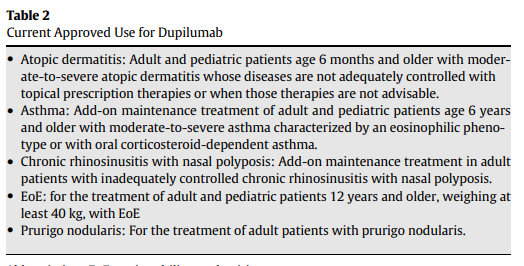
“This review summarizes the data leading to FDA approval for dupilumab and provides a practical approach for clinical use of dupilumab.” Dupilumab, a humanized monoclonal antibody that blocks interleukin (IL)-4 receptor alpha, is currently the only FDA-approved medication for EoE. It is noted that in the trials leading to FDA approval, all patients were PPI refractory and ~70% had received topical steroids (with about half either intolerant or nonresponsive).
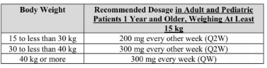
Dosing: 300 mg weekly injection with a single-dose prefilled autoinjector pen or a syringe with a needle shield. It is recommended that refrigerated medicine is brought to room temperature for at least 45 minutes prior to injection. It “can remain unrefrigerated up to 14 days.”
In Figure 1, the articles details positioning of use of dupilumab in EoE management algorithm:
New diagnosis, patient preference
Additional atopic condition with approved dupilumab use (strong indication)
Lack of response to current treatment (diet, PPI, swallowed steroids) or adverse effects from current treatment (strong indications)
“It is reasonable to repeat endoscopy with biopsy 24 weeks after initiation of dupilumab in many patients…However, endoscopy may be completer earlier” in selected patients.
At least 5 other biologics are in phase 2 or phase 3 studies (listed in Table 1).
My take: EoE is increasing in prevalence and new therapies (often expensive) are emerging.
Also, there is a fairly good patient education 7-page pamphlet from the makers of Dupixent encouraging patients with symptoms suggestive of EoE to speak with their physicians.
Disclaimer: This blog, gutsandgrowth, assumes no responsibility for any use or operation of any method, product, instruction, concept or idea contained in the material herein or for any injury or damage to persons or property (whether products liability, negligence or otherwise) resulting from such use or operation. These blog posts are for educational purposes only. Specific dosing of medications (along with potential adverse effects) should be confirmed by prescribing physician. Because of rapid advances in the medical sciences, the gutsandgrowth blog cautions that independent verification should be made of diagnosis and drug dosages. The reader is solely responsible for the conduct of any suggested test or procedure. This content is not a substitute for medical advice, diagnosis or treatment provided by a qualified healthcare provider. Always seek the advice of your physician or other qualified health provider with any questions you may have regarding a condition.
NASPGHAN has developed a useful ~6 minute video for families reviewing the treatments (diet and medications) for eosinophilic esophagitis. When I visited the site, it had not garnered much traction yet (very few views). I would recommend this video to families:
This is a terrific review of the dysphagia and the multidisciplinary approach to management. Many pearls are in this article. For example, laryngo-tracheo-esophageal cleft (LTEC), “while rare, 1 in 10,000-20,000 live births, the incidence of LTEC is higher (7.6%-22%) in children with aerodigestive issues such as a chronic cough.” [As an aside, this should be repeated given the changing population of patients being seen.]
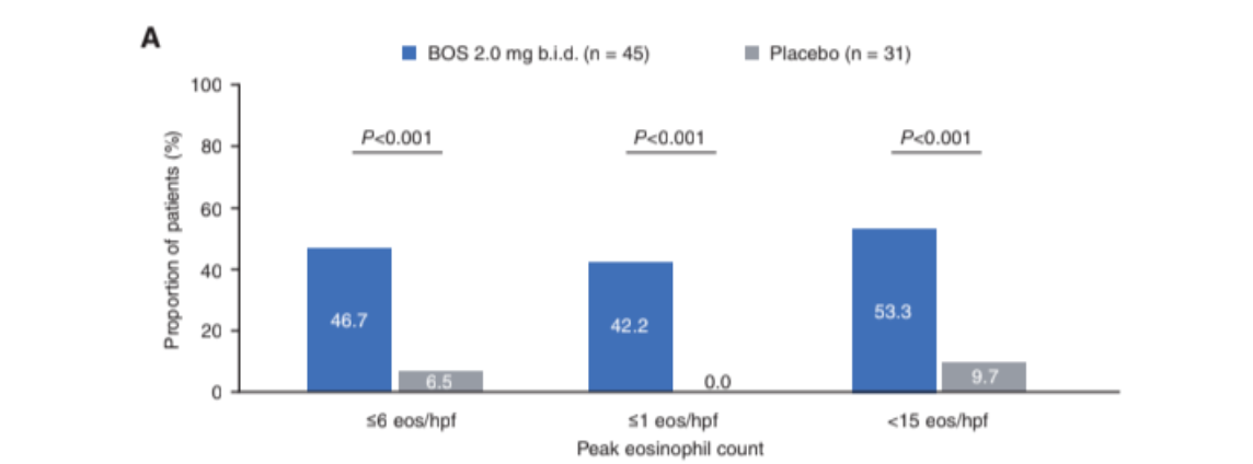
Key finding: Significantly more patients who received BOS (2mg BID) than placebo achieved histologic responses (≤6 eos/hpf: 46.7% vs 6.5%; ≤1 eos/hpf: 42.2% vs 0.0%; <15 eos/hpf: 53.3% vs 9.7%; P < 0.001)
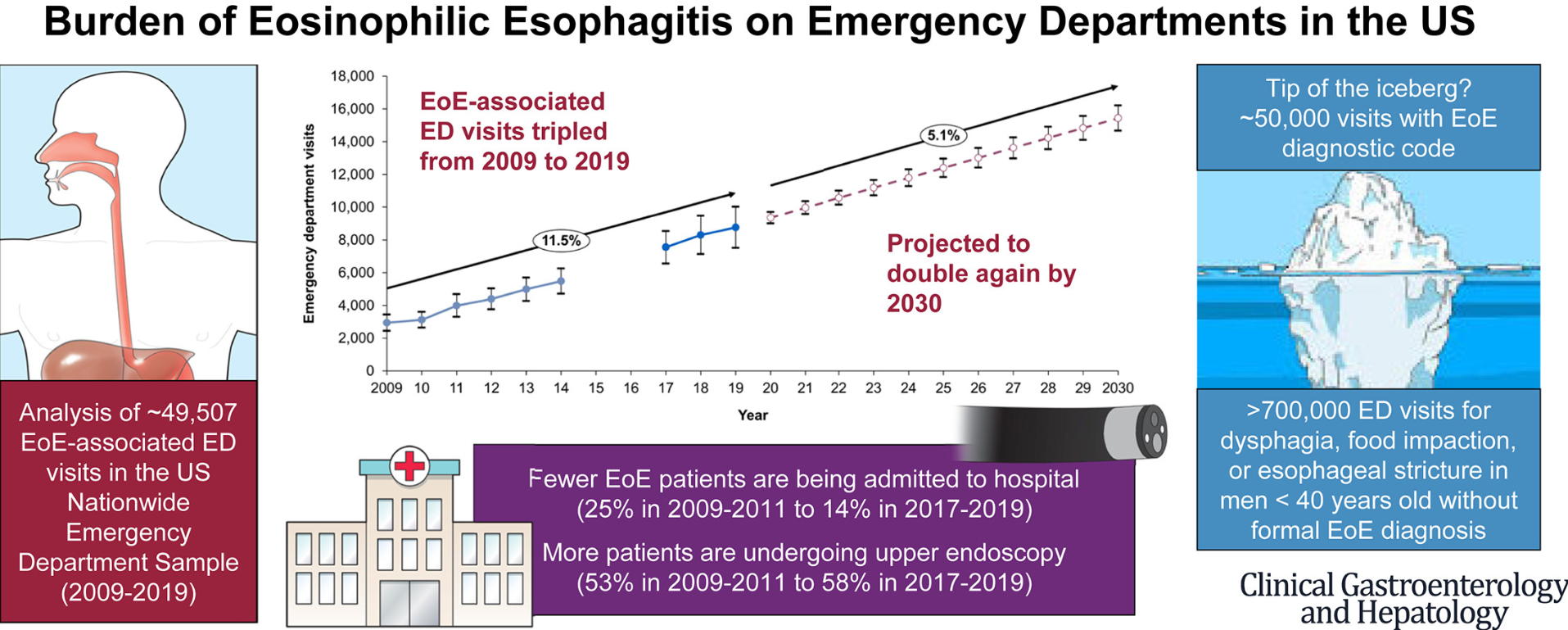
Methods: Data from the US Nationwide Emergency Department Sample (NEDS) were used to estimate weighted annual EoE-associated ED visits from 2009 to 2019. Autoregressive integrated moving average (ARIMA) models were used to project EoE-associated ED visits to 2030. NEDS is a large, publicly available, all-payer ED database in the United States, approximating a 20% stratified sample of US hospital-based EDs.
Key points:
There has been a near tripling of the frequency of EoE-associated ED visits over the course of the past decade which is correlated with an increasing prevalence of EoE. The annual volume of EoE-associated ED visits increased from 2934 in 2009 to 8765 in 2019, and is projected to reach 15,445 by 2030.
Without new interventions, this article projects further increases with doubling again by 2030 (using conservative estimates).
Increasingly EoE is being managed without admission, though average charges associated with ED visits for EoE have tripled since 2009. Total mean inflation-adjusted charges for an EoE-associated ED visit were $9025 US dollars in 2019.
Half of EoE patients presenting to the ED required an endoscopy and 40% required a foreign body/food impaction removal.
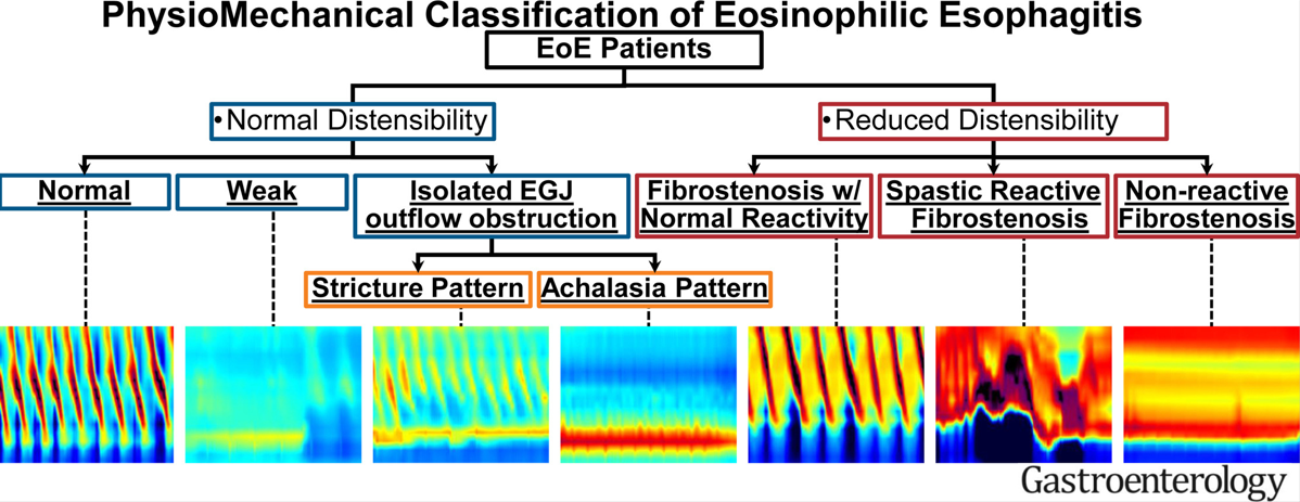
215 adults with EoE who completed FLIP during endoscopy were included in a cross-sectional study. FLIP helped separate the physiomechanical properties of esophageal function in this cohort. The criteria used to define the PhysioMechanical classification in EoE with a representative FLIP panometry image for each classification. Normal compliance was defined as a DP >17 mm and body compliance >450 mm3/mm Hg; reduced compliance (fibrostenosis) was defined by DP ≤17 mm or compliance ≤450 mm3/mm Hg. Normal EGJ opening was defined as a maximum EGJ diameter ≥16 mm; reduced as maximum EGJ diameter <16 mm. ∗Spastic-reactive contractile response (SRCR) with normal body distensibility and normal EGJ opening was assigned as “achalasia pattern” (n = 1 in this cohort).
Key findings:
FLIP was normal in 50 (23%), weak pattern in 7 (3%), IsoEGJOO stricture pattern in 27 (13%), IsoEGJOO achalasia pattern in 26 (12%), Fibrostenosis with normal reactivity in 61 (28%), spastic reactive fibrostenosis with normal reactivity in 30 (14%), and noreactive fibrostenosis in 14 (7%)
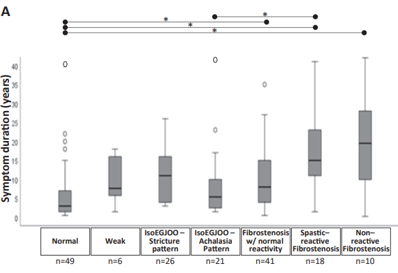
My take: FLIP testing helps define the mechanism of esophageal dysfunction in patients with EoE. Longer duration of symptoms was associated with more severe esophageal dysfunction.
“That’s all well and good in practice… but how does it work in theory?” I saw this quote many years ago when I was visiting the University of Chicago.
This quote came to mind as I was reading this article which showed relatively little change in the efficacy between more and less stringent elimination diets for eosinophilic esophagitis. This meta-analysis included 915 children and 847 adults and assessed the efficacy rates of 4 major dietary treatment regimens in eosinophilic esophagitis: 6-food (SFED), 4-food (FFED), 1-food (OFED), and a targeted elimination diet (TED).
Key findings:
The overall rate of histologic remission was 53.8% and in the individual dietary groups was 61.3% for SFED, 49.4% for FFED, 51.4% for OFED, and 45.7% for TED.
The overall rate of clinical response was 80.8%, with response rates of 92.8% for SFED, 74.1% for FFED, 87.1% for OFED, and 69.0% for TED.
Percentage of food antigen triggers identified via endoscopic and clinical evaluation after food re-introduction.
My take: It is clear to me that more restrictive diets can yield better response rates; however, in clinical practice they are difficult to maintain and this study shows that the improvement with more food restrictions may be quite limited.
Another reference on eosinophilic esophagitis: CJ Ketchem et al. Clin Gastroenterol Hepatol. 2023 Aug;21(9):2252-2259. Open Access! Higher Body Mass Index Is Associated With Decreased Treatment Response to Topical Steroids in Eosinophilic Esophagitis. Key finding: Histologic response (n=296) to topic steroids was higher for those who were nonobese compared with obese at fewer than 15 eosinophils per high-power field (61% vs 47%; P = .049); in addition, nonobese patients had significantly greater endoscopic and symptomatic responses.
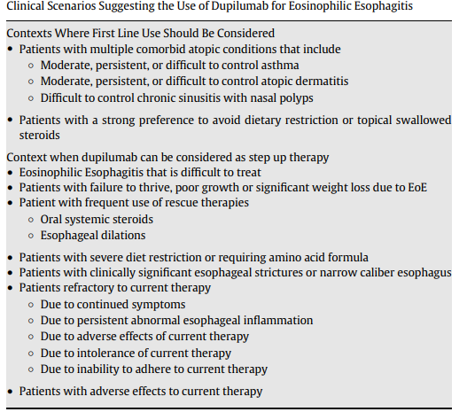
This article summarizes updated recommendations for eosinophilic esophagitis from the Joint Task Force for the American Academy of Allergy Asthma Immunology and American College of Allergy Asthma Immunology and the American Gastroenterology Association (JTF-AGA). It offers a good number of recommendations regarding when using dupilumab should be considered.
Other Key Points:
“Dupilumab can be considered as first-line therapy in patients presenting with severe EoE”and in patients with multiple atopic diseases.
In addition, it recommends “performing a repeat EGD, along with obtaining biopsies, 5 to 6 months after either starting dupilumab therapy or whenever adjusting the dupilumab dose.” In some cases, like stricture dilatation, the authors indicate that earlier EGD may be appropriate.
The advantages/disadvantages of current treatment options are summarized in Table 3. For dupiliumab, the disadvantages include its high price of dupilumab and weekly injections. Conjunctivitis has been an adverse effect identified in its usage with other indications.
My take: Dupilumab is a major advance for patients with EoE. Due to the need for weekly injections and its costs, it is likely a 2nd line agent for most kids with EoE.
Key findings: HEV Ag was specifically taken up by renal cells and was disposed into urine, during which the level of Ag was concentrated >10‐fold, resulting in the higher diagnosing sensitivity of urine Ag than serum Ag. Moreover, Ag in urine appeared 6 days earlier, lasted longer than viremia and antigenemia, and showed good concordance with fecal RNA in a rabbit model.
Background: Autoimmune enteropathy (AIE) is a severe form of enteropathy characterized by chronic diarrhea refractory to any exclusion diet and associated with autoimmunity…In a recent cohort of 40 AIE patients, anti-enterocyte antibodies were reported in only 14% (4/28) of the cases, likely caused by the high frequency of patients with primary hypogammaglobulinemia…30%–50% of adult AIE can display anti-transglutaminase antibodies. The common histopathologic presentation of AIE includes intestinal villous atrophy with variable lymphocytic infiltration and various features of follicular lymphoid hyperplasia, cryptitis, graft-versus-host disease-like lesions, and loss of Paneth and goblet cells.
Key findings: Pathogenic variants were identified in 20/48 adult patients (41.6%); most common variants: CTLA4, LRBA, STAT3, and STAT1; 12/20; 60% of those with variants. Thus, specific therapeutics were available for more than half of the patients who received a molecular diagnosis
Representative endoscopic aspects in patients with CTLA4 variants and AIE.
Key finding: In this prospective pediatric cohort (n=59) with EoE, distensibility index (DI) <4.5 mm2/mmHg predicted grade 2 rings on endoscopy. Lower DI was associated with increased risk of food impaction but did not correlate with eosinophilic count. DI was “superior to diameter in assessing fibrostenotic severity.”
Background: “Chronic inflammation can lead to tissue remodeling in the esophagus with fibrosis in the lamina propria that is partially responsible for symptoms and complications of EoE.3,4 At times, a firmness to the esophagus can be appreciated with a noticeable force required to obtain biopsies from EoE. This sensation has been described as the “tug” or “pull” sign.5,6“
Methods: in this prospective study with 159 patients (128 pediatric, 31 adult), the authors devised a digital force gauge to measure the force required to take biopsy specimens. The study included 88 patients with EoE and 71 controls.
Key finding:
EoE patients showed an increase in the mean force required to obtain biopsies: 14.9 Newton (N) compared to 11.6 in control group
Peak force was greater in EoE patients: 20.4 N compared to 15 N in control group.
The pediatric subgroup had higher peak force in EoE patients: 22.4 N compered to 16.1 N for control group
My take: I had not heard of the term “Tug” sign for EoE, though it is something that is intuitive for GI providers who care for these patients. This study quantifies this problem.
“When you can measure what you are speaking about and express it in numbers, you know something about it; but when you cannot express it in numbers, your knowledge is of a meagre and unsatisfactory kind” –Lord Kelvin 1883