New Trend: Oral Medicines Replacing Injections

  • R Bissonnette et al. NEJM 2025; 393: 1784-1795. Oral Icotrokinra for Plaque Psoriasis
  • RS Stern. NEJM 2025; 303: 1854-1855. Oral Psoriasis Therapy — For Whom and at What Cost and Risk?
  • S Wharton et al. NEJM 2025; 303: 1796-1806. Orforglipron, an Oral Small-Molecule GLP-1 Receptor Agonist for Obesity Treatment

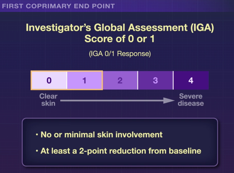
In the ICONIC-LEAD study (Bissonnette et al), 684 adolescents and adults participated in a DBPC trial with an oral peptide, icotrokinra, which binds the IL-23 receptor. This medication is of interest as there are ongoing trials with it for inflammatory bowel disease. Other injectable medications targeting IL-23 are already approved for IBD.

Key Findings:

The associated editorial notes that this new therapy is likely to cost ~$70,000 per year. The cost of psoriasis care has increased more than 2000% since 1997. “Because of these high prices, rebates and discounts to pharmacy benefit managers that often guide formulary preferences are likely to govern clinician’s selection of immune-based oral and parenteral therapies for psoriasis.”

In the ATTAIN-1 Trial (Wharton et al), the authors share the results of an oral GLP-1 Receptor Agonist, Orforglipron, monotherapy for obesity.

Key findings:

My take: There are similar injectable alternatives to each of these medications for psoriasis, obesity and diabetes. The availability of oral medications could reduce one barrier to treatment. Cost barriers may preclude their use in many patients when they become available. In addition, long-term outcome data are still needed.

Related blog posts:

Paradoxical Immune Mediated Disorders Associated with TNF Inhibitors

Previously, it has been noted that several immune mediated problems paradoxically can be triggered by the use of TNF inhibitors (eg. infliximab, adalimumab) even though these medications are often used to treat these problems (see posts below).

Using 2 nationwide cohorts (Danish & French), Ward et al (Clin Gastroenterol Hepatol 2024; 22: 135-143. Open Access! Tumor Necrosis Factor Inhibitors in Inflammatory Bowel Disease and Risk of Immune Mediated Inflammatory Diseases) report on the associated risk of developing a number of additional immune mediated inflammatory diseases (IMIDs) after treatment with anti-TNF agents for inflammatory bowel disease (IBD). The Danish and French cohorts comprised 18,258 and 88,786 subjects with IBD. Key findings:

  • Anti-TNF therapy was associated with an increased risk of rheumatoid arthritis, psoriasis, and hidradenitis suppurativa in both the Danish (HR, 1.66) and the French cohort (HR, 1.78), with a pooled HR of 1.76
  • The absolute risk of IMIDs in the Danish cohort was 5.3/1000 person years compared to 3.8/1000 PY those who had not received anti-TNFs; in the French cohort, the rate in anti-TNF exposed was 5.4/1000 PY compared to 3.0/1000 PY in the unexposed group.
  • Anti-TNF therapy was also associated with an increased risk of the IMIDs when compared with azathioprine (pooled HR, 2.94).

The results suggest that anti-TNFs paradoxically increase the risk of IMIDs; however, individuals receiving anti-TNFs are likely at higher risk for these disorders and this could be difficult to control for in a retrospective study.

My take: While anti-TNF agents have been a tremendous advance in the treatment of IBD, in a small number of individuals, these agents appear to trigger a paradoxical reaction.

Related blog posts:

Chattahoochee River at Island Ford. Sandy Springs

Ustekinumab Efficacy in Crohn’s Disease With Concurrent Autoimmune Skin Disease

E Fradkov et al. Inflamm Bowel Dis 2022; 28: 895-904. Efficacy of Ustekinumab in Crohn’s Disease With and Without Concurrent Autoimmune Skin Disease

This retrospective study reviewed 395 CD patients received ustekinumab therapy (79 CD-ASD (autoimmune skin disease), 316 CD-none). ASD included atopic dermatitis, eczema, psoriasis/psoriaform dermatitis and alopecia. The skin disease group also included those with cutaneous manifestations of Crohn’s disease: erythema nodosum, pyoderma gangrenosum, pyostomatitis vegetans, Sweet’s syndrome, granulomatous vasculitis, and leukocytoclastic vasculitis. 55 of the 79 with CD-ASD had psoriatic disease, 20 had eczema, 11 had erythema nodosum, 8 had pyoderma gangrenosum.

Key findings:

  • Ustekinumab had greater efficacy in CD-ASD when evaluated by fecal calprotectin (P = .0337) and CRP (P = .078). For calprotectin, the values decreased by 61% after at least 5 months of therapy (394 to 164) in the CD-ASD group compared to 11% in the group without skin disease (365 to 265)
  • The CD-ASD group also showed better outcomes in Likert scores of endoscopy (P = .016), histopathology (P = .074), and imaging (P = .094). 

My take: Ustekinumab appears to be particularly effective in patients with concurrent skin disease.

Related posts:

Chattahoochee River near Morgan Falls

Psoriasis Due to Infliximab –Latest Data

Briefly noted: O Courbette et al. JPGN 2019; 69: 189-93. In a retrospective review, among 147 children with inflammatory bowel disease treated with infliximab (IFX) (123 CD, 24 UC), 20 patients (13.6%) developed psoriaform rashes.  14 of 20 were in remission when skin rashes (especially on scalp) occurred and rash developed at median of 355 days.  In this cohort, all were controlled by local steroids; no patients required IFX discontinuation.

Related blog posts:

Wahkeena Falls Trail, OR

IBD Shorts -September 2018

S Sridhar et al. Inflamm Bowel Dis 2018; 24: 2086-92.  This retrospective pediatric study with 409 patients examined dermatologic manifestations on anti-TNF therapy.  47 (11.4%) had dermatologic findings recorded including 33 with psoriasis, 28 with infections, and 10 with eczema (some had multiple skin findings). The majority were able to continue with current anti-TNF regmimen, including 60% of those with psoriasis.

Related blog posts:

NA Rozette et al. Inflamm Bowel Dis 2018; 24: 2007-14. This study with 50 subjects showed good safety of rapid versus standard infliximab infusions. One interesting aspect of their study which included a retrospective arm (standard infusion) and a prospective arm (rapid infusion) was a declining use of premedication, though even in their prospective group 60% received premedication including the combination of acetaminophen, benadryl, and methylprednisolone in 30%.  There were two patients in the rapid infusion with mild reactions who reverted to standard infusion rates.

CJ Moran, JL Kaplan, HS Winter. Inflamm Bowel Dis 2018; 24: 2048-52. This study with 199 subjects with active Crohn’s disease (CD) (21-86 years) –had their BioBank blood tested for 5 common CRP genetic variants.  Some specific variants,  rs2794520TT & rs1800947, were associated with lower CRP levels. This study helps explain why CRP is not a useful marker in some patients with CD.

Sunshine Meadows, Banff

Characteristics of Skin Lesions Associated with Anti-Tumor Necrosis Factor Therapy

As noted in previous blog posts (see below), anti-tumor necrosis factor (anti-TNF) therapy has been associated with skin problems.  The following study/abstract elaborate on this issue further and indicate that while ~30% of patients with IBD may develop skin reactions, only 28 of 917 (3%) patients required anti-TNF therapy to be discontinued due to skin reactions.

I Cleynen et al. Ann Intern Med. Published online December 2015 doi:10.7326/M15-0729  Characteristics of Skin Lesions Associated With Anti–Tumor Necrosis Factor Therapy in Patients With Inflammatory Bowel DiseaseA Cohort Study ONLINE FIRST

 Background: A subgroup of patients with inflammatory bowel disease (IBD) treated with anti–tumor necrosis factor (TNF) antibodies develop skin lesions, but the lesions and their clinical course are not well-characterized.

Objective: To describe patients treated with anti-TNF antibodies who did and did not develop skin lesions.

Design: Retrospective cohort.

Setting: Single IBD tertiary referral center.

Patients: 917 consecutive patients with IBD who initiated anti-TNF therapy.

Measurements: Skin lesions, patient demographic characteristics, treatments, clinical course, and serologic and genetic markers.

Results: During a median follow-up of 3.5 years (interquartile range [IQR], 0.5 to 7.4 years), skin lesions associated with the use of anti-TNF therapy developed in 264 of 917 (29%) patients (psoriasiform eczema, 30.6%; eczema, 23.5%; xerosis cutis, 10.6%; palmoplantar pustulosis, 5.3%; psoriasis, 3.8%; other, 26.1%). Lesions typically developed at flexural regions, genitalia, and the scalp, especially the psoriasiform lesions. Thirty-one percent of women and 26% of men developed lesions. Median cumulative doses (2864 mg/y [IQR, 2203 to 3819 mg/y] and 2927 mg/y [IQR, 2377 to 3667 mg/y]) and trough levels (4.2 µg/mL [IQR, 2.6 to 5.8 µg/mL] and 4.0 µg/mL [IQR, 1.6 to 5.9 µg/mL]) of infliximab were similar in patients with and without lesions. All but 28 patients (11%) were successfully managed without needing to stop therapy because of lesions.

Limitation: Retrospective nature and no matched control group of patients not receiving anti-TNF therapy.

Conclusion: Skin lesions occur frequently in association with anti-TNF therapy but rarely require discontinuation of therapy. Close surveillance and early referral to a dedicated dermatologist are recommended.

Related blog posts:

Immune-Mediated Reactions to Anti-TNFs and What to Do About Them

Statue

A recent review article (Feuerstein JD et al. Inflamm Bowel Dis 2015; 21: 1176-86) serves as a useful reference regarding immune-mediated reactions to anti-tumor necrosis factor (anti-TNF) medications used in inflammatory bowel disease (IBD).

Background:

  • All anti-TNF agents induce antidrug antibodies (ADAs).
  • With regard to infliximab (IFX) which has the most literature, it is well-recognized that combination therapy with an immunomodulator reduces the risk of antibodies to infliximab (ATIs).  For example, in the SONIC study, ATIs were noted in 0.9% of those with combination therapy compared with 14.6% of those receiving monotherapy with IFX.  With the UC-SUCCESS, the rates were 19% and 3% respectively.

Acute Infusion Reactions -Key points:

  • Acute infusion reactions (IRs) are more common in patients with ADAs.  IRs can be categorized as acute (w/in 24h) and chronic (2-14 d after infusion).
  • Acute IRs can be mild (dizziness, flushing, nausea, palpitation), moderate (chest pain, hypertension [SBP increase of more than 20], hypotension, fevers urticaria, mild dyspnea, chills, rash) or severe (severe hypertensions [SBP increase of more than 40] , severe hypotension, significant dyspnea with brochospasm, stridor, and rigors)
  • The authors provide a treatment algorithm (Figure 1) based on severity of acute IR.  All reactions are initially treated by stopping infusion, but many can be restarted at a low rate after administration of acetaminophen (mild & moderate), normal saline (mild & moderate), diphenhydramine (moderate), and possibly hydrocortisone (if needed in moderate cases).  While the algorithm suggests the possibility of restarting infusion reaction in severe cases without anaphylaxis, if this is considered, it may be worthwhile to attempt in a hospital setting.
  • Typically if infusions are restarted, the rates are 10 mL/hr x 15 minutes –>20 mL/hr x 15 minutes–> 40 mL/hr x 15 minutes –>80 mL/hr x 15 minutes –>100 mL/hr x 15 minutes–>125 mL/hr until completion.
  • Following an IR, the authors recommend checking for ATIs and for IFX level.
  • Prophylaxis for mild IRs includes the use of acetaminophen and antihistamines (2nd generation antihistamine daily for 5 days prior or first generation antihistamine an hour prior to infusion).  In addition, the infusion should be started at 10 mL/hr
  • Prophylaxis for moderate IRs includes the use of acetaminophen and antihistamines and steroids (prednisone 50 mg q12 hr x 3 doses prior or hydrocortisone 100 mg (or equivalent) 20 minutes prior to infusion).  In addition, the infusion should be started at 10 mL/hr
  • The authors recommend against premedication in those who have not had IRs. Use of premedication may cause a paradoxical increase in IRs due to symptoms induced by the antihistamine.

Autoimmune Complications:

  • Autoantibodies: anti-nuclear antibody (ANA), anti-double-stranded DNA antibody (anti-dsDNA), anti-cardiolipin antibody, antihistone antibody
  • Drug-Induced Lupus Erythematosus (DILE) -“the most frequently presenting symptoms, seen in 90% of cases, is symmetric arthralgias.”  Systemic involvement of the kidneys or central nervous system is rare. Treatment is cessation of the offending medication.
  • Vasculitis -likely due to the development of circulating immune complexes that deposits into smaller capillaries–>result in a type III hypersensitivity reaction.  The most common manifestation would be palpable purpura due to a leukocytoclastic vasculitis.
  • While autoimmune complications can be a class effect, many patients have been able to switch to a different anti-TNF.

Dermatologic Complications:

The authors review both anti-TNF induced psoriasis and eczema.  Treatment should be in conjunction with dermatology.  For psoriasis that involves >5% of body surface area, this could require changing to a different anti-TNF or a different drug class.  For severe cases, “anti-TNF therapy should be discontinued.”

Related blog posts:

Disclaimer: These blog posts are for educational purposes only. Specific dosing of medications/diets (along with potential adverse effects) should be confirmed by prescribing physician/nutritionist.  This content is not a substitute for medical advice, diagnosis or treatment provided by a qualified healthcare provider. Always seek the advice of your physician or other qualified health provider with any questions you may have regarding a condition.

Sunflowers

IBD Shorts -Skin, Adalimumab Kinetics

From the IMAgINE study with 192 pediatric patients (Sharma S et al. Inflamm Bowel Dis 2015; 21: 783-92), the authors determined levels for adalimumab, that at week 52, were associated with remission and response.  A cutoff level of 3.6 mcg/mL had a sensitivity of 32.7%  and specificity of 88.6% for predicting remission; the same cutoff had a sensitivity of 46.2% and specificity of 83% for predicting a response.  Overall, the authors noted dose proportionality with patients who received higher (or more frequent) doses with higher serum levels.

Related posts:

“Concomitant Use of Azathioprine/6-Mercaptopurine Decreases the Risk of Anti-TNF-Induced Skin Lesions” (Soh JS et al. Inflamm Bowel Dis 2015; 21: 832-9). Among a cohort of 500 Korean patients, the incidence of psoriaform and eczematiform lesions was 6.2%.  Concomitant use of a thiopurine was associated with a hazard ratio of 0.452 (lower risk) for developing these adverse skin reactions.

Related posts:

Four IBD Articles –Four Teaching Points

  1. Inflamm Bowel Dis 2014; 20: 1891-1901
  2. Inflamm Bowel Dis 2014; 20: 1996-2003
  3. Inflamm Bowel Dis 2014; 20: 2013-21.
  4. Inflamm Bowel Dis 2014; 20: 2056-66.

The first article is listed as a ‘basic science’ article.  However, it has direct relevance to the clinical problem of anti-TNF-induced psoriasiform skin lesions.  The article notes that this problem affects about 5% of patients treated with anti-TNF agents.  The authors found that IL-36γ and IL-17C are increased in anti-TNF-induced psoriasiform skin lesions of patients with Crohn’s disease (n=13 patients).  An important clinical point was that 7 of these patients with “severe anti-TNF induced skin lesions were successfully treated with the IL-12/IL-23 neutralizing antibody ustekinumab.”  This was superior to topical steroids or topical tacrolimus.

The second article is a proof-in-principle type article showing that proactive measurements of infliximab (IFX) levels may improve outcomes.  This retrospective observational study examined 48 patients who had proactive IFX levels. In 12 of 48, IFX dosing was escalated after the first proactive monitoring.  In addition, over the study period, 15% of patients had their dosing lowered. In those with proactive monitoring, the probability of remaining on IFX was >80%.  Those patients who achieved trough levels >5 mcg/mL had >90% probability of remaining on IFX therapy.  The authors hypothesize that better IFX levels may reduce anti-infliximab levels.

The third article examines carbohydrate intake in relation to the development of Crohn’s disease (CD) and ulcerative colitis (UC). The authors utilized the “EPIC” cohort (European Prospective Investigation into Cancer and Nutrition) (Public Health Nutr 2002; 5: 1113-24).  among 401,326 enrollees at recruitment, the dietary intakes of carbohydrates were measured using validated food frequency questionnaires. In this cohort, 110 developed CD and 244 developed UC during followup. Key finding: there was no significant risk for IBD based on total carbohydrate intake.  This study does not exclude the possibility that specific carbohydrates could have an etiological role.

The fourth article, a case-control study (2002-2011),  examines risk factors for endoscopy-associated perforation and perforation-associated complications (PAC) in patients with and without IBD. n=217,334 lower endoscopies (with 9518 in IBD patients).  Perforation rates: 18.91 per 10,000 and 2.50 per 10,000 for IBD and non-IBD endoscopy respectively.  The use os systemic corticosteroids at the time of endoscopy was associated with a 13 times greater risk for PAC.

Related blog posts:

Pattern of Skin Reactions to Anti-TNF Agents

A recent study indicated a high rate of skin reactions to anti-TNFα in their pediatric inflammatory bowel disease (IBD) population (Inflamm Bowel Dis 2014; 20: 1309-15).

In a two-year prospective study, 84 children with IBD (64 with Crohn’s) who were receiving infliximab infusions were screened for skin changes and had labwork (blood tests and stool calprotectiin).

Key findings:

  • 40 (47.6%) had chronic skin reactions and half of these were considered severe. However, when looking at the “severe” lesions shown in Figure 2, one might question the characterization.
  • Ear lobes and scalp were affected most frequently with psoriasis-like manifestations, followed by eyelids, perioral and pubic areas, trunk, and extremities.
  • Skin reactions were more common in those with a low degree of intestinal inflammation based on calprotectin levels: 133 mg/g in those with skin changes compared with 589 in those without.
  • Seven patients (8.3% of entire cohort) discontinued therapy due to skin reaction.
  • Most patients responded well to topical potent corticosteroids.

Take-home message: In this prospectively-followed cohort, there was a surprisingly high rate of skin reactions.  In patients receiving anti-TNFα therapy, it is a good idea to look closely at their ears and scalp.

Related blog post:

TNF Antagonists and Psoriasis | gutsandgrowth